. 2013 Apr;4(2):245-52.
doi: 10.1007/s13244-012-0205-9. Epub 2012 Dec 15.

An overview of thoracic actinomycosis: CT features

Affiliations

An overview of thoracic actinomycosis: CT features

Ji-Yeon Han et al. Insights Imaging. 2013 Apr.

Abstract

Background: Thoracic actinomycosis is an uncommon, chronic suppurative bacterial infection caused by actinomyces species, especially Actinomyces israelii.

Methods: It is usually seen in immunocompetent patients with respiratory disorders, poor oral hygiene, alcoholism and chronic debilitating diseases.

Results: We illustrate the radiological manifestations of thoracic actinomycoses in various involved areas in the thorax.

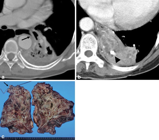
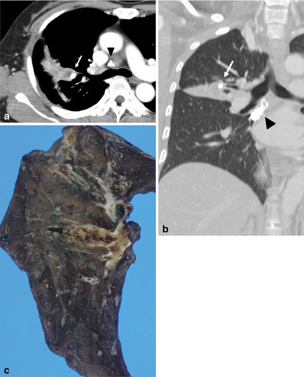
Conclusion: Thoracic actinomycosis can be radiologically divided into the parenchymal type, the airway type including bronchiectasis, the endobronchial form, and the mediastinum or chest wall involvement type.

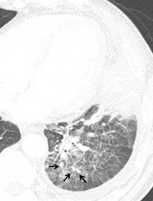
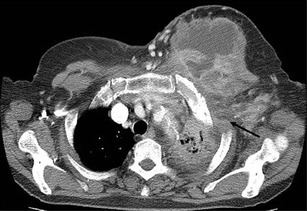
Teaching points: • Important risk factors for thoracic actinomycosis are underlying respiratory disorders such as emphysema and chronic bronchitis. • Different CT patterns can be distinguished in thoracic actinomycosis: parenchymal, bronchiectatic, endobronchial and extrapulmonary. • Typical CT findings in the parenchymal pattern are a central low density within the parenchymal consolidation and adjacent pleural thickening.


Figures

Fig. 1
Fig. 1
Typical parenchymal actinomycosis in a 58-year-old man with haemoptysis for a month. The CT scan in the mediastinal window setting shows a 3-cm mass-like consolidation with central, low-density peripheral rim enhancement in the right lower lobe and adjacent pleural thickening with subpleural fat infiltration (arrow)
Fig. 2
Fig. 2
Early stage parenchymal actinomycosis in a 44-year-old man who was a heavy alcohol drinker. The CT image displayed on lung window settings shows focal consolidation surrounded by ill-defined, peripheral pulmonary nodules (arrows), which suggests bronchogenic spread of the disease and interlobular septal thickening
Fig. 3
Fig. 3
Parenchymal actinomycosis with abscess formation in a 51-year-old man with cough and dyspnoea who had a history of alcoholism. Chest CT scan displayed on a mediastinal setting (a) and thin-section CT (b) show peripheral consolidation with multiple internal cavities, a central low-attenuation area and adjacent pleural thickening in the right upper lobe
Fig. 4
Fig. 4
Transfissural extension of parenchymal actinomycosis in a 43-year-old man with high fever for 10 days manifesting as chronic necrotising pneumonia. Chest CT scan in mediastinal window setting shows multifocal lobar consolidation containing a large low attenuation area in the right lower and middle lobe, which demonstrates extension across the fissure
Fig. 5
Fig. 5
Bronchiectatic form of actinomycosis in a 44-year-old man with underlying bronchiectasis. Chest CT scan displayed on the mediastinal (a) and lung (b) window setting shows bronchial dilatation with wall thickening and focal consolidation. Note the adjacent pleural thickening in the right lower lobe
Fig. 6
Fig. 6
Co-infection of bronchiectatic actinomycosis with Aspergillus in a 52-year-old man. Chest CT scan in lung window setting obtained with an axial scan (a) and a coronal reformatted image (b) shows focal bronchiectatic changes with intraluminal nodules in the right upper lobe. The presumptive diagnosis was aspergilloma within bronchiectasis. Low-power photomicrography of the surgical specimen from the right upper lobe (c) (original magnification, ×20; haematoxylin-eosin stain) demonstrates an Actinomyces colony (star) within the ectatic bronchus (arrows). Medium-power photomicrograph (d) (original magnification, ×400; Gomori methenamine-silver stain) shows a nodule within bronchiectasis composed of central aggregation of Aspergillus (arrow) and surrounding Actinomyces colonies (star)
Fig. 7
Fig. 7
Mixed parenchymal, bronchiectatic and mediastinal actinomycosis in a 69-year-old woman with haemoptysis. a Chest CT scan with a modified window setting shows lobar consolidation with cavitary change in the left lower lobe. Note the distended oesophagus with a beak-like appearance that extends toward the consolidation of the left lower lobe (arrow). This lesion was confirmed to be an oesophagobronchial fistula after lobectomy. b Chest CT scan with the mediastinal window setting at the level below (a) shows calcified brocholith (arrowhead) within the dilated tubular mucus retained bronchus. c Left lower lobectomy specimen showed cystic dilation of thickened bronchi, filled with greyish, fragile and granular material with a foul odour. A connection between the oesophagus and bronchus (oesophagobronchial fistula, arrow) was found where the steel rod is pointing (arrow)
Fig. 8
Fig. 8
Endobronchial actinomycosis with broncholithiasis in a 53-year-old woman. ab The axial and coronal chest CT scans in the lung window setting show a small broncholith (arrow) obstructing the lumen of the subsegmental bronchus of the anterior segment and distal subsegmental consolidation within the internal necrotic portion. Broncholiths were confirmed as granular colonies of Actinomyces. Calcified mediastinal lymph nodes are also noted, suggesting previous tuberculosis infection (arrowheads). c Photographs of the right upper lobectomy specimen show dilatation of central bronchi filled with yellowish-grey granular and fragile material
Fig. 9
Fig. 9
Actinomycosis involving the chest wall in a 75-year-old woman, manifesting as a palpable mass on the left chest wall. Axial CT image shows a heterogeneous mass with central low attenuation and peripheral enhancement on the left chest wall, contiguous with consolidation (arrow) in lung parenchyma, and pleural effusion
Fig. 10
Fig. 10
Actinomycosis with oesophagobronchial fistula in a 44-year-old woman who complained of a repetitive cough with food intake. a Barium oesophagography depicts the fistula tract (arrowheads) between the oesophagus and right bronchus intermedius. b Chest CT scan shows a segmental consolidation and bronchiectasis containing a broncholith (arrow) in the right lower lobe of the superior segment. Consolidation extends to the mediastinum and oesophagus

References

    1. Yildiz O, Doganay M. Actinomycoses and nocardia pulmonary infections. Curr Opin Pulm Med. 2006;12:228–234. doi: 10.1097/01.mcp.0000219273.57933.48. - DOI - PubMed
    1. Russo TA. Agents of actinomycosis. In: Mandell GL, Bennet JE, Dolin R, editors. Principles and practice of infectious disease. 6. Philadelphia: Churchill Livingstone; 2005. pp. 2924–2934.
    1. Mabeza GF, Macfarlane J. Pulmonary actinomycosis. Eur Respir J. 2003;21:545–551. doi: 10.1183/09031936.03.00089103. - DOI - PubMed
    1. Andreani A, Cavazza A, Marchioni A, Richeldi L, Paci M, Rossi G. Bronchopulmonary actinomycosis associated with hiatal hernia. Mayo Clin Proc. 2009;84(2):123–128. doi: 10.4065/84.2.123. - DOI - PMC - PubMed
    1. Bartlett AH, Rivera AL, Krishnamurthy R, Baker CJ. Thoracic actinomycosis in children: case report and review of the literature. Pediatr Infect Dis J. 2008;27(2):165–169. - PubMed

LinkOut - more resources