

Association of facet tropism with lumbar disc herniation
- PMID: 23242621
- PMCID: PMC3657057
- DOI: 10.1007/s00586-012-2612-5
Association of facet tropism with lumbar disc herniation
Abstract
Purpose: Facet tropism is defined as asymmetry between left and right facet joints and is postulated as a possible cause of disc herniation. In the present study, the authors used a 3-T MRI to investigate the association between facet tropism and lumbar disc herniation at a particular motion segment. They also examined whether the disc herniated towards the side of the more coronally oriented facet joint.
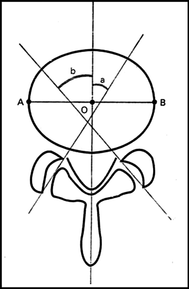
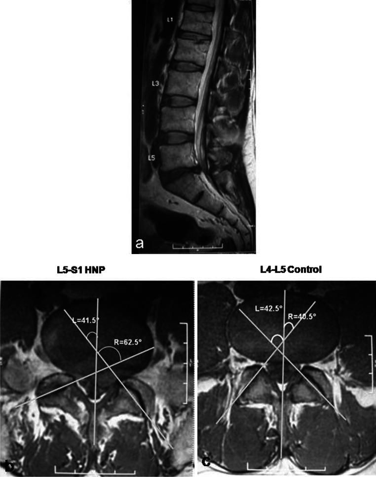
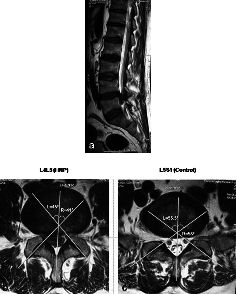
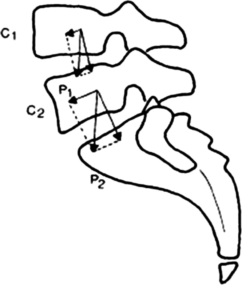
Methods: Sixty patients (18-40 years) with single level disc herniation (L3-L4, L4-L5, or L5-S1) were included in the study. Facet angles were measured using MRI of 3-T using the method described by Karacan et al. Facet tropism was defined as difference of 10° in facet joint angles between right and left sides. Normal disc adjacent to the herniated level was used as control. We also examined if disc herniated towards the side of more coronally oriented facet.
Results: Twenty-five herniations were at L4-L5 level and 35 at L5-S1. Statistical analysis was performed using the Fischer Exact Test. At L4-L5 level, 6/25 cases had tropism compared to 3/35 controls (p = 0.145). At L5-S1 level, 13/35 cases had tropism as compared to 1/21 controls (p = 0.0094). Of 19 cases having tropism, the disc had herniated towards the coronally oriented facet in six (p = 0.11).
Conclusion: The findings of the study suggest that facet tropism is associated with lumbar disc herniation at the L5-S1 motion segment but not at the L4-L5 level.
Figures
References
-
- Brailsford JF. Deformities of the lumbosacral region of the spine. Br J Surg. 1928;16:562–627. doi: 10.1002/bjs.1800166405. - DOI
-
- Badgley CE. The articular facets in relation to low-back pain and sciatic radiation. J Bone Jt Surg. 1941;23:481–496.
-
- Farfan HF, Sullivan JD. The relation of facet orientation to intervertebral disc failure. Can J Surg. 1967;10:179–185. - PubMed
-
- Ferguson AB. The clinical and roentgenographic interpretation of lumbosacral anomalies. Radiology. 1934;22:548–558.
MeSH terms
LinkOut - more resources
Full Text Sources
Medical
