. 2013 Feb 1;73(3):1168-79.
doi: 10.1158/0008-5472.CAN-12-2496. Epub 2012 Dec 12.

Trask loss enhances tumorigenic growth by liberating integrin signaling and growth factor receptor cross-talk in unanchored cells

Affiliations

Trask loss enhances tumorigenic growth by liberating integrin signaling and growth factor receptor cross-talk in unanchored cells

Danislav S Spassov et al. Cancer Res. .

Abstract

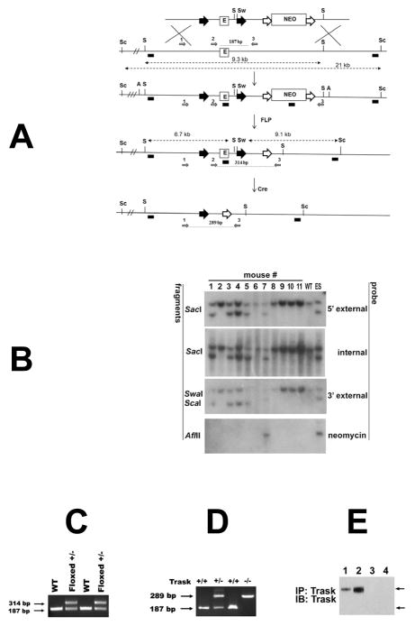
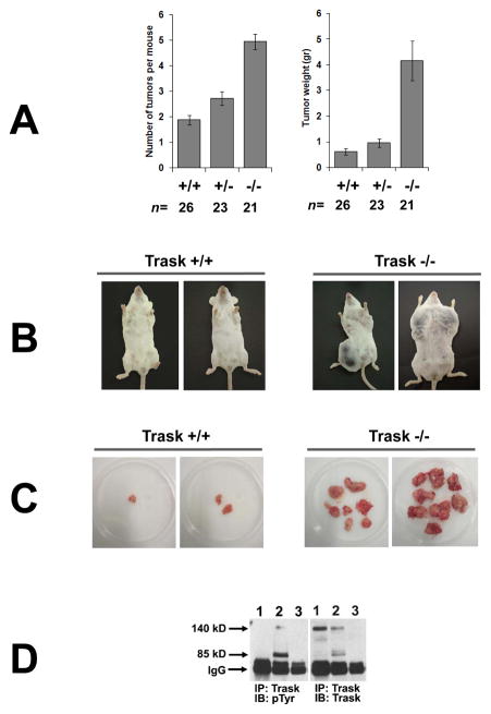
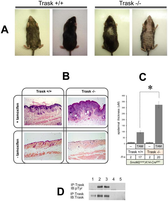
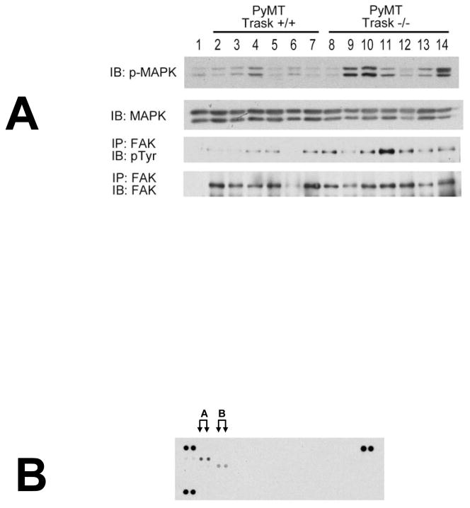
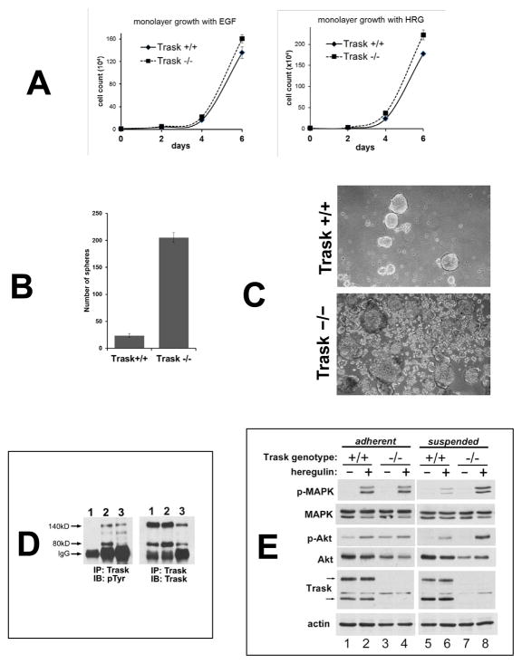
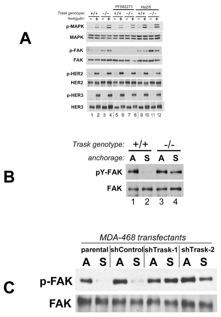
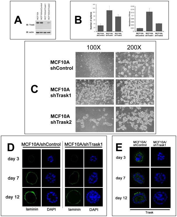
The cell surface glycoprotein Trask/CDCP1 is phosphorylated during anchorage loss in epithelial cells in which it inhibits integrin clustering, outside-in signaling, and cell adhesion. Its role in cancer has been difficult to understand, because of the lack of a discernible pattern in its various alterations in cancer cells. To address this issue, we generated mice lacking Trask function. Mammary tumors driven by the PyMT oncogene and skin tumors driven by the SmoM2 oncogene arose with accelerated kinetics in Trask-deficient mice, establishing a tumor suppressing function for this gene. Mechanistic investigations in mammary tumor cell lines derived from wild-type or Trask-deficient mice revealed a derepression of integrin signaling and an enhancement of integrin-growth factor receptor cross-talk, specifically in unanchored cell states. A similar restrictive link between anchorage and growth in untransformed epithelial cells was observed and disrupted by elimination of Trask. Together our results establish a tumor-suppressing function in Trask that restricts epithelial cell growth to the anchored state.


Conflict of interest statement

Conflicts of interest:

The authors have no conflicts to disclose

Figures

Figure 1
Figure 1. Targeting of the murine Trask locus
(A) Diagrams of the targeted locus and construct used for the generation of Trask floxed and null mice. The loxp sites are shown as big black arrowheads that flank 1.4 kb fragment containing exon 1 (E) and core promoter sequences (not shown). Neomycin cassette (Neo) is flanked by FRT sites (big white arrows). The SacI (S), SwaI (Sw), Sca1 (Sc) and AflII (A) restriction site used for Southern analysis are indicated. The probes used for analysis include 5′ external, internal (exon1), 3′ external and Neo are depicted as small black boxes. The diagnostic SacI and Sca-1 SwaI fragments are shown with punctuated lines with double arrowheads. The removal of the Neo was achieved by crossing with actin-FLP mice, and generation of the null allele by crossing with EIIa-Cre transgenic mice. The PCR primers used for genotyping of floxed and null mice are depicted as small grey arrows and numbered 1, 2 and 3. (B) Southern analysis for germline transmission and Neo excision. Genomic DNA from the progeny of Trask chimeric mice and actin-FLP mice were digested with the indicated restriction enzymes and analyzed with 5′ extrenal, internal, 3′ external and Neo probes. DNA from wild type (WT) mouse and targeted ES clone were used as controls. Mice 1, 3, 4, and 5 are floxed +/− and used as founders for subsequent crossing. (C) Genotyping with primers 2 and 3 gives 187 bp product from the wt template and 314 bp product from the floxed allele. (D) PCR analysis of tail DNA with primers 1,2 and 3 were used to genotype Trask +/+, Trask +/− and Trask −/− mice. (E) The expression of Trask protein was assayed by immunoprecipitation and immunoblotting of lysates in mouse skin (1,3) and intestine (2,4) in Trask +/+ (1,2) and Trask −/− (3,4) mice. Only the 140 kD Trask is expressed in these mouse tissues.
Figure 2
Figure 2. Inactivation of Trask accelerates PyMT induced tumorigenesis
(A) Mammary tumorigenesis in mice carrying an MMTV-PyMT transgene was compared among mice carrying the indicated Trask genotypes. Trask −/− mice have both an increase in the number of detectable tumors and in the weight of the tumors. The difference between +/+ and −/− genotypes is highly significant in both tumor number (p<0.001) and tumor weight (p<0.001). The difference between +/+ and +/− genotypes is more modest, but also significant in both tumor number (p<0.005) and tumor weight (p<0.04). (B) Pictures of representative age-matched tumor-bearing mice are shown for the Trask+/+ (left) and Trask −/− (right) genotypes. (C) Representative ex vivo images of PyMT mammary tumors from Trask +/+ and Trask−/− mice. Each dish contains the entire tumor burden of a single mouse. Trask null mice have both an increase in the number of tumors and in the size of the tumors. (D) Lysates from mouse tissue were assayed by immunoprecipitation. Lanes correspond to 1) normal mammary gland from Trask +/+ mouse, 2) mammary tumor from PyMT, Trask +/+ mouse, 3) mammary tumor from MMTV-PymT, Trask −/− mouse. Trask is phosphorylated in PyMT tumors, but not in the normal mouse mammary gland.
Figure 3
Figure 3. Inactivation of Trask accelerates SmoM2 induced neoplasia
(A) Conditional SmoM2 mice in the indicated Trask genotypes were treated with tamoxifen to induce the oncogenic SmoM2 allele. Dorsal and abdominal view of representative Trask +/+ (left) and Trask −/− (right) mice are shown 12 weeks after tamoxifen treatment. Note more severe hair loss, scaling and skin thickening in Trask null mice. (B) Representative H&E staining of skin samples from K14-CreERT SmoM2 Trask +/+ and K14-CreERT SmoM2 Trask −/− mice treated with or without tamoxifen. Skin samples were taken 12 weeks after tamoxifen injection. (C) Quantification of epidermal thickness (reflecting tumor burden) from histopathology sections showing much thicker tumors in the Trask −/− mice (p<0.0001). This analysis was performed on 17 K14-CreERT SmoM2 Trask +/+ and 20 K14-CreERT SmoM2 Trask −/− mice 12 weeks after tamoxifen treatment. Control mice were not injected with tamoxifen. (D) The expression and phosphorylation of Trask was assayed as indicated from mouse skin by immunoprecipitation. Lanes correspond to 1) normal mouse skin, 2,3) skin from two Trask +/+ mice bearing SmoM2-induced skin tumors, 4) skin from a Trask −/− mouse bearing an SmoM2-induced skin tumor, 5) IgG immunprecipitate from a tumor-bearing Trask +/+ mouse skin. The mouse skin and SmoM2 induced tumors express only the full-length 140 kD Trask form. Trask is phosphorylated in SmoM2 induced tumors, but not in the normal mouse skin.
Figure 4
Figure 4. Integrin and growth factor signaling in PyMT tumors in vivo
(A) Lysates from PyMT Trask +/+ and PyMT Trask −/− tumors were analyzed by immunoblotting. Phosphorylation of MAPK (T202/Y204), tyrosine phosphorylation of FAK and their total protein expressions are shown. Each lane corresponds to a tumor derived from a different mouse. (B) The Trask +/+ tumor lysates were used in the R&D Systems p-RTK profiling. This analysis revealed that the predominant active RTK in these tumors is HER2 (labeled A) and to a lesser extent PDGFR (labeled B).
Figure 5
Figure 5. Loss of Trask augments growth factor signaling in PyMT tumor derived cell lines
(A) Tumor cell lines from MMTV-PyMT mice in the indicated Trask genotypes were grown in the presence of either epidermal growth factor or heregulin for six days and counted. (B) Tumor cell lines from PyMT-driven mouse mammary tumors originating from Trask+/+ or Trask−/− mice were cultured in non-adherent plates for 7 days and the number of spheroids counted manually under microscopic examination. (C) Phase contrast microscope images of the spheroids are shown here. (D) Cell lysates were assayed for the expression and phosphorylation of Trask by immunoprecipitation. Lanes correspond to 1) PyMT tumor cell line cultured in the adherent state, 2) PyMT tumor cell line cultured in the suspended state, 3) in vivo PyMT tumor from a mouse. Trask is phosphorylated during tumor growth in vivo, resembling the suspended state when grown in culture. (E) Serum starved PyMT cell lines from the indicated genotypes were grown in adherent or suspended form were stimulated with heregulin. The growth factor-induced activation of MAPK is suppressed in the suspended Trask+/+ cells. This suppression does not occur in Trask−/− cells. There is a more subtle enhancement of Akt signaling in the Trask−/− cells. The arrows indicate the 140 and 80kD forms of Trask. The phospho-immunoblots reflect the T202/Y204 phosphorylation of MAPK and the S473 phosphorylation of Akt.
Figure 6
Figure 6. Trask inhibits integrin signaling and disrupts integrin-growth factor receptor crosstalk
(A) PyMT cell lines were serum starved for 24 hrs, detached and incubated in suspended conditions. Cells were incubated with FAK inhibitor PF562271 or β1-integrin activating antibody Ha2/5 for 2 hrs, followed by stimulation with herugulin for 10 min as indicated. Phosphorylation of MAPK (T202/Y204), FAK (all tyrosines), HER2 (Y1248) and HER3 (Y1289) and their total expression levels was determined by immunoblotting. (B) PyMT tumor cell lines from Trask +/+ and −/− mice were assayed while adherent (A), or after 2 hours of culture in suspension in non-adhering plates (S). The phosphorylation state of FAK was assayed by IP/pTyr analysis. FAK is normally dephosphorylated when anchorage is lost (lane 2). But in Trask −/− cells, there is abnormal persistence of FAK phosphorylation in the unanchored state (lane 4). (C) The same analysis was done in MDA-468 breast cancer cells looking at p-FAK in suspension using either of two Trask shRNA knockdown pools compared with control cells (parental or non-silencing shRNA transfectant).
Figure 7
Figure 7. Anchorage-independent growth in the presence or absence of Trask
(A) MCF10A cells were engineered to stably express either of two Trask-targeting shRNAs or a non-silencing shRNA and the loss of Trask expression in these cells was confirmed by immunoblotting. MCF10A/shTrask1 cells have a complete loss of Trask while MCF10A/shTrask2 cells have a partial loss of Trask expression. (B) The indicated cell types were cultured in non-adherent plates for 6 days and the number of spheroids counted manually by microscopic examination in triplicate wells (left). The total number of cells in each well was also compared using the MTT assay (right). (C) The appearance of the spheroids was captured by phase contrast microscopy at the indicated magnifications for the three cell types. (D) MCF10A/shControl and MCF10A/shTrask1 cells were grown on matrigel for the indicated number of days and the cellular architecture, density, and expression of laminin were determined by immunofluorescence staining and confocal microscopy. Laminin was stained using green-fluorescent reagents and DAPI staining was used to stain the cell nuclei. (E) Additional sections were stained with anti-Trask antibodies in green as shown.

Similar articles

Cited by

References

    1. Goel HL, Languino LR. Integrin signaling in cancer. Cancer Treat Res. 2004;119:15–31. - PubMed
    1. Rathinam R, Alahari SK. Important role of integrins in the cancer biology. Cancer Metastasis Rev. 2010;29:223–37. - PubMed
    1. Alam N, Goel HL, Zarif MJ, Butterfield JE, Perkins HM, Sansoucy BG, et al. The integrin-growth factor receptor duet. J Cell Physiol. 2007;213:649–53. - PubMed
    1. Streuli CH, Akhtar N. Signal co-operation between integrins and other receptor systems. Biochem J. 2009;418:491–506. - PubMed
    1. Soung YH, Clifford JL, Chung J. Crosstalk between integrin and receptor tyrosine kinase signaling in breast carcinoma progression. BMB Rep. 2010;43:311–8. - PubMed

Publication types

MeSH terms