. 2013 Jan 15;73(2):1020-30.
doi: 10.1158/0008-5472.CAN-12-2862. Epub 2012 Dec 14.

ΔNp63α-mediated activation of bone morphogenetic protein signaling governs stem cell activity and plasticity in normal and malignant mammary epithelial cells

Affiliations

ΔNp63α-mediated activation of bone morphogenetic protein signaling governs stem cell activity and plasticity in normal and malignant mammary epithelial cells

Amanda L Balboni et al. Cancer Res. .

Abstract

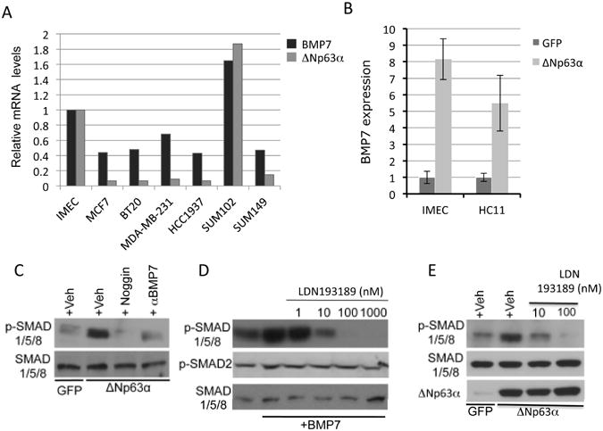
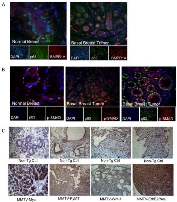
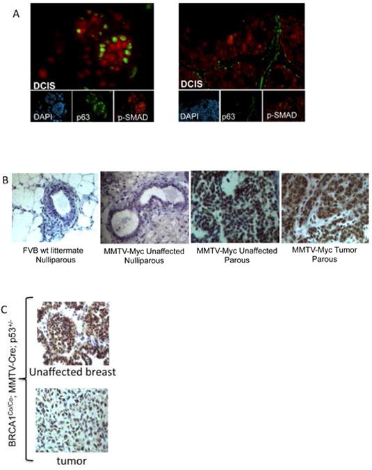
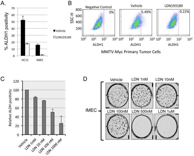
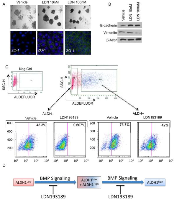
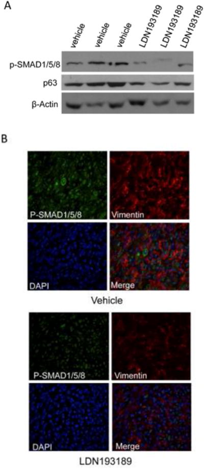
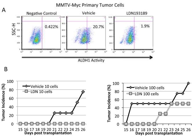
Genetic analysis of TP63 indicates that ΔNp63 isoforms are required for preservation of regenerative stasis within diverse epithelial tissues. In squamous carcinomas, TP63 is commonly amplified, and ΔNp63α confers a potent survival advantage. Genome-wide occupancy studies show that ΔNp63 promotes bidirectional target gene regulation by binding more than 5,000 sites throughout the genome; however, the subset of targets mediating discreet activities of TP63 remains unclear. We report that ΔNp63α activates bone morphogenic proteins (BMP) signaling by inducing the expression of BMP7. Immunohistochemical analysis indicates that hyperactivation of BMP signaling is common in human breast cancers, most notably in the basal molecular subtype, as well as in several mouse models of breast cancer. Suppression of BMP signaling in vitro with LDN193189, a small-molecule inhibitor of BMP type I receptor kinases, represses clonogenicity and diminishes the cancer stem cell-enriched ALDH1(+) population. Importantly, LDN193189 blocks reconstitution of mixed ALDH1(+)/ALDH1(-) cultures indicating that BMP signaling may govern aspects of cellular plasticity within tumor hierarchies. These results show that BMP signaling enables reversion of committed populations to a stem-like state, potentially supporting progression and maintenance of tumorigenesis. Treatment of a mouse model of breast cancer with LDN193189 caused reduced expression of markers associated with epithelial-to-mesenchymal transition (EMT). Furthermore, in vivo limiting dilution analysis assays revealed that LDN193189 treatment suppressed tumor-initiating capacity and increased tumor latency. These studies support a model in which ΔNp63α-mediated activation of BMP signaling governs epithelial cell plasticity, EMT, and tumorigenicity during breast cancer initiation and progression.


Conflict of interest statement

The authors declare that no conflicts of interest exist.

Figures

Figure 1
Figure 1. ΔNp63α induces canonical BMP signaling in mammary epithelial cells via induction of the BMP7 ligand
A. Quantitative PCR analysis of ΔNp63 and BMP7 expression in human breast cancer cell lines. Expression levels are normalized to normal hTERT-immortalized human mammary epithelial cells (IMECs) and to GAPDH. B. Ectopic ΔNp63α induces the expression of BMP7 mRNA in IMECs and HC11 cells. C. Western blots were prepared with extracts from IMECs infected with adenoviral ΔNp63α or GFP as a control, and treated with either vehicle, rhNoggin (250 ng/ml), or BMP7-neutralizing antibodies. Blots were probed with antibodies againstP-SMAD1/5/8 (identifying active BMP signaling), and total SMAD1/5/8 as a loading control. D. Western blot analysis of IMECs pre-treated with vehicle or LDN193189 for 10 minutes, followed by stimulation with vehicle orrhBMP7 (50ng/ml) for 45 minutes. Blots were probed with antibodies directed against P-SMAD1/5/8 and P-SMAD2. Total SMAD1/5/8 is used as a loading control. E. IMECs were infected with adenoviruses programmed to express GFP (control) or ΔNp63α. Following infection, cells were treated with vehicle (control) or the indicated doses of LDN193189. Blots were probed with antibodies directed against P-SMAD1/5/8. β-Actin is used as a loading control. Data is presented as mean values of triplicate points ± S.E.
Figure 2
Figure 2. BMP Signaling is elevated in basal subtype breast tumors and in mouse models of human breast cancer
A. BMP type 1 Receptor A (BMPR1A) expression (red), and p63 expression (green) detected by immunofluorescence in normal breast and triple-negative type tumor tissue. B. Phosphorylated-SMAD1/5/8 (red) and p63 (green) expression detected by immunofluorescence in normal breast and triple negative tumor tumors; N=5 per group. Nuclei are stained with DAPI. C. Immunohistochemical staining of phospho-SMAD1/5/8 on MMTV-Myc, MMTV-PyMT, MMTV-Wnt-1, and MMTV-ErbB2/Neu mouse tumor tissue and non-transgenic normal tissue controls; sections were counterstained with hematoxylin; N=3 per group.
Figure 3
Figure 3. BMP Signaling is elevated in mouse models of breast premalignancy and is upregulated at early stages of breast cancer progression
A. Phosphorylated-SMAD1/5/8 (red) and p63 (green) expression detected by immunofluorescence in ductal carcinoma in situ (DCIS); N=10. B. Phospho-SMAD1/5/8 expression detected by immunohistochemical analysis in mammary tissue sections derived from mice at various stages during MMTV-Myc tumor progression. Sections were counterstained with hematoxylin. C. Phospho-SMAD1/5/8 expression, detected by immunohistochemical analysis, in tumors and unaffected mammary glands from tumor bearing mice with the BRCA1Co/Co; MMTV-Cre; p53+/-genotype.
Figure 4
Figure 4. Pharmacologic inhibition of BMP signaling reduces normal and cancer stem cell populations in vitro
A. Graphic display of FACS-based analysis of ALDH1 activity in HC11 cells and IMECs treated with vehicle or 1uM LDN193189 for 48 hours. Cells were incubated with DEAB, an ALDH1 inhibitor, and used as a negative control. Gates were set according to the negative control. B. FACS analysis of ALDH1 activity in primary MMTV-Myc tumor cells treated with vehicle or 1μM LDN193189 for 48 hours. Cells were incubated with DEAB, an ALDH1 inhibitor, and used as a negative control. Gates were set according to the negative control. C. HC11 cells were treated with the indicated doses of LDN193189 for 48 hours and assayed for ALDH1 activity by flow cytometry. Data are expressed as the percentage of ADLH1+ cells relative to vehicle-treated controls. D. Colony formation assay in IMECs treated with vehicle control and the indicated concentrations of LDN193189. Cells were stained with crystal violet 14 days after plating at 500 cells/well in 6-well plates.
Figure 5
Figure 5. LDN193189 promotes Mesenchymal to Epithelial Transition (MET) and restricts cellular plasticity
A, top. IMECs were cultured at clonogenic density and treated with the indicated concentrations of LDN193189. Colonies were stained with crystal violet 10-14 days after plating. A, bottom. Immunofluorescent analysis of the tight junction protein ZO-1 in colonies formed in the presence or absence of LDN193189. B. Western blot analysis of E-cadherin and vimentin levels indicate that treatment of IMECs with LDN193189 causes changes that are consistent with MET. C. HC11 cells were FACS-segregated based on ALDH1 activity and re-cultured in the presence of vehicle or LDN193189 for 72 hours. Cells were then reassayed for ALDH1 activity to determine the ability of each fraction to reconstitute the mixed parental ALDH1+/ALDH1- culture. D. Schematic summarizing the results of data shown in C.
Figure 6
Figure 6. LDN193189 exhibits pharmacologic activity in vivo and causes reversion of EMT
One thousand primary MMTV-Myc tumor cells were embedded in matrigel and transplanted into the dorsal right flanks of female syngeneic (FVB/N) wt recipients; N=40. At two days post transplant, mice were injected intraperitoneally with 2.5 mg/kg LDN193189 or vehicle control daily for 16 days and then sacrificed two hours after the last injection. A. Western blot analysis of 3representative tumor lysates from vehicle and LDN193189 treatment groups probing for phospho-SMAD1/5/8 and p63. β-Actin is used as a loading control. B. Representative images of 2-color Immunofluorescent analysis of mouse tumor tissue probing for P-SMAD1/5/8 (green) and Vimentin (red), a marker of EMT. Nuclei are stained with DAPI.
Figure 7
Figure 7. Limiting dilution analysis reveals that LDN193189 reduces tumor initiating ability and increases tumor latency
A. Primary MMTV-Myc tumor cells were cultured ex vivo in the presence or absence of 1μM LDN193189 for 48 hours and subjected to FACS-based analysis of ALDH1 activity. Cells were incubated with DEAB, an ALDH1 inhibitor, and used as a negative control. Gates were set according to the negative control. The cells were subsequently transplanted by limiting dilution into the dorsal right flanks of FVB wild-type female recipient mice. Mice were treated two days post transplantation daily for 24 days with 2.5 mg/kg LDN193189 or vehicle control by IP injection. B. Tumor latency is plotted as time to palpitation for each group of mice. N=4 for each group.

References

    1. Cerami E, Gao J, Dogrusoz U, Gross BE, Sumer SO, Aksoy BA, et al. The cBio Cancer Genomics Portal: An Open Platform for Exploring Multidimensional Cancer Genomics Data. Cancer discovery. 2012;2:401–4. - PMC - PubMed
    1. Mills AA, Zheng B, Wang XJ, Vogel H, Roop DR, Bradley A. p63 is a p53 homologue required for limb and epidermal morphogenesis. Nature. 1999;398:708–13. - PubMed
    1. Yang A, Schweitzer R, Sun D, Kaghad M, Walker N, Bronson RT, et al. p63 is essential for regenerative proliferation in limb, craniofacial and epithelial development. Nature. 1999;398:714–8. - PubMed
    1. Romano RA, Smalley K, Magraw C, Serna VA, Kurita T, Raghavan S, et al. DeltaNp63 knockout mice reveal its indispensable role as a master regulator of epithelial development and differentiation. Development. 2012;139:772–82. - PMC - PubMed
    1. Yang A, Kaghad M, Wang Y, Gillett E, Fleming MD, Dotsch V, et al. p63, a p53 homolog at 3q27-29, encodes multiple products with transactivating, death-inducing, and dominant-negative activities. Molecular cell. 1998;2:305–16. - PubMed

Publication types

MeSH terms