

Case Reports
doi: 10.1093/icvts/ivs517.
Epub 2012 Dec 12.
A novel method for the treatment of dysphagia lusoria due to aberrant right subclavian artery
Affiliations
- PMID: 23243037
- PMCID: PMC3568821
- DOI: 10.1093/icvts/ivs517
Item in Clipboard
Case Reports
A novel method for the treatment of dysphagia lusoria due to aberrant right subclavian artery
Interact Cardiovasc Thorac Surg.
2013 Mar.
Abstract
Dysphagia lusoria occurs secondary to an aberrant right subclavian artery coursing posterior to the oesophagus. Open ligation and transposition to the right carotid artery via a right supraclavicular approach has been described as a minimally invasive method. However, approaching the origin of the aberrant right subclavian artery through this incision can be extremely challenging. A persistent aberrant right subclavian artery stump may account for postoperative residual dysphagia. This article describes a safe, effective and reproducible surgical approach to dysphagia lusoria due to a non-aneurysmal aberrant right subclavian artery.
Figures
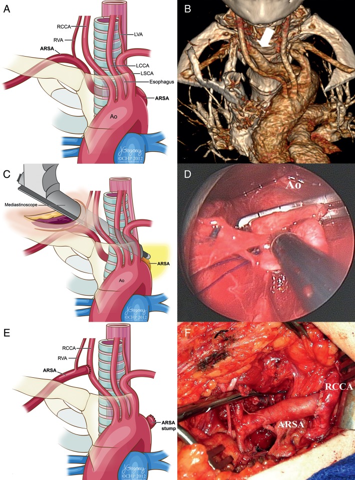
(A) Illustration of the ARSA. The ARSA is coursing posteriorly behind the oesophagus. ARSA: aberrant right subclavian artery; RCCA: right common carotid artery; LCCA: left common carotid artery; LSCA: left subclavian artery, RVA: right vertebral artery; LVA: left vertebral artery; Ao: Aorta. (B) Preoperative 3D computed tomography reconstruction (anteroposterior view with cranial angulation) showing the origin of the ARSA from the posterior wall of the aortic arch (arrow). (C) Operative illustration showing the mediastinoscope-assisted dissection using the suction cautery device. (D) Operative image from the mediastinoscope demonstrating the aortic wall and the ARSA stump. This plane was created by the use of mediastinoscopic blunt dissection. (E and F) The ARSA was ligated at its origin and reimplanted to the RCCA.
References
-
- Ota T, Okada K, Takanashi S, Yamamoto S, Okita Y. Surgical treatment for Kommerell's diverticulum. J Thorac Cardiovasc Surg. 2006;131:574–8. doi:10.1016/j.jtcvs.2005.10.012. - DOI - PubMed
-
- Edwards JE. Congenital malformations of the heart and great vessels. Section H. Malformations of the thoracic aorta. In: Gould SE, editor. Pathology of the Heart. 2nd edn. Springfield, IL: Charles C. Thomas; 1960. pp. 391–462.
-
- Janssen M, Baggen MG, Veen HF, Smout AJ, Bekkers JA, Jonkman JG, et al. Dysphagia lusoria: clinical aspects, manometric findings, diagnosis, and therapy. Am J Gastroenterol. 2000;95:1411–6. doi:10.1111/j.1572-0241.2000.02071.x. - DOI - PubMed
-
- Stone WM, Ricotta JJ, 2nd, Fowl RJ, Garg N, Bower TC, Money SR. Contemporary management of aberrant right subclavian arteries. Ann Vasc Surg. 2011;25:508. doi:10.1016/j.avsg.2011.02.012. - DOI - PubMed
Publication types
MeSH terms
LinkOut - more resources
Full Text Sources
Medical
