. 2012 Dec 19;104(24):1879-87.
doi: 10.1093/jnci/djs451. Epub 2012 Dec 12.

Prognostic impact of (18)FDG-PET-CT findings in clinical stage III and IIB breast cancer

Affiliations

Prognostic impact of (18)FDG-PET-CT findings in clinical stage III and IIB breast cancer

David Groheux et al. J Natl Cancer Inst. .

Abstract

Background: This study prospectively evaluated the yield of fluorodeoxyglucose positron emission tomography/computed tomography ((18)FDG-PET-CT) in patients with clinical stages II and III breast cancer and the impact of PET-CT results on prognosis.

Methods: In the course of 71 months, 254 consecutive patients with clinical stages II and III breast cancer (based on clinical examination, mammography, breast magnetic resonance imaging, and locoregional ultrasonography) underwent (18)FDG-PET-CT. The yield was assessed in the whole population and for each American Joint Committee on Cancer subgroup. The prognostic impact of PET-CT findings was analyzed. Tests of statistical significance were two-sided.

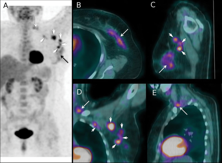
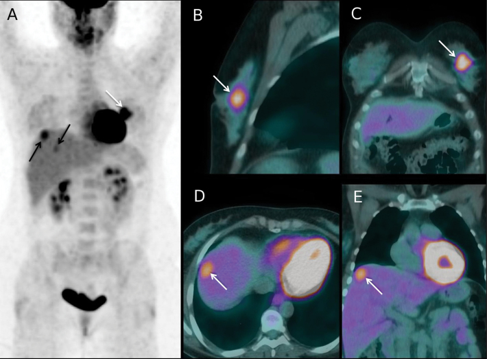
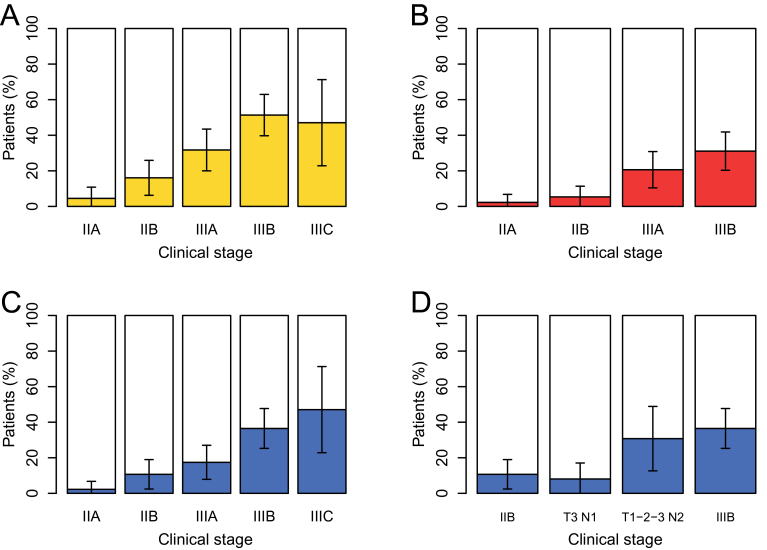
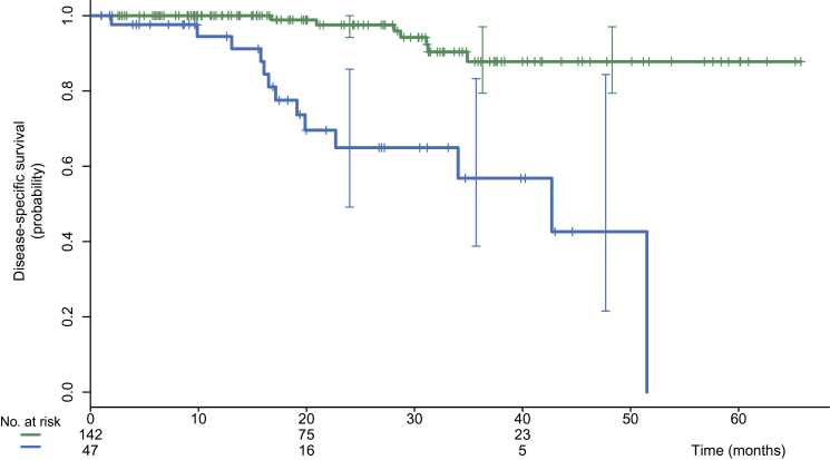
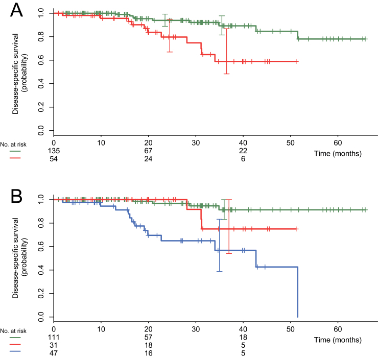
Results: (18)FDG-PET-CT changed the clinical stage in 77 of 254 patients (30.3%; 95% confidence interval [CI] = 25.0% to 36.2%). It showed unsuspected N3 disease (infraclavicular, supraclavicular, or internal mammary nodes) in 40 patients and distant metastases in 53. PET-CT revealed distant metastases in 2.3% (1 of 44) of clinical stage IIA, 10.7% (6 of 56) of stage IIB, 17.5% (11 of 63) of stage IIIA, 36.5% (27 of 74) of stage IIIB, and 47.1% (8 of 17) of stage IIIC patients. Among 189 patients with clinical stage IIB or higher disease and adequate follow-up, disease-specific survival was statistically significantly shorter in the 47 patients scored M1 on (18)FDG-PET-CT in comparison with those scored M0, with a three-year disease-specific survival of 57% vs 88% (P < .001). In multivariable analysis, only distant disease on PET-CT and triple-negative phenotype were statistically significant prognostic factors. The relative risk of death was 26.60 (95% CI = 6.60 to 102.62) for M1 vs M0 patients.

Conclusions: The yield of (18)FDG-PET-CT appeared substantial in patients with clinical stage IIB or higher breast cancer. In these patients, (18)FDG-PET-CT provided powerful prognostic stratification.


Figures

Figure 1.
Figure 1.
Fluorodeoxyglucose positron emission tomography/computed tomography (18FDG-PET-CT) findings in a 63-year-old woman with a 90-mm invasive mixed ductal/lobular carcinoma of the left breast (tumor phenotype = estrogen receptor positive, human epidermal growth factor receptor 2 negative, grade 2) and matted lymph nodes in axilla level 1. Based on clinical examination and loco-regional workup, the patient was clinically staged IIIA (cT3 cN2a cM0). A) Maximum intensity projection image PET showing 18FDG uptake in the primary tumor (black arrow), in lymph nodes at level 1 and 2 of axilla (white arrowheads), and in supraclavicular lymph nodes (white arrow). B) Axial projection PET-CT fusion images through the left breast showing the primary tumor (white arrow). C) PET-CT sagittal projection showing the primary tumor (white arrow) and three level 1 axillary lymph nodes (white arrowheads). D) PET-CT coronal section showing 18FDG uptake in level 1 and level 2 nodes (white arrowheads) and in a supraclavicular node (white arrow). E) PET-CT sagittal section showing 18FDG uptake in one supraclavicular node (white arrow). After PET-CT, cancer was classified cT3 cN3c cM0 (stage IIIC). The neoadjuvant chemotherapy regimen was performed as planned (4 cycles of epirubicin 75mg/m² plus cyclophosphamide 750mg/m² followed by 4 courses of docetaxel 100mg/m²). After completion of neoadjuvant chemotherapy, the patient underwent mastectomy, axillary lymph node dissection, and, because of data from the initial PET-CT scan, additional dissection of the supraclavicular area. Pathological examination showed residual tumor in breast with >50% pathological response, six axillary metastases (with evidence of therapeutic effect), and tumor-involved supraclavicular nodes (with evidence of therapeutic effect). Radiation therapy to the chest wall and locoregional basins was performed, and the patient received adjuvant hormone therapy. At 24 months of follow-up, no recurrence was observed.
Figure 2.
Figure 2.
Fluorodeoxyglucose positron emission tomography/computed tomography (18FDG-PET-CT) findings at initial staging in a 45-year-old woman with a 60-mm invasive ductal carcinoma of the left breast (tumor phenotype = estrogen receptor positive, human epidermal growth factor receptor 2 negative, grade 2). The patient had no regional lymph nodes metastases at clinical and ultrasound examination (cT3 cN0 cM0; stage IIB). A) Maximum intensity projection PET image showing 18FDG uptake in the primary tumor (white arrow), and also FDG uptake foci in the liver (black arrows). B, C) Sagittal and coronal projections PET-CT fusion images through the left breast showing the primary tumor (white arrows). D) Axial projection PET-CT fusion image through the liver dome showing a 18FDG-avid liver lesion (white arrow). E) Coronal projection image showing the same metastasis (white arrow). No metastases had been depicted on initial liver ultrasound but abdominal contrast-enhanced CT performed after PET-CT confirmed liver metastases. The patient was upstaged to cT3 cN0 cM1 (stage IV). Chemotherapy regimen was adapted to metastatic disease. Following primary chemotherapy, mastectomy and axillary clearance showed a residual breast primary with pathological response >50% and no metastases in the 14 removed nodes. Then, right liver surgery was performed and showed residual metastases. Radiation therapy to the chest wall only (without regional nodal basins irradiation) was performed, and the patient received adjuvant hormone therapy. In the 7 months since liver surgery, no recurrence was documented.
Figure 3.
Figure 3.
Yield of fluorodeoxyglucose positron emission tomography/computed tomography (18FDG-PET-CT) according to initial clinical American Joint Committee on Cancer subgroup and tumor nodes metastases subset. A) Percentage of stage modification. B) Percentage of N3 disease uncovered by PET-CT. All patients with clinical N3 (stage IIIC) were also confirmed on PET-CT and are not represented here. C) Percentage of patients with distant metastases uncovered by PET-CT. D) Percentage of patients with distant metastases uncovered by PET-CT for specific tumor node metastases subsets of stage IIIA. The yield in T3 N1 patients is similar to that of stage IIB, whereas the yield in patients with clinical N2 disease is similar to that in stage IIIB patients (D). Error bars on each graph show the 95% confidence interval.
Figure 4.
Figure 4.
Kaplan–Meier disease-specific survival for 189 patients with clinical stages IIB, IIIA, IIIB, and IIIC disease and adequate follow-up. Comparison of patients with and without distant metastases on fluorodeoxyglucose positron emission tomography/computed tomography (18FDG-PET-CT). The upper curve (in green color online) shows patients without distant metastases (n = 142; events = 7). The lower curve (in blue color online) shows patients with distance metastases detected by PET-CT (n = 47; events = 13). Log-rank P less than .001.
Figure 5.
Figure 5.
Kaplan–Meier disease-specific survivals for the 189 patients with clinical stages IIB, IIIA, IIIB, and IIIC disease. Comparison of patients with and without N3 disease (infraclavicular, supraclavicular, or internal mammary lymph nodes) shown by fluorodeoxyglucose positron emission tomography/computed tomography. A) Comparison of patients with and without N3 disease. The upper curve (in green color online) shows patients without N3 disease (n =135; events = 9). The lower curve (in red color online) shows patients with N3 disease (n = 54; events = 11). Log-rank P equals .001. B) Women with M1 disease separated from others. The upper curve (in green color online) shows patients without N3 disease and without distant metastases (n = 111; events = 4). The intermediate curve (in red color online) shows patients with N3 disease and without distant metastases (n = 31; events = 3). The lower curve (in blue color online) shows patients with distant metastases (n = 47; events = 13). Log-rank P equals .16 between the two upper curves.

Comment in

References

    1. Edge SB, Byrd DR, Compton CC, Fritz AG, Greene FL, Trotti A. AJCC Cancer Staging Manual. 7thed. New York: Springer; 2010;.
    1. Groheux D, Hindié E, Rubello D, et al. Should FDG PET/CT be used for the initial staging of breast cancer?. Eur J Nucl Med Mol Imaging. 2009; 36(10):1539–1542 - PubMed
    1. Hindié E, Groheux D, Brenot-Rossi I, Rubello D, Moretti JL, Espié M. The sentinel node procedure in breast cancer: nuclear medicine as the starting point. J Nucl Med. 2011; 52(3):405–414 - PubMed
    1. Yang WT, Le-Petross HT, Macapinlac H, et al. Inflammatory breast cancer: PET/CT, MRI, mammography, and sonography findings. Breast Cancer Res Treat. 2008; 109(3):417–426 - PubMed
    1. Carkaci S, Macapinlac HA, Cristofanilli M, et al. Retrospective study of 18F-FDG PET/CT in the diagnosis of inflammatory breast cancer: preliminary data. J Nucl Med. 2009; 50(2):231–238 - PubMed

MeSH terms