. 2013 Jan 29;127(4):486-99.
doi: 10.1161/CIRCULATIONAHA.112.116988. Epub 2012 Dec 14.

A crucial role for p90RSK-mediated reduction of ERK5 transcriptional activity in endothelial dysfunction and atherosclerosis

Affiliations

A crucial role for p90RSK-mediated reduction of ERK5 transcriptional activity in endothelial dysfunction and atherosclerosis

Nhat-Tu Le et al. Circulation. .

Abstract

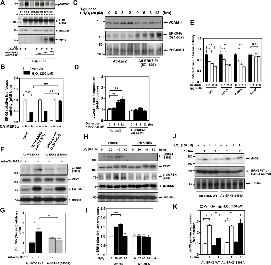
Background: Diabetes mellitus is a major risk factor for cardiovascular mortality by increasing endothelial cell (EC) dysfunction and subsequently accelerating atherosclerosis. Extracellular-signal regulated kinase 5 (ERK5) is activated by steady laminar flow and regulates EC function by increasing endothelial nitric oxide synthase expression and inhibiting EC inflammation. However, the role and regulatory mechanisms of ERK5 in EC dysfunction and atherosclerosis are poorly understood. Here, we report the critical role of the p90 ribosomal S6 kinase (p90RSK)/ERK5 complex in EC dysfunction in diabetes mellitus and atherosclerosis.

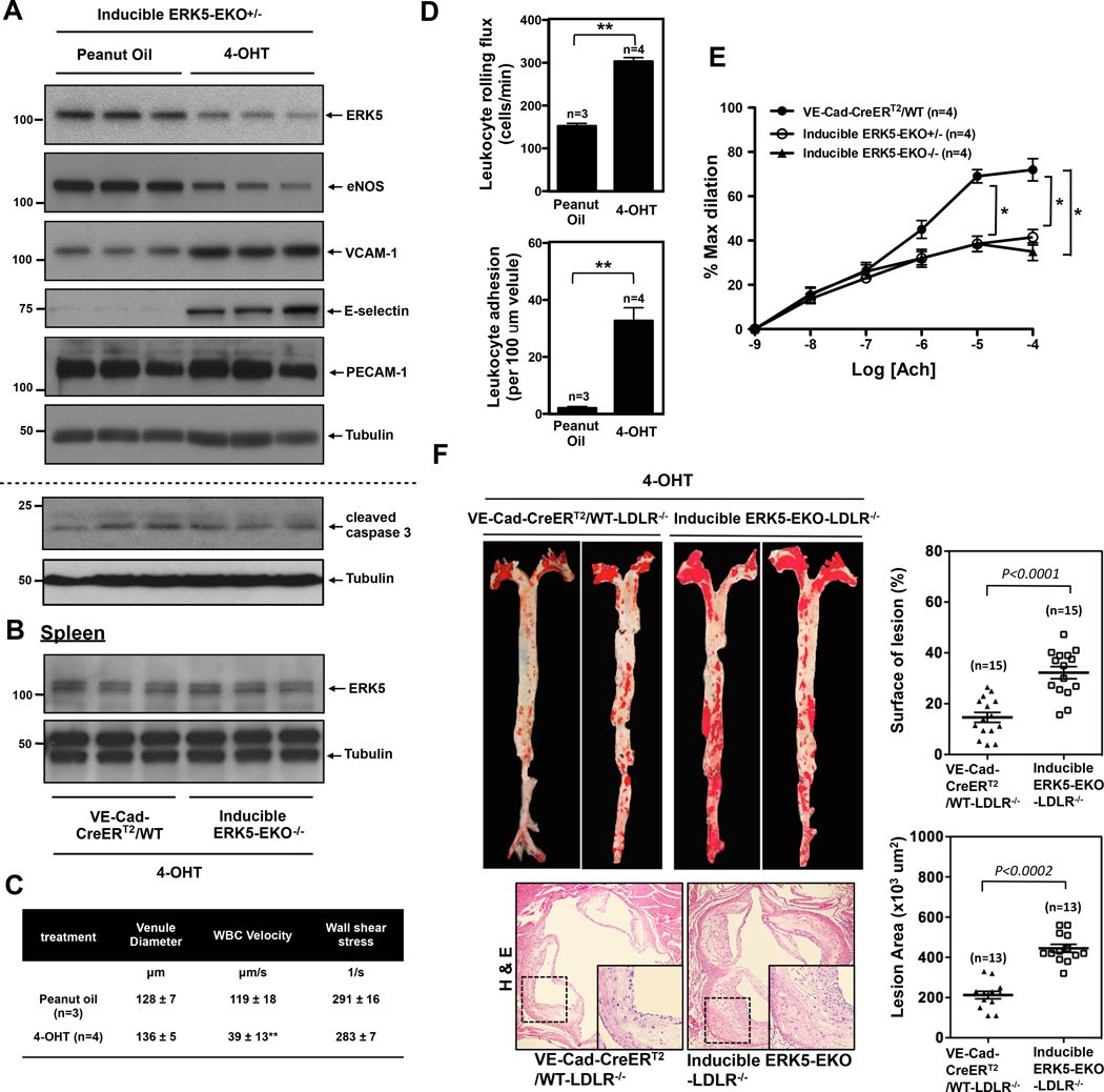
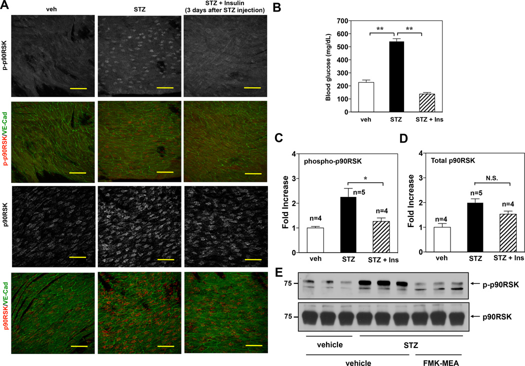
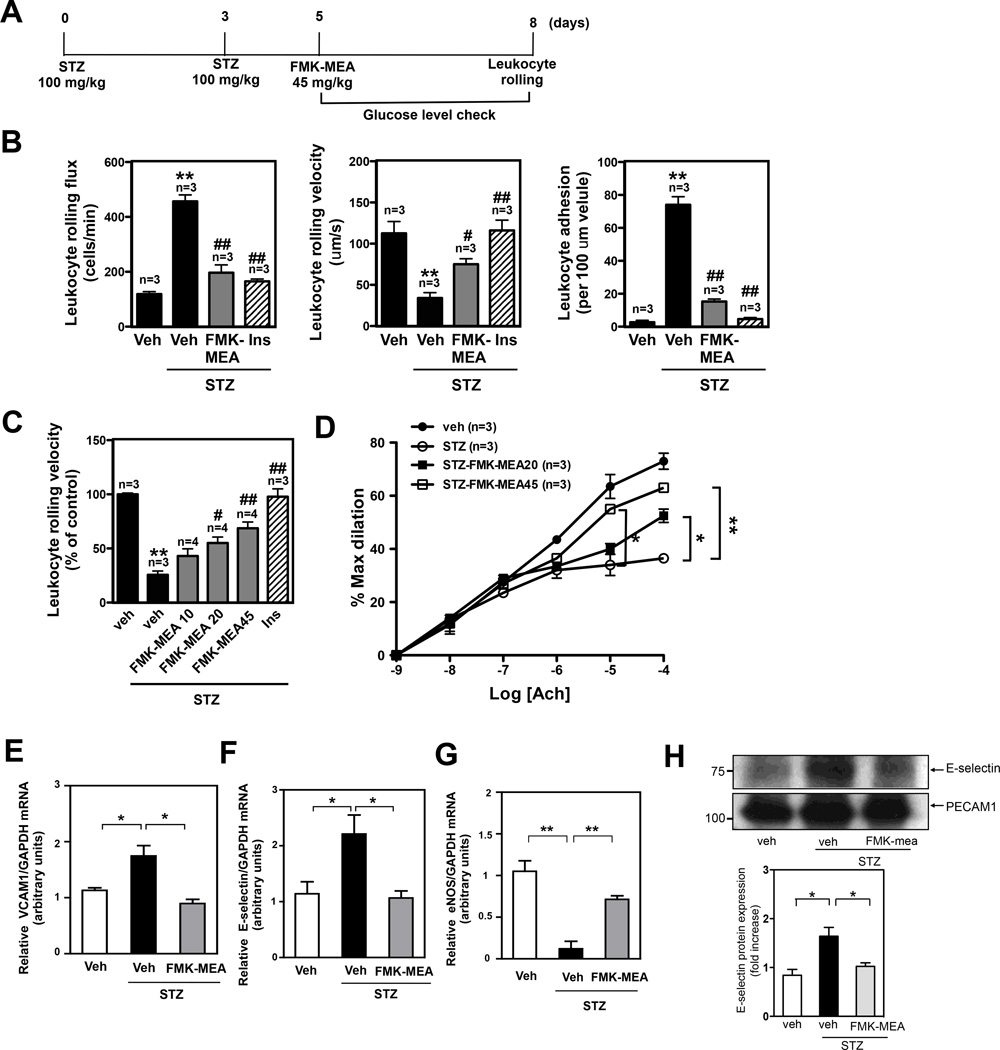
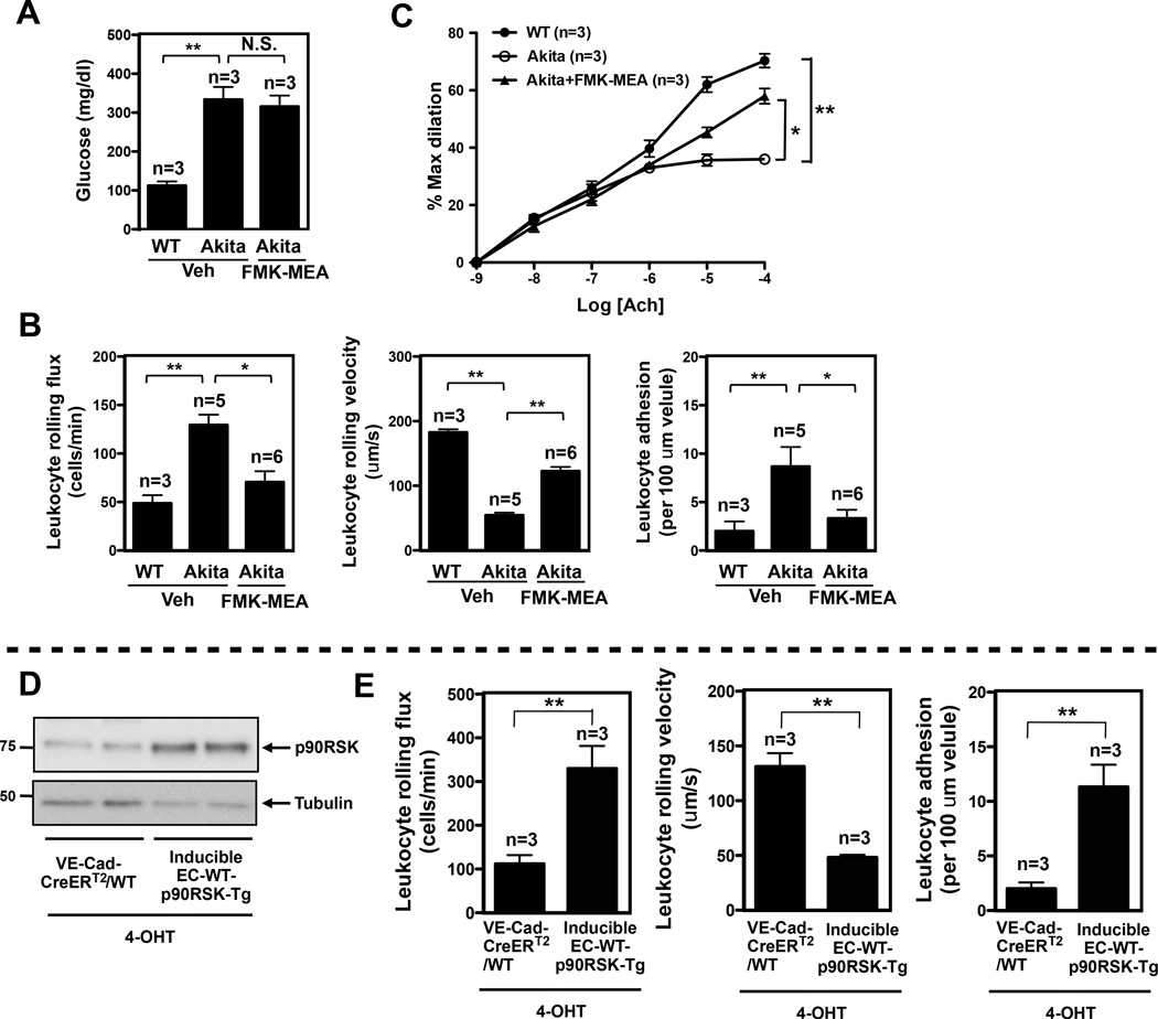
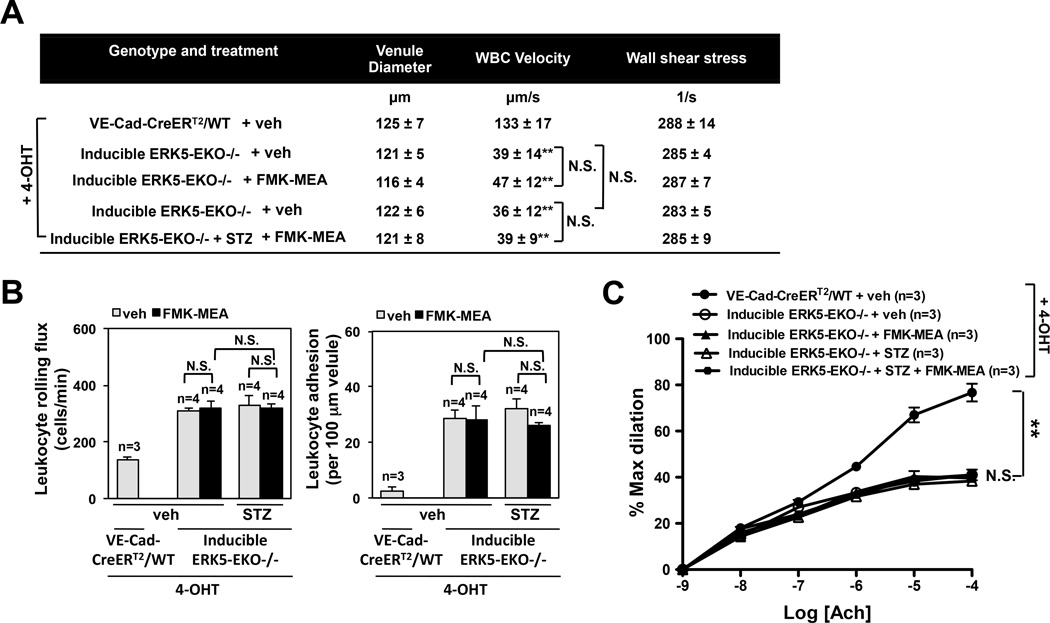
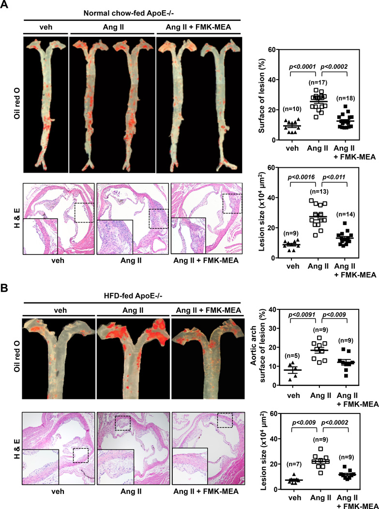
Methods and results: Inducible EC-specific ERK5 knockout (ERK5-EKO) mice showed increased leukocyte rolling and impaired vessel reactivity. To examine the role of endothelial ERK5 in atherosclerosis, we used inducible ERK5-EKO-LDLR(-/-) mice and observed increased plaque formation. When activated, p90RSK associated with ERK5, and this association inhibited ERK5 transcriptional activity and upregulated vascular cell adhesion molecule 1 expression. In addition, p90RSK directly phosphorylated ERK5 S496 and reduced endothelial nitric oxide synthase expression. p90RSK activity was increased in diabetic mouse vessels, and fluoromethyl ketone-methoxyethylamine, a specific p90RSK inhibitor, ameliorated EC-leukocyte recruitment and diminished vascular reactivity in diabetic mice. Interestingly, in ERK5-EKO mice, increased leukocyte rolling and impaired vessel reactivity were resistant to the beneficial effects of fluoromethyl ketone-methoxyethylamine, suggesting a critical role for endothelial ERK5 in mediating the salutary effects of fluoromethyl ketone-methoxyethylamine on endothelial dysfunction. Fluoromethyl ketone-methoxyethylamine also inhibited atherosclerosis formation in ApoE(-/-) mice.

Conclusions: Our study highlights the importance of the p90RSK/ERK5 module as a critical mediator of EC dysfunction in diabetes mellitus and atherosclerosis formation, thus revealing a potential new target for therapeutic intervention.


Conflict of interest statement

Conflict of Interest Disclosures: J.T. is a cofounder of Principia Biopharma, which has licensed FMK-MEA.

Figures

Figure 1
Figure 1
p90RSK activation inhibits ERK5 transcriptional activation and regulates subsequent KLF2-eNOS expression and VCAM-1 expression. (A) HUVECs were treated with 200µM of H2O2 for indicated times. Cell lysate were immunoprecipitated with anti-ERK5 or IgG as a negative control. ERK5-bound p90RSK was detected by Western blotting using anti-p90RSK antibody (top). p-p90RSK, p90RSK, p-ERK5, ERK5, p-ERK1/2, and tubulin were detected using each specific antibody. (B, C, D) Densitometric quantification of: ERK5-bound-p90RSK (B), phosphorylation of p90RSK (C) and ERK5 (D). Results were normalized to the lowest association (B) phosphorylation level (C, D) within each set of experiments. Results are expressed relative to untreated cells (0 min), and relative to the corresponding band intensity of p90RSK (3rd from top) (C) or ERK5 (5th from top) (D) at each time point. Statistical significance was determined by comparing the average level of the control group with that of each experimental data point. Experiments were carried out in triplicate using 3 different batches of HUVECs (B-D, mean ± SEM, n=3, *P<0.05, **P<0.01 compared to control). (E) HUVECs were co-transfected with an empty vector or plasmids encoding p90RSK-WT, Gal4-ERK5, and the Gal4-responsive luciferase reporter pG5-Luc. Four hours later, Opti-MEM was replaced by supplemented M200 medium. Eight hours post transfection, cells were stimulated with 200µM H2O2 for 16hrs, and ERK5 transcriptional activity was assayed by the dual-luciferase reporter assay. p90RSK overexpression inhibited ERK5 transcriptional activity in a dose-dependent manner, and also enhanced H2O2-mediated reduction in ERK5 transcriptional activity. Luciferase activity was normalized relative to Renilla luciferase activity. The mean luciferase activity of an empty vector (vehicle treatment) was set as 1 (mean ± SEM, n=3). (F, G) HUVECs were co-transfected with plasmids encoding Gal4-ERK5 and the Gal4-responsive luciferase reporter pG5-Luc (F) or KLF2 promoter with luciferase (KLF2-Luc) and ERK5 wild type (G). Cells were also transfected with an empty vector or CA-MEK5α as indicated. Eight hours post transfection, cells were treated with FMK (10µM) for 3hrs, followed by stimulation with 200µM H2O2 for 16hrs. Finally, ERK5 transcriptional activity (F) and KLF2 promoter activity (G) were assayed by a dual-luciferase reporter assay (F-G, mean ± SEM, n=3). (H, I) MECs were pretreated with FMK-MEA (10µM, 3hrs), followed by stimulation with either HG/low H2O2 or Mannitol/low H2O2 as a control and (H) KLF2 mRNA and (I) VCAM-1 mRNA level were detected by qRT-PCR as described in methods (H-I, mean ± SEM, n=3). (J, K) HUVECs were transduced with either adenovirus vector containing DN-p90RSK or LacZ and 24hrs later, exposed to s-flow for 24 hrs in the presence or absence of a combination of high glucose (25mM) and low dose H2O2 (20µM). eNOS, p90RSK, and tubulin expressions were detected by Western blotting with specific antibodies (J). eNOS expression was quantified (mean ± SEM, n=3) (K). (L, M) MECs (passage 3) were transduced with either adenovirus vector containing DN-p90RSK or LacZ and 24hrs later, treated with a combination of high glucose (25mM) and a low dose of H2O2 (20µM) for indicated times. VCAM-1, PECAM-1 and p90RSK expressions were detected by Western blotting with specific antibodies (L). VCAM1 expression was quantified (mean ± SEM, n=3) (M).
Figure 2
Figure 2
p90RSK interacts with ERK5, phosphorylates ERK5-Ser496, and inhibits ERK5 transcriptional activity. (A) HeLa cells were co-transfected with Flag-tagged ERK5 wild type, Xpress-tagged wild type p90RSK, and a VP16-tagged ERK5 fragment (aa571-807) for 24hrs. Cell lysates were immunoprecipitated with anti-Flag, followed by immunoblotting with anti-p90RSK to determine p90RSK-ERK5 association (top). The expression of p90RSK, ERK5 and the ERK5 fragment were detected by immunoblotting with specific antibodies. The VP16-tagged ERK5 fragment inhibited p90RSK-ERK5 association. Data is a representative of 3 independent experiments. (B) HUVECs were co-transfected with an empty vector or plasmids encoding an ERK5 fragment (aa571-807) or (aa412-577), Gal4-ERK5, and the Gal4-responsive luciferase reporter pG5-Luc. Cells were also transfected with an empty vector or CA-MEK5α as indicated. Twelve hours post transfection, cells were stimulated by 200µM H2O2 for 16hrs, and then ERK5 transcriptional activity was assayed by the dual-luciferase reporter assay as described above. Luciferase activity was normalized relative to Renilla luciferase activity, and the mean luciferase activity at the condition of CA-MEK5α transfection for vehicle treatment was set at the same level. Data are representative of three independent experiments (mean ± SEM, n=3). (C, D) MECs (passage 3) were transduced with either adenovirus vector containing ERK5 fragment aa571-806 (Ad-ERK5-Fr(571–807)) or Ad-LacZ. After 24hrs of transduction, cells were treated with a combination of high glucose (25mM) and a low dose of H2O2 (20µM) for indicated times. VCAM-1, PECAM-1 and ERK5 expressions were detected by Western blotting with specific antibodies (C). Densitometric quantification of VCAM-1 expression is shown (mean ± SEM, n=3). (D). (E) Transcriptional activity of the ERK5 S496A mutant cannot be inhibited by p90RSK. HUVECs were co-transfected for 24hrs with an empty vector or plasmids encoding p90RSK-WT with Gal4-ERK5-WT or a Gal4-ERK5-T475A, -S486A, or -S496A mutant, and a Gal4-responsive luciferase reporter. ERK5 transcriptional activity was assayed by a dual-luciferase reporter assay. Luciferase activity was normalized relative to Renilla luciferase activity, and the mean luciferase activity of an empty vector (p90RSK=0) was set as 1. Data are representative of three independent experiments (mean ± SEM, n=3). (F, G) HUVECs were transduced with either Ad-WT-ERK5 or Ad-ERK5 S496A mutant with Ad-p90RSK or Ad-LacZ as a control for 9hrs. ERK5-S496 phosphorylation, ERK5, p90RSK, and tubulin expression were detected by Western blotting with specific antibodies (F). Phosphorylation level of ERK5-S496 was densitometrically quantified (mean ± SEM, n=3) (G). (H, I) H2O2 increases ERK5-S496 phosphorylation via p90RSK activation. HUVECs were pre-treated with FMK-MEA (10µM) for 3hrs, then stimulated with H2O2 for indicated times. ERK5-S496 phosphorylation, ERK5, S380 p90RSK phosphorylation, p90RSK, and tubulin expression were detected by Western blotting with specific antibodies (H). Densitometric quantification was shown (mean ± SEM, n=3) (I). (J, K) HUVECs were transduced with either adenovirus vector containing ERK5 wild type (Ad-ERK5-WT) or ERK5-S496A mutant (Ad-ERK5-S496A). After 24hrs of transduction, cells were treated with or without H2O2 (400µM) and then exposed to s-flow for 24 hrs. eNOS, ERK5, and tubulin expressions were then detected by Western blotting with their specific antibodies (J). Densitometric quantification was shown (mean ± SEM, n=3) (K).
Figure 3
Figure 3
Reduced ERK5 expression in ECs in inducible ERK5-EKO mice increases leukocytes rolling, inhibits endothelium-dependent vasodilatation, and accelerates atherosclerosis formation in LDLR−/− mice. (A) The expression of ERK5, eNOS, VCAM-1, E-selectin, PECAM-1, and tubulin in cultured MECs isolated from two weeks of peanut oil or tamoxifen (4-OHT) treated inducible heterozygous ERK5-EKO+/− mice. MEC lysates were probed by Western blotting. (B) The expression of ERK5 and tubulin in the cell lysate obtained from the spleen of either VE-Cad-CreERT2-WT or inducible ERK5-EKO−/− mice after two weeks of 4-OHT injections are shown. (C, D) Leukocyte rolling in vivo. Mice were anesthetized and injected with rhodamine 6G to label leukocytes. The mesentery was externalized, and then Leukocyte rolling in venules 120 to 150µm in diameter was recorded with a digital video camera. Shown are quantified data of leukocyte rolling velocity (C), leukocyte rolling flux (D, top), and leukocyte adhesion (D, bottom). To analyze these parameters, image analysis software (NIS elements, Nikon) was used (n=3–4, mean ± SEM; **P<0.01 versus controls, non-parametric Wilcoxon T test). (Supplemental video 1 and Fig.3D). (E) Endothelium-dependent relaxation to Ach. In tamoxifen treated VE-Cad-CreERTR/wild type mice, endothelium-dependent vasodilatation to Ach was observed, but in the tamoxifen-treated inducible heterozygous ERK5-EKO+/− or homozygous ERK5−/− mice, vasodilatation was significantly reduced. The number of venules used per animals was 5 for each group. Data are shown as mean ± SEM, n=4 mice. (F) At the end of 16-week high-cholesterol diet, VE-Cad-CreERT2/WT-LDLR−/− and inducible ERK5-EKO−/−; LDLR−/− mice (Top, Left panel) showed increased oil red O–stained atherosclerotic lesions in the aorta and (Bottom, Left panel) increased H&E stained atherosclerotic lesions in the cross section of aortic valves. Each right panel graph presents mean ± SEM, (n=13–15, P value was calculated by non-parametric Wilcoxon T test).
Figure 4
Figure 4
p90RSK activation in mouse aortic arch and in MECs under hyperglycemia. (A) Confocal images of the endothelium of the greater curvature in mouse aortic arch of twelve-week-old C57BL/6 wild type. The en face specimen was double-stained with anti-VE-Cadherin (green) and anti-phospho-p90RSK (upper two panels, grey and red (merge)) or anti-total p90RSK (lower two panels, grey and red (merge)). Individual red signals as well as merged images are shown as indicated. Two weeks after STZ injection, blood glucose level was measured. Insulin (Ins) (Humalin N, twice daily, 5IU/kg) treatment was started after 3 days of STZ injection. Scale bars, 50µm. (B) Glucose tests in the blood of male mice after insulin treatment followed by injection of vehicle or STZ. (C, D) En face confocal images of diabetic and non-diabetic mice in s-flow area (greater curvature of aortic arch area) were obtained using the same image acquisition setting, and fluorescence intensity was quantified. Data are shown as means ± SEM. (E) C57BL/6 wild type mice were intra-peritoneally injected for three days with vehicle or STZ (100mg/kg/day), followed by vehicle or FMK-MEA injection as described in Fig.5A. After four days of FMK-MEA injection, MECs were isolated from the lungs of these mice as described in methods. Immediately, RIPA buffer was added to lyse the cells, then p-p90RSK (upper) and total p90RSK (lower) protein expressions were assayed by Western blotting using each specific antibody (n=3).
Figure 5
Figure 5
The DM condition in STZ treated mice increases leukocyte rolling and reduces vasodilatation, and FMK-MEA treatment improves EC dysfunction with inhibition of EC inflammation and up-regulation of eNOS expression in these mice in vivo. (A) Scheme for STZ and FMK-MEA treatment. (B) Leukocyte rolling in vivo. Insulin (Humalin N, twice daily, 5IU/kg) treatment was stated after 4 days of first STZ injection. Mice were anesthetized and injected with rhodamine 6G to label leukocytes. Leukocyte rolling was imaged with a digital video camera for 2 min (n=3–4). Quantification of leukocyte rolling flux (number of rolling leukocytes passing a perpendicular line placed across the observed vessel in one minute), leukocyte rolling velocity, and leukocyte adhesion in vivo. To analyze these parameters, image analysis software (NIS elements, Nikon) was used (mean ± SEM, n=4 mice; **P<0.01 compared to vehicle with no STZ treatment group. #P<0.05, ##P<0.01 compared with vehicle with STZ treatment group.). (Supplemental video 2) (C) Effects of FMK-MEA (10- 45 mg/kg/day, i.p.) and insulin on leukocyte rolling. Results are expressed relative to vehicle with no STZ treatment (100%). Shown is mean ± SEM (n = 3–4 mice); **P<0.01 compared to vehicle with no STZ treatment group. #P<0.05, ##P<0.01 compared to vehicle with STZ treatment group.) (D) Endothelium-dependent relaxations to Ach. Compared to -non-diabetic mice, vasodilatation in STZ-treated diabetic mice was significantly diminished. FMK treatment improved vessel reactivity to Ach in STZ-treated diabetic mice. Data are shown as means ± SEM, **P<0.01 compared to STZ treatment group (n=3). (E-H) C57BL/6 wild type mice were intraperitoneally injected for three days with vehicle or STZ (100mg/kg/day), followed by vehicle or FMK-MEA injection as described in Fig. 5A. After four days of FMK-MEA injection, MECs were isolated from the lungs of these mice and then (for E-G) RNA was immediately extracted for real-time PCR (E) VCAM-1 mRNA (F) E-selectin mRNA (G) eNOS mRNA levels were detected by qRT-PCR as described in Methods. (H) RIPA buffer was added to lyse the cells, then E-selectin (upper) and PECAM-1 (lower) protein expressions were assayed by Western blotting using each specific antibody. Quantification of E-selectin protein was expressed as the relative ratio compared to PECAM-1 bands. Data (E-H) are shown as means ± SEM, n=3.
Figure 6
Figure 6
DM condition in Akita mice and inducible endothelial specific wild type p90RSK transgenic mice increase leukocyte rolling and reduce vasodilatation. (A) Blood glucose levels in Akita male mice after vehicle and FMK-MEA treatment (45mg/kg/day, i.p.) for 4 days (mean ± SEM, n=3). (B) Quantification of leukocyte rolling flux, leukocyte rolling velocity, and leukocyte adhesion in vivo. To analyze these parameters, image analysis software (NIS elements, Nikon) was used (mean ± SEM, n=3–6). (Supplemental video 3) (C) Endothelium-dependent relaxations to Ach. FMK treatment improved vessel reactivity to Ach in Akita diabetic mice (means ± SEM, n=3 mice). (D) The expression of p90RSK and tubulin in cultured MECs isolated from two weeks of peanut oil- or 4-OHT-treated inducible EC-WT-p90RSK-Tg or VE-Cad-CreERT2-WT mice. MEC lysates were probed by Western blotting. (E) Quantification of leukocyte rolling flux, leukocyte rolling velocity, and leukocyte adhesion in the 4-OHT treated inducible EC-WT-p90RSK-Tg or VE-Cad-CreERT2-WT mice (mean ±SEM, n=3; **P<0.01 based on non-parametric Wilcoxon T test). (Supplemental video 4)
Figure 7
Figure 7
FMK-MEA is ineffective in tamoxifen treated homozygous inducible ERK5-EKO−/− mice. (A) After 2 weeks of tamoxifen (4-OHT) treatment, inducible ERK5-EKO−/− mice were made hyperglycemia by intraperitoneal injection (100mg/kg) of streptozotocine (STZ) twice at days 0 and 3 (Fig.5A). At the third day of vehicle or STZ injection, both VE-Cad-CreERTR/wild type and inducible ERK5-EKO−/− were injected with vehicle or FMK-MEA (45mg/kg/day) for three more days, and leukocyte rolling was examined. Quantification of leukocyte rolling velocity (A), rolling flux (B, left), and adhesion (B, right) in vivo is shown. To analyze these parameters, image analysis software (NIS elements, Nikon) was used (n = 3–4, mean ± SEM; ** P<0.01 versus controls). (Supplemental video 5) (C) Endothelium-dependent vasodilation by Ach. In the 4-OHT treated VE-Cad-CreERTR/wild type mice, vasodilation by Ach was observed. However, in the 4-OHT-treated inducible homozygous ERK5-EKO−/− mice, vasodilation by Ach was significantly dampened. Both in the presence and in the absence of STZ injection, FMK-MEA failed to improve the vessel reactivity. For each group, 5 venules per animals were used for analysis (means ± SEM, n=3–5).
Figure 8
Figure 8
FMK-MEA treatment inhibits atherosclerosis formation. (A, B) ApoE-KO mice receiving vehicle, AngII, or AngII + FMK-MEA under (A) normal chow diet or (B) high-fat diet for 28 days show (Top, Left panel) atherosclerotic lesions in the aorta with oil red O staining and (Bottom, Left panel) atherosclerotic lesions in the cross section of aortic valves with H&E staining. Each right panel graph presents mean ± SEM values (n=5–14).

Similar articles

Cited by

References

    1. Geraldes P, King GL. Activation of protein kinase c isoforms and its impact on diabetic complications. Circ Res. 2010;106:1319–1331. - PMC - PubMed
    1. Kim JA, Montagnani M, Koh KK, Quon MJ. Reciprocal relationships between insulin resistance and endothelial dysfunction: Molecular and pathophysiological mechanisms. Circulation. 2006;113:1888–1904. - PubMed
    1. Cai H, Harrison DG. Endothelial dysfunction in cardiovascular diseases: The role of oxidant stress. Circ Res. 2000;87:840–844. - PubMed
    1. Yao D, Brownlee M. Hyperglycemia-induced reactive oxygen species increase expression of the receptor for advanced glycation end products (rage) and rage ligands. Diabetes. 2010;59:249–255. - PMC - PubMed
    1. Lusis AJ. Atherosclerosis. Nature. 2000;407:233–241. - PMC - PubMed

Publication types

MeSH terms