. 2013 Feb 7;121(6):996-1007.
doi: 10.1182/blood-2012-06-436022. Epub 2012 Dec 13.

Retinoid agonist Am80-enhanced neutrophil bactericidal activity arising from granulopoiesis in vitro and in a neutropenic mouse model

Affiliations

Retinoid agonist Am80-enhanced neutrophil bactericidal activity arising from granulopoiesis in vitro and in a neutropenic mouse model

Wanjing Ding et al. Blood. .

Abstract

Despite advances in the therapeutic use of recombinant granulocyte colony-stimulating factor (G-CSF) to promote granulopoiesis of human hematopoietic stem cells (HSCs), neutropenia remains one of the most serious complications of cancer chemotherapy. We discovered that retinoid agonist Am80 (tamibarotene) is more potent than G-CSF in coordinating neutrophil differentiation and immunity development. Am80-induced neutrophils (AINs) either in vitro or in neutropenic mouse model displayed strong bactericidal activities, similar to those of human peripheral blood neutrophils (PBNs) or mouse peripheral blood neutrophils (MPBNs) but markedly greater than did G-CSF–induced neutrophils (GINs). In contrast to GINs but similar to PBNs, the enhanced bacterial killing by AINs accompanied both better granule maturation and greater coexpression of CD66 antigen with the integrin β2 subunit CD18. Consistently, anti-CD18 antibody neutralized Am80-induced bactericidal activities of AINs. These studies demonstrate that Am80 is more effective than G-CSF in promoting neutrophil differentiation and bactericidal activities, probably through coordinating the functional interaction of CD66 with CD18 to enhance the development of neutrophil immunity during granulopoiesis. Our findings herein suggest a molecular rationale for developing new therapy against neutropenia using Am80 as a cost-effective treatment option.


Figures

Figure 1
Figure 1
Am80 promotes neutrophil differentiation more effectively than G-CSF while stimulating proliferation and preventing cell death similar to G-CSF. (A) Better granulocytic induction associated with lower monocytic induction in CD34+ cells treated with G-CSF for 6 versus 9 or 12 days. (B-C) Reduced concentration of Am80 (2.5nM) leads to more effective induction of granulocytic differentiation with negligible monocytic induction compared with G-CSF (B). Similar to G-CSF, 2.5nM of Am80 displayed capacity of stimulating proliferation while preventing cell death (C). (D) Overall comparison of Am80 and G-CSF under conditions found to be optimal for granulocytic differentiation. Granulopoiesis from CD34+ cells was assessed by light microscopy; arrows indicate examples of cells at their differentiation states. MB indicates myeloblast; PM, promyelocyte; MC, myelocyte; MMC, metamyelocyte; B, band neutrophils; S, segmented neutrophils; and MNC, monocyte (*band neutrophils, P < .03; segmented neutrophils, P < .0025).
Figure 2
Figure 2
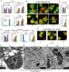
AINs produce and secrete granules more effectively than do GINs. (A-C) Ultrastructural images of GINs (A), AINs (B), or freshly isolated PBNs (C) with electron microscope. The vesicles in GINs appeared to contain less dense and amorphous material, whereas those in AINs showed dense material or both dense and amorphous material. AINs appeared to have more primary and secondary-like granules than do GINs. Arrows indicate examples of vesicles and granules. Ve indicates vesicle; Pg, primary granule; and Sg, secondary granule. (D) NE activity challenged by fMLP stimuli. CB indicates cytochalasin B. (E) AINs had a greater production and secretion of the cleaved 38-kDa product of MPO upon E coli stimuli. Images at the top of the horizontal line were derived from 8% gel, whereas images under the horizontal line from 12% gel. The band intensities of Actin detected in 12% gel are similar to those in 8% gel (data not shown). (F) Effective secretion of lactoferrin by AINs versus GINs upon E coli stimuli. To make the sample orders in the medium section match those in the lysate section, the positions between nonstimulated and stimulated samples in the medium section have been switched, as indicated by a vertical line. (G) Greater production and degranulation of LL37 by AINs versus GINs. (H) Increased abundance of intracellular MMP-9 upon E coli stimuli but insufficient degranulation in both GINs and AINs. To make the sample orders in the medium section match those in the lysate section, the positions between nonstimulated and stimulated samples in the medium section have been switched, as indicated by a vertical line. In addition, the samples loaded in lysate and medium sections in panels E through H were originally separated by a molecular weight (MW) marker. This MW marker was deleted so that a blank space is shown between lysate and medium samples.
Figure 3
Figure 3
Both unsorted and sorted AINs display a higher capacity for clearance of bacteria than do unsorted and sorted GINs. (A) Morphology of GINs and AINs before and after sorting with anti-CD15 antibody. Arrows indicate examples of monocytes. (B) Morphology of freshly isolated PBNs. (C-D) Quantification of monocytes (C) and band/segmented neutrophils (D) in panels A and B. (E-F) Comparison of the effect of unsorted and sorted GINs and AINs on the clearance of intracellular bacteria. Clearance efficiency was determined from the numbers of viable bacteria recovered from the intracellular compartment after infection. E coli DH5α at an MOI of 100 were used to infect sorted GINs, sorted AINs, and PBNs (F). (G) In situ labeling of bacteria with anti-OmpA antibodies in unsorted GINs and AINs.
Figure 4
Figure 4
AINs possess significantly higher phagocytotic and bactericidal activities than do GINs. (A) Phagocytotic and bactericidal activities against E coli infection were determined by the number of extracellular bacteria, phagocytized bacteria, recovered intracellular bacteria, and killed bacteria. There was a 1.26- ± 0.01-fold increase in bacterial numbers over the 45 minutes of the experiment (*GINs versus AINs, P < .03 at least). (B) Confocal fluorescence microscopy of surviving and dead bacteria in GINs, AINs and PBNs. White arrow indicates surviving (green) or dead bacteria (red), whereas black arrow indicates example of neutrophil nucleus stained in red simultaneously by PI fluorescent dye. Representative confocal fluorescence images of bacteria were reproduced at 60× magnification. (C) Quantification of both extracellular bacteria after infection and in situ killed bacteria in panel B (*GINs versus AINs or PBNs, P < .04 at least). (D-E) Similar to panels A and B, phagocytic and bactericidal activities against S aureus infection were determined in GINs, AINs, and PBNs (*GINs versus AINs, P < .04 at least). (F-H) Ultrastructural images of bacterial infection of GINs (F), AINs (G), and PBNs (H) with electron microscope. Lb indicates living bacteria; Db, suggestive of dead bacteria; Pg, primary granule; and Sg, secondary granule.
Figure 5
Figure 5
Granulocytes induced by Am80 coexpress CD66 and CD18 surface markers at a higher level than do those induced by G-CSF. (A-D) Flow cytometric analyses of G-CSF– versus Am80-induced granulocytes in coexpression of CD66a-CD18 and CD66a-CD11b surface markers (*GINs versus AINs. CD66a-CD18, P < .003; CD66a-CD11b, P < .028). (E-H) Increased induction of both CD66b-CD18 and CD66b-CD11b surface markers in Am80 versus G-CSF–induced granulocytes (*GINs versus AINs; CD66b-CD18, P < .047; CD66b-CD11b, P < .033). Corresponding isotypes and nonstaining (blank cells) served as controls.
Figure 6
Figure 6
Anti-CD18 antibody neutralizes Am80-enhanced neutrophil bactericidal activities. (A) Effects of anti-CD18 antibody on neutralization of AINs, GINs, and PBNs phagocytic and bactericidal activities against E coli (**GINs versus AINs without Abs, P < .037 at least; *AINs versus AINs+Abs, P < .036 at least; GINs versus GINs+Abs, P < .006 at least; PBNs versus PBNs+Abs, P < .007 at least). Abs indicates antibodies. (B) In situ bacterial killing imaged by confocal microscopy. White arrow indicates surviving (green) or dead bacteria (red), whereas black arrow indicates example of neutrophil nucleus stained in red simultaneously by PI fluorescent dye. (C) Quantification of both extracellular bacteria after infection and in situ killed bacteria in panel B (**GINs versus AINs or PBNs, P < .006 at least; *AINs+IgG versus AINs+Abs, P < .008; PBNs+IgG versus PBNs+Abs, P < .03). (D-E) Effects of anti-CD18 antibody on neutralization of AINs, GINs, and PBNs phagocytic and bactericidal activities against S aureus (**GINs versus AINs or PBNs, P < .03 at least; *AINs+IgG versus AINs+Abs, P < .007 at least; PBNs+IgG versus PBNs+Abs, P < .02 at least).
Figure 7
Figure 7
Neutrophils mobilized by Am80 in neutropenic mice display greater bactericidal activity than those by G-CSF. Twenty C57BL6/J mice were randomly divided into 4 groups for the experiments. (A) G-CSF induced a remarkably accelerated neutrophil recovery compared with mice treated with Am80 or vehicle at day 5. (B) Morphologic analysis of PB neutrophils at day 5 (*G-CSF versus Am80, P < 1.2 × 10⁻⁶; G-CSF versus control, P < 4.0 × 10⁻⁵; G-CSF versus vehicle, P < 8.2 × 10⁻⁸; Am80 versus vehicle, P < 1.3 × 10⁻⁶; Am80 versus control, P < .016. (C) Representative purity of PB neutrophils, as shown by freshly isolated GINs from mice. (D) Phagocytotic and bactericidal activities of neutrophils, isolated from PB of different mice, were reflected by the number of extracellular bacteria, phagocytized bacteria, and killed bacteria. *Extracellular: AINs versus GINs, P < .043; AINs versus C-MPBNs, P < 1.1 × 10⁻⁴; MPBNs versus C-MPBNs, P < 3.7 × 10⁻⁴; GINs versus C-MPBNs, P < .049. *Phagocytosis: AINs versus GINs, P < .026; AINs versus C-MPBNs, P < .02; MPBNs versus GINs, P < .042; MPBNs versus C-MPBNs, P < .003; GINs versus C-MPBNs, P < .009. *Killing: AINs versus GINs, P < .026; AINs versus C-MPBNs, P < .005; MPBNs versus GINs, P < .015; MPBNs versus C-MPBNs, P < .003; GINs versus C-MPBNs, P < .009. (E) Accelerated recoveries of WBC and neutrophil were ceased at day 7 after 96 hours of stimuli with G-CSF or Am80 or vehicle. (F) Morphologic analysis of PB neutrophils at day 9 (*G-CSF versus control, P < .005; Am80 versus control, P < 3.9 × 10⁻⁴; Am80 versus vehicle, P < .03; Vehicle versus control, P < .03). (G) AINs possessed significantly higher phagocytotic and bactericidal activities than do GINs 48 hours after cessation of accelerated neutrophil recovery (E). *Extracellular: AINs versus GINs, P < .02; AINs versus C-MPBNs, P < .007; MPBNs versus GINs, P < .04; MPBNs versus C-MPBNs, P < .02. *Phagocytosis: AINs versus C-MPBNs, P < .034; MPBNs versus C-MPBNs, P < .043. *Killing: AINs versus C-MPBNs, P < .034; MPBNs versus C-MPBNs, P < .043.

Similar articles

Cited by

References

    1. Gurion R, Belnik-Plitman Y, Gafter-Gvili A, et al. Colony-stimulating factors for prevention and treatment of infectious complications in patients with acute myelogenous leukemia. Cochrane Database Syst Rev. 2012;6:CD008238. - PMC - PubMed
    1. Smith TJ, Khatcheressian J, Lyman GH, et al. 2006 update of recommendations for the use of white blood cell growth factors: an evidence-based clinical practice guideline. J Clin Oncol. 2006;24(19):3187–3205. - PubMed
    1. Hartmann LC, Tschetter LK, Habermann TM, et al. Granulocyte colony-stimulating factor in severe chemotherapy-induced afebrile neutropenia. N Engl J Med. 1997;336(25):1776–1780. - PubMed
    1. Kuderer NM, Dale DC, Crawford J, Cosler LE, Lyman GH. Mortality, morbidity, and cost associated with febrile neutropenia in adult cancer patients. Cancer. 2006;106(10):2258–2266. - PubMed
    1. Sung L, Nathan PC, Alibhai SM, Tomlinson GA, Beyene J. Meta-analysis: effect of prophylactic hematopoietic colony-stimulating factors on mortality and outcomes of infection. Ann Intern Med. 2007;147(6):400–411. - PubMed

Publication types

MeSH terms