. 2013 Mar;195(5):977-89.
doi: 10.1128/JB.01274-12. Epub 2012 Dec 14.

Bacillus anthracis acetyltransferases PatA1 and PatA2 modify the secondary cell wall polysaccharide and affect the assembly of S-layer proteins

Affiliations

Bacillus anthracis acetyltransferases PatA1 and PatA2 modify the secondary cell wall polysaccharide and affect the assembly of S-layer proteins

J Mark Lunderberg et al. J Bacteriol. 2013 Mar.

Abstract

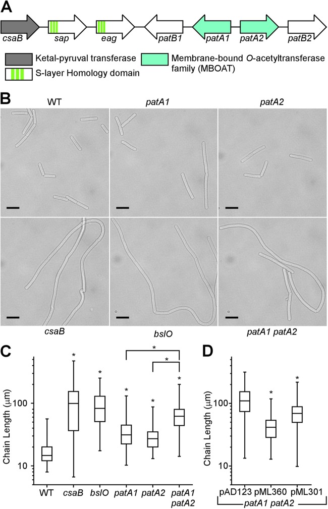
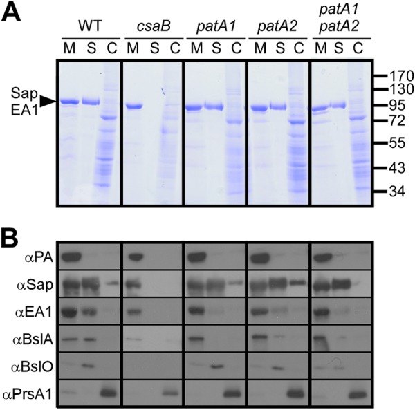
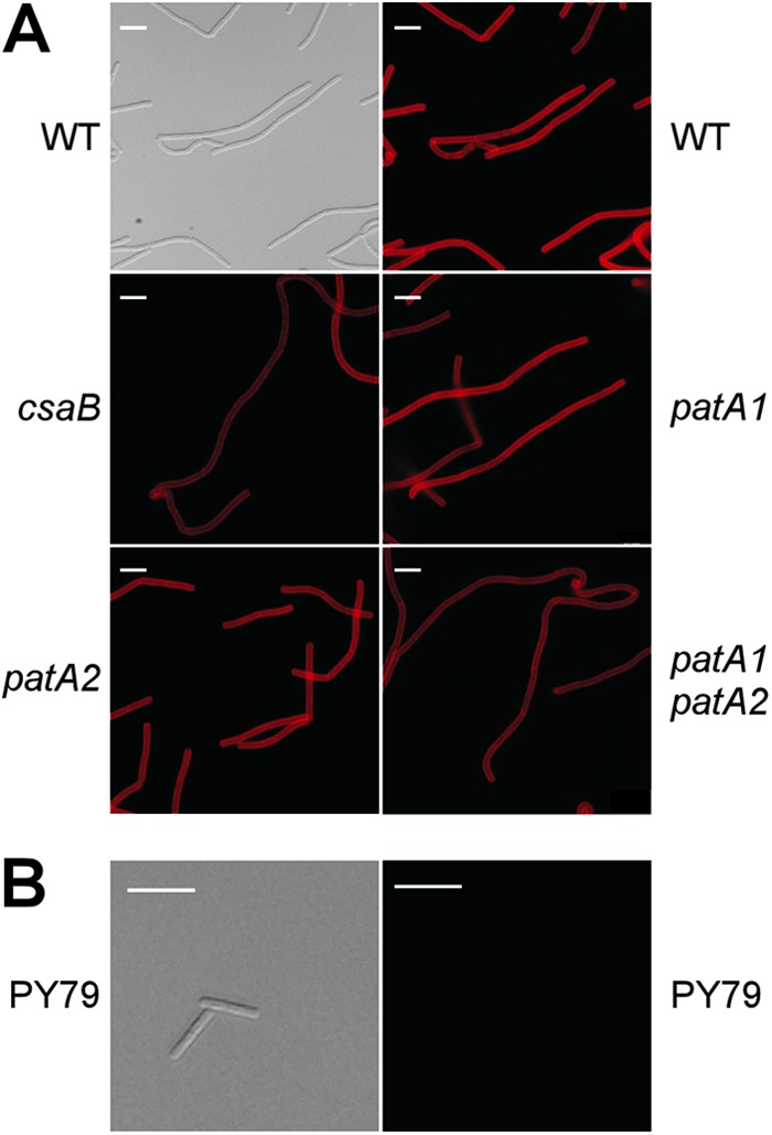
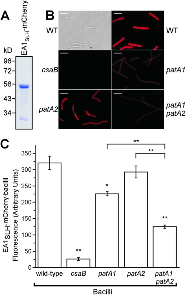
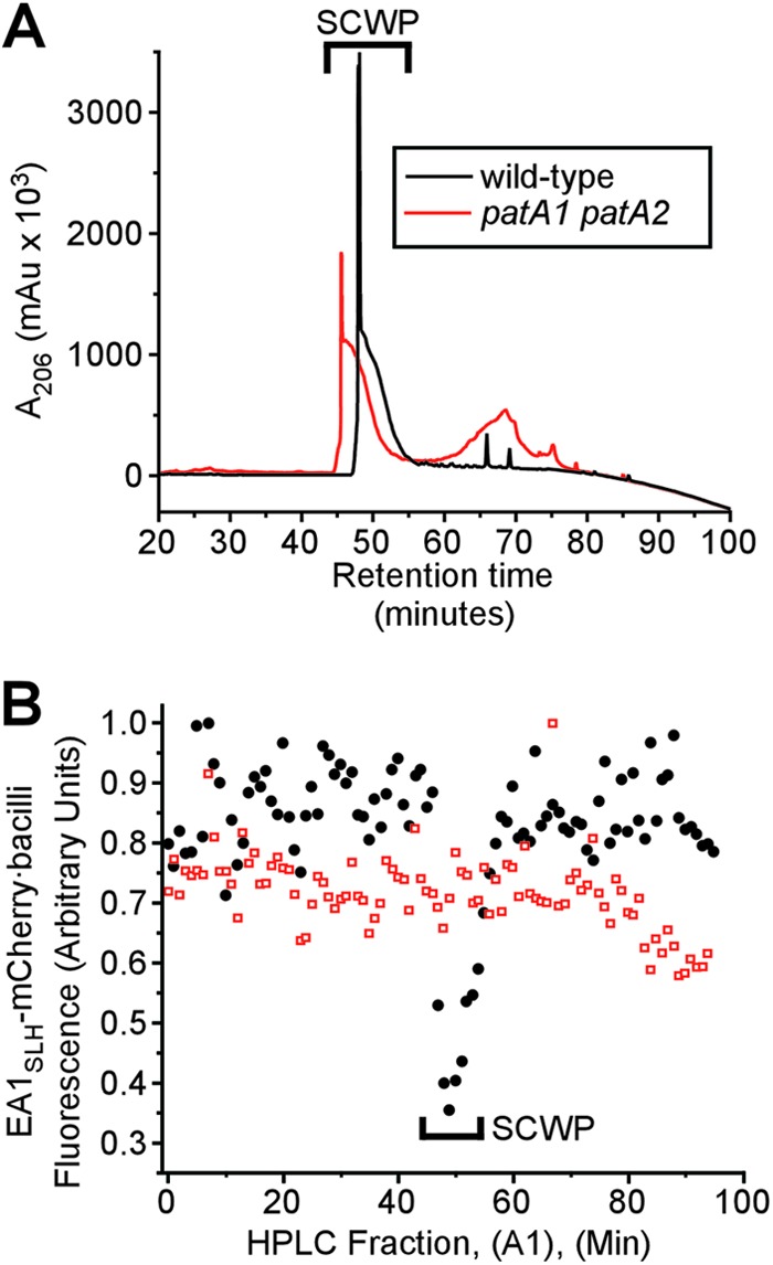
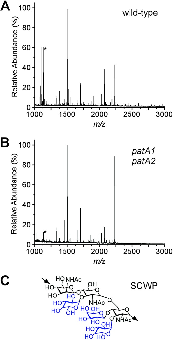
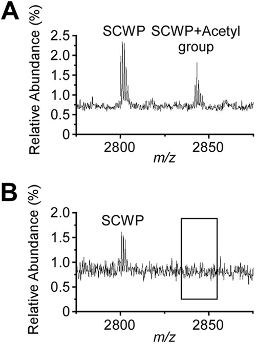
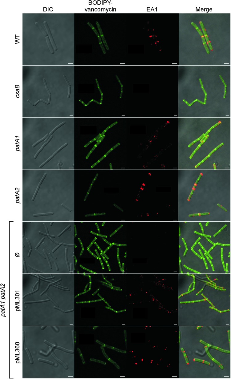
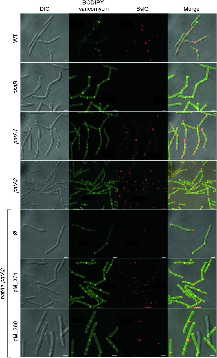
The envelope of Bacillus anthracis encompasses a proteinaceous S-layer with two S-layer proteins (Sap and EA1). Protein assembly in the envelope of B. anthracis requires S-layer homology domains (SLH) within S-layer proteins and S-layer-associated proteins (BSLs), which associate with the secondary cell wall polysaccharide (SCWP), an acetylated carbohydrate that is tethered to peptidoglycan. Here, we investigated the contributions of two putative acetyltransferases, PatA1 and PatA2, on SCWP acetylation and S-layer assembly. We show that mutations in patA1 and patA2 affect the chain lengths of B. anthracis vegetative forms and perturb the deposition of the BslO murein hydrolase at cell division septa. The patA1 and patA2 mutants are defective for the assembly of EA1 in the envelope but retain the ability of S-layer formation with Sap. SCWP isolated from the patA1 patA2 mutant lacked acetyl moieties identified in wild-type polysaccharide and failed to associate with the SLH domains of EA1. A model is discussed whereby patA1- and patA2-mediated acetylation of SCWP enables the deposition of EA1 as well as BslO near the septal region of the B. anthracis envelope.


Figures

Fig 1
Fig 1
Mutations in the patA1 and patA2 genes cause chain length phenotypes in Bacillus anthracis vegetative forms. (A) Genes for the PatA1 and PatA2 acetyltransferases are located on the Bacillus anthracis chromosome immediately adjacent to genes for the major S-layer proteins (sap and eag) and to csaB, which encodes a ketal pyruvyl transferase essential for S-layer formation. (B) DIC microscopy images of vegetative forms germinated from B. anthracis wild-type and csaB, bslO, patA1, patA2, and patA1 patA2 mutant spores. Scale bar, 10 μm. (C) The chain lengths of 100 vegetative bacilli were quantified for each strain; the data are presented as a box-and-whiskers plot, where the size of each box is defined by the boundaries of the 25th and 75th percentiles, the whiskers are the maximum and minimum measured lengths, and the center bar indicates the median length. Data were examined with an unpaired two-tailed Student's t test. A significant difference (P < 0.0001) relative to wild-type is indicated with an asterisk (*). The patA1 patA2 mutant was also compared to mutants with singular deletions of patA1 and patA2 genes. (D) The B. anthracis patA1 patA2 mutant strain was transformed with pAD123, i.e., the empty vector, pML360, a plasmid expressing patA1-patB1, or pML301, a plasmid expressing patA2-patB2. A significant difference (P < 0.0001) between the empty vector and the pML360 and pML301 strains was observed after data sets were compared with a Student t test.
Fig 2
Fig 2
Impact of patA1 and patA2 mutations on S-layer protein and S-layer-associated protein trafficking in B. anthracis. (A) B. anthracis vegetative cultures were fractionated into medium (M), S-layer (S), and cell (C) fractions, and proteins in each sample were analyzed by Coomassie-stained SDS-PAGE gels. The arrowhead denotes the migratory position of the S-layer proteins Sap and EA1, and the numbers indicate the position of molecular mass markers (in kDa). (B) Samples generated in the experiment shown in panel A were examined by immunoblotting with antibodies raised against protective antigen (PA), a secreted protein and component of anthrax toxins (42), and PrsA1, a peptidylprolyl isomerase and membrane lipoprotein (43), as well as S-layer proteins (Sap and EA1) and S-layer-associated proteins (BslA and BslO). α, anti.
Fig 3
Fig 3
Mutation in csaB, patA1, or patA2 does not alter the abundance of the SCWP in B. anthracis. (A) Vegetative forms derived from wild-type, csaB, patA1, patA2, and patA1 patA2 mutant B. anthracis spores were stripped of their S-layers via treatment with 3 M urea. Bacilli were subsequently fixed with 4% paraformaldehyde and subjected either to DIC microscopy or to immunofluorescence microscopy with rabbit polyclonal antibodies raised against purified SCWP-BSA conjugate followed by secondary antibody conjugated to DyLight594. Scale bar, 10 μm. (B) B. subtilis strain PY79, a strain that does not elaborate an SCWP, does not show immunoreactivity toward the SCWP antibody. A refreshed overnight growth was fixed with 4% paraformaldehyde and subjected to DIC microscopy and immunofluorescence microscopy in the same manner as the B. anthracis strains. Scale bar, 10 μm.
Fig 4
Fig 4
The SLH domain of EA1 displays reduced binding to the SCWP from patA1 patA2 mutant B. anthracis. (A) Coomassie-stained SDS-PAGE gel of the purified fusion protein EA1SLH-mCherry. Numbers indicate the positions of molecular mass markers (in kDa). (B) Vegetative forms of B. anthracis strains were stripped of their S-layers via treatment with 3 M urea. Differential interference contrast (DIC) and fluorescence microscopy images of the wild type and fluorescence microscopy images of csaB, patA1, patA2, and patA1 patA2 mutant B. anthracis strains incubated with EA1SLH-mCherry are shown. Scale bar, 15 μm. (C) The fluorescence intensity of EA1SLH-mCherry binding to the envelope of S-layer-stripped B. anthracis strains was quantified in three independent experimental determinations. Data were averaged and examined with an unpaired two-tailed Student's t test. *, P < 0.01; **, P < 0.001.
Fig 5
Fig 5
SCWP isolated from wild-type and patA1 patA2 mutant B. anthracis strains. (A) The SCWP was released via hydrofluoric acid (HF) treatment from murein sacculi that had been isolated from either wild-type or patA1 patA2 mutant vegetative forms. Ethanol-precipitated SCWP was subjected to rpHPLC, and the eluate's absorbance at 206 nm (A206) was recorded in milli-arbitrary units (mAu). (B) Collected rpHPLC fractions from panel A (A1), were tested for competitive inhibition of EA1SLH-mCherry binding to the SCWP of wild-type bacilli. SCWPs from wild-type but not SCWPs from patA1 patA2 mutant bacilli displayed competitive inhibitor activities toward the binding of bacilli to EA1SLH-mCherry.
Fig 6
Fig 6
MALDI-TOF mass spectrometry of SCWP isolated from wild-type and patA1 patA2 mutant B. anthracis strains. (A) rpHPLC-purified SCWP from wild-type B. anthracis was subjected to MALDI-TOF mass spectrometry, and ion spectra were recorded. m/z, mass-to-charge ratio. The ion signal marked with an asterisk (*) identifies the sodiated adduct of a compound with the predicted structure [β-ManNAc-(1→4)-β-GlcNAc(Gal)-(1→6)-α-GlcNAc(Gal)2]. (B) rpHPLC-purified SCWP from patA1 patA2 mutant B. anthracis was analyzed by MALDI-TOF mass spectrometry, and ion spectra were recorded. (C) The SCWP is comprised of the repeating unit [→4)-β-ManNAc-(1→4)-β-GlcNAc-(1→6)-α-GlcNAc-(1→]n (printed in black), where α-GlcNAc is replaced with α-Gal and β-Gal at O-3 and O-4, respectively, and the β-GlcNAc is replaced with α-Gal at O-3 (galactosyl substitutions printed in blue) (16).
Fig 7
Fig 7
Ion signals for acetylated SCWP in wild-type but not in patA1 patA2 mutant B. anthracis cells. (A) Ion signals from the wild-type SCWP were detected with MALDI-TOF mass spectrometry; m/z 2,800.7 was interpreted as the sodiated ion [ManNAc-GlcNAc2(Gal3)]2-ManNac-GlcNAc(Gal), and m/z 2,842.8 was interpreted as its acetylated variant. (B) Ion signals from the patA1 patA2 mutant SCWP were detected with MALDI-TOF mass spectrometry; m/z 2,800.7 was interpreted as the sodiated ion [ManNAc-GlcNAc2(Gal3)]2-ManNac-GlcNAc(Gal). m/z 2,842.7 was not detected in the spectrum. Of note, although mass spectrometry data are in agreement with the proposed compound structures, they do not constitute experimental proof for the structural assignments.
Fig 8
Fig 8
Localization of the S-layer protein EA1 in the envelope of wild-type, csaB, patA1, patA2, and patA1 patA2 B. anthracis strains. Spores from wild-type and mutant B. anthracis strains were diluted into BHI broth, and germinated bacilli were incubated for 3 h. Vegetative forms were fixed in 4% buffered formalin. Differential interference contrast (DIC) and fluorescence microscopy with BODIPY-vancomycin or rabbit polyclonal antibodies staining against the S-layer protein EA1 followed by secondary antibody DyLight594 conjugate were used to acquire images. Data sets were merged to reveal the location of cell wall septa (BODIPY-vancomycin) and S-layer protein EA1 in wild-type and mutant bacilli. The patA1 patA2 mutant strain was transformed with plasmid pML301, expressing patA1 and patB1, or pML360, expressing patA2 and patB2, or left untransformed (Ø). Scale bar, 1 μm.
Fig 9
Fig 9
Localization of the S-layer protein EA1 in the envelope of wild-type, csaB, patA1, patA2, and patA1 patA2 B. anthracis strains. Spores from wild-type and mutant B. anthracis strains were diluted into BHI broth, and germinated bacilli were incubated for 3 h. Vegetative forms were fixed in 4% buffered formalin. Differential interference contrast (DIC) and fluorescence microscopy with BODIPY-vancomycin or rabbit polyclonal antibodies staining against the S-layer-associated protein BslO followed by secondary antibody DyLight594 conjugate were used to acquire images. Data sets were merged to reveal the location of cell wall septa (BODIPY-vancomycin) and the S-layer-associated protein BslO in wild-type and mutant bacilli. The patA1 patA2 mutant strain was transformed with plasmid pML301, expressing patA1 and patB1, or pML360, expressing patA2 and patB2, or left untransformed (Ø). Scale bar, 1 μm.

References

    1. Koch R. 1876. Die Ätiologie der Milzbrand-Krankheit, begründet auf die Entwicklungsgeschichte des Bacillus anthracis. Beitr. Biol. Pflanz. 2: 277– 310
    1. Marraffini LA, Schneewind O. 2006. Targeting proteins to the cell wall of sporulating Bacillus anthracis. Mol. Microbiol. 62: 1402– 1417 - PubMed
    1. Candela T, Fouet A. 2006. Poly-gamma-glutamate in bacteria. Mol. Microbiol. 60: 1091– 1098 - PubMed
    1. Richter GS, Anderson VJ, Garufi G, Lu L, Joachimiak A, He C, Schneewind O, Missiakas D. 2009. Capsule anchoring in Bacillus anthracis occurs by a transpeptidation mechanism that is inhibited by capsidin. Mol. Microbiol. 71: 404– 420 - PMC - PubMed
    1. Anderson VJ, Kern JW, McCool JW, Schneewind O, Missiakas DM. 2011. The SLH domain protein BslO is a determinant of Bacillus anthracis chain length. Mol. Microbiol. 81: 192– 205 - PMC - PubMed

Publication types

MeSH terms

LinkOut - more resources