. 2013 Feb 15;288(7):4744-54.
doi: 10.1074/jbc.M112.408039. Epub 2012 Dec 14.

Crystal structures of the DNA-binding domain tetramer of the p53 tumor suppressor family member p73 bound to different full-site response elements

Affiliations

Crystal structures of the DNA-binding domain tetramer of the p53 tumor suppressor family member p73 bound to different full-site response elements

Abdul S Ethayathulla et al. J Biol Chem. .

Abstract

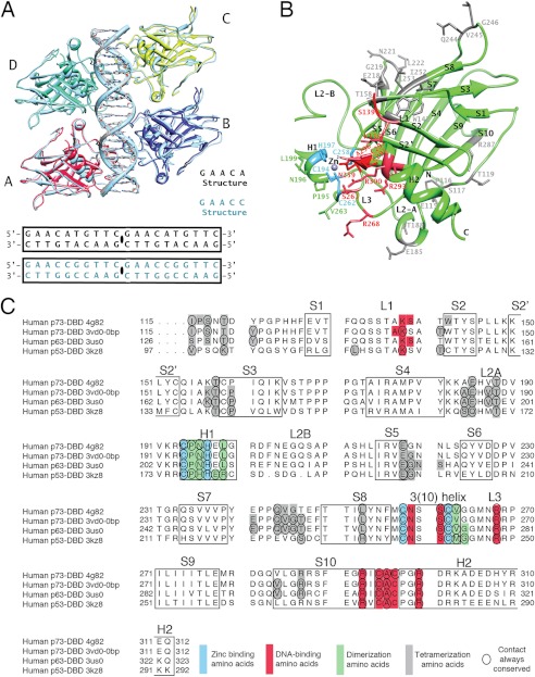
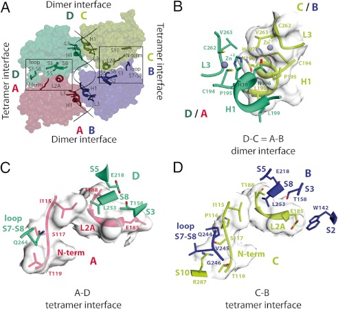
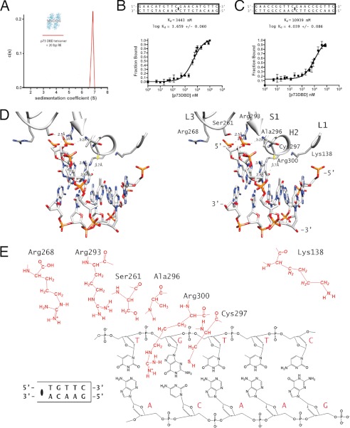
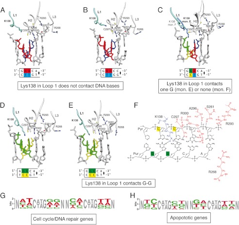
How cells choose between developmental pathways remains a fundamental biological question. In the case of the p53 protein family, its three transcription factors (p73, p63, and p53) each trigger a gene expression pattern that leads to specific cellular pathways. At the same time, these transcription factors recognize the same response element (RE) consensus sequences, and their transactivation of target genes overlaps. We aimed to understand target gene selectivity at the molecular level by determining the crystal structures of the p73 DNA-binding domain (DBD) in complex with full-site REs that vary in sequence. We report two structures of the p73 DBD bound as a tetramer to 20-bp full-site REs based on two distinct quarter-sites: GAACA and GAACC. Our study confirms that the DNA-binding residues are conserved within the p53 family, whereas the dimerization and tetramerization interfaces diverge. Moreover, a conserved lysine residue in loop L1 of the DBD senses the presence of guanines in positions 2 and 3 of the quarter-site RE, whereas a conserved arginine in loop 3 adapts to changes in position 5. Sequence variations in the RE elicit a p73 conformational response that might explain target gene specificity.


Figures

FIGURE 1.
FIGURE 1.
Structures of the p73 DBD tetramer in complex with a full-site RE. A, GAACA and GAACC tetramer structures. Monomers A and B in complex with half of the 20-bp DNAs used for crystallization form the asymmetric unit. Monomers C and D and the other half of the 20-bp DNA are related by translation and are identical to the content of the asymmetric unit. B, p73 DBD monomer. The secondary structure elements are labeled following the established nomenclature for the p53 DBD (17). There are 10 β-strands (S1–S10), two helices (H1 and H2), and three long loops (L1, L2A/L2B, and L3). Residues involved in dimerization (green), tetramerization (gray), zinc binding (blue), and DNA binding (red) are labeled. C, sequence alignment of the DBDs of the three members of the p53 protein family. Amino acids forming the secondary structure elements are boxed. Colored boxes indicate the residues involved in dimerization (green), tetramerization (gray), zinc binding (blue), and DNA binding (red) for four structures that have a similar tetrameric arrangement (4G82 described in this study and 3VD0 Model 1 for the p73 DBD-DNA complex (27), 3US0 for the p63-DNA complex (26), and 3KZ8 for the p53-DNA complex (21)). Residues where the same contact is conserved for all of the monomers of the tetramer are circled.
FIGURE 2.
FIGURE 2.
Dimerization and tetramerization interfaces of the p73 DBD tetramer bound to a full-site RE. A, surface area model of the protein tetramer showing the two dimerization interfaces (AB and DC) and the two tetramerization interfaces (AD and CB). B, the DC (green/yellow) and AB (red/blue) dimerization interfaces are identical due to crystal symmetry. The same two secondary structure elements, helix H2 and loop L3, are involved in the DC and AB dimerization interfaces. Three of the four residues that coordinate the zinc ion are displayed (Cys-194, His-197, and Cys-262), together with the four residues involved in the dimerization (Pro-195, Asn-196, Leu-199, and Val-263). C, atomic details of the AD tetramerization interface. Residues in the N terminus and loop L2A of monomer A (red) contact residues in the S7–S8 loop and strands S3, S5, and S8 of monomer D (green). D, atomic details of the CB tetramerization interface. Residues in the N terminus and loop L2A of monomer C (yellow) contact residues in the S7–S8 loop and strands S2, S3, S5, and S8 of monomer B (blue).
FIGURE 3.
FIGURE 3.
Binding of the p73 DBD to a full-site RE. A, sedimentation coefficient distribution of the p73 DBD bound to a 20-bp DNA containing the same full-site RE used in crystallization experiments obtained in a sedimentation velocity experiment. B, binding affinity constant of the p73 DBD measured by fluorescence anisotropy using fluorescein-labeled DNA for the full-site RE used in crystallization of the GAACA structure. C, binding affinity constant of the p73 DBD measured by fluorescence anisotropy using fluorescein-labeled DNA for the full-site RE used in crystallization of the GAACC structure. D, stereo view of the residues of monomer A from the GAACA crystal structure that have been described as contacting DNA (27). E, schematic diagram of the atomic interactions between the p73 DBD and a quarter-site RE of the GAACA structure.
FIGURE 4.
FIGURE 4.
Comparison of the DNA-binding residues of five structures of the p73 DBD in complex with DNA. A and B, residues in the p73 DBD that contact the DNA bases and backbone for the quarter-site REs (5′-GAACA-3′ and 5′-GAACC-3′). Lys-138 in loop L1 is far from the DNA bases and does not contact the DNA. C, residues in the p73 DBD that contact the DNA bases and backbone for the quarter-site RE (5′-GGACA-3′). Lys-138 in loop L1 acquires two conformations. Monomer (mon.) E does not contact the bases; monomer F is far from the DNA bases and does not contact the DNA. D and E, residues in the p73 DBD that contact the DNA bases and backbone for the quarter-site REs (5′-AGGCA-3′ and 5′-GGGCC-3′). In both structures, Lys-138 reaches the DNA bases in the major groove forming two hydrogen bonds with the O2 carbonyl oxygens of the guanines in positions 2 and 3 of the quarter-site RE. F, schematic diagram of the atomic interactions between the p73 DBD and DNA with guanines in positions 2 and 3 of the consensus quarter-site RE. G, WebLogo diagram of positions 2 and 3 of each quarter-site in the 20-bp REs of cell cycle and DNA repair genes found in microarrays to be activated by p73 (39). H, WebLogo diagram of positions 2 and 3 of each quarter-site in the 20-bp REs of apoptotic genes found in microarrays to be activated by p73 (39).

Similar articles

Cited by

References

    1. Riley T., Sontag E., Chen P., Levine A. (2008) Transcriptional control of human p53-regulated genes. Nat. Rev. Mol. Cell Biol. 9, 402–412 - PubMed
    1. Menendez D., Inga A., Resnick M. A. (2009) The expanding universe of p53 targets. Nat. Rev. Cancer 9, 724–737 - PubMed
    1. Yang A., Walker N., Bronson R., Kaghad M., Oosterwegel M., Bonnin J., Vagner C., Bonnet H., Dikkes P., Sharpe A., McKeon F., Caput D. (2000) p73-deficient mice have neurological, pheromonal and inflammatory defects but lack spontaneous tumours. Nature 404, 99–103 - PubMed
    1. Kommagani R., Whitlatch A., Leonard M. K., Kadakia M. P. (2010) p73 is essential for vitamin D-mediated osteoblastic differentiation. Cell Death Differ. 17, 398–407 - PMC - PubMed
    1. Mills A. A., Zheng B., Wang X.-J., Vogel H., Roop D. R., Bradley A. (1999) p63 is a p53 homologue required for limb and epidermal morphogenesis. Nature 398, 708–713 - PubMed

Publication types

MeSH terms