

Primary myeloid sarcoma masquerading as an obstructing duodenal carcinoma
- PMID: 23243527
- PMCID: PMC3517833
- DOI: 10.1155/2012/490438
Primary myeloid sarcoma masquerading as an obstructing duodenal carcinoma
Abstract
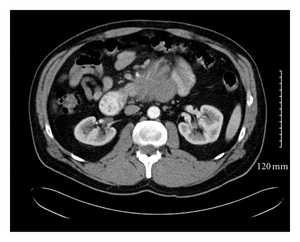
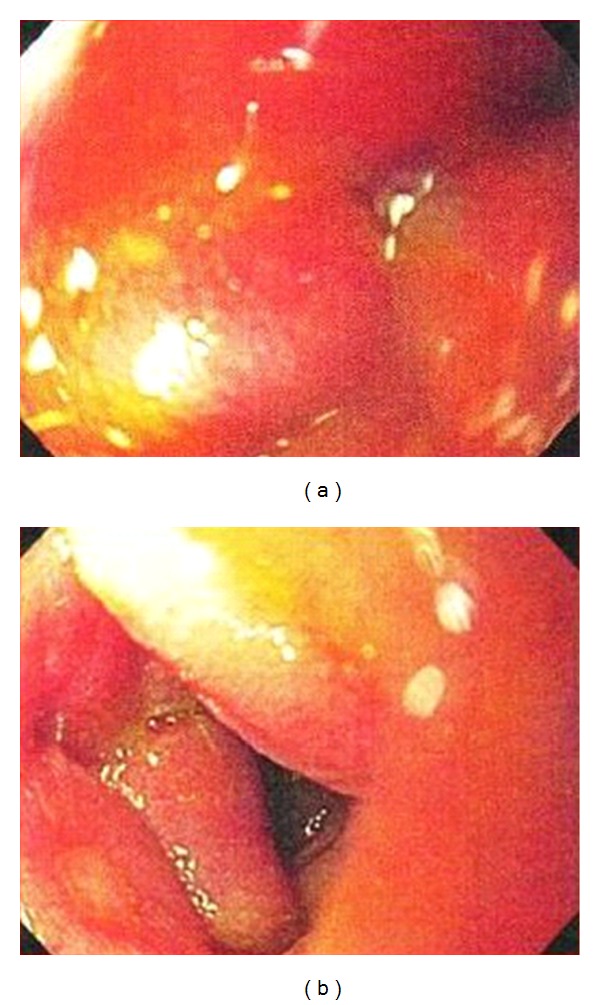
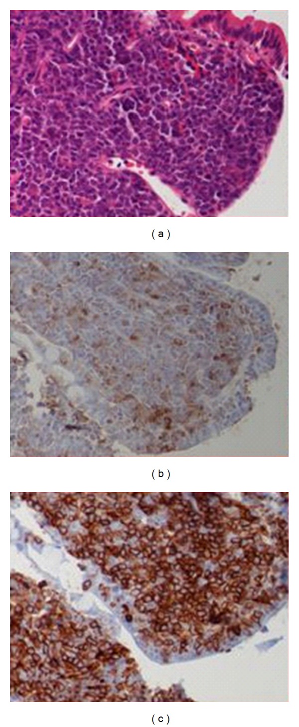
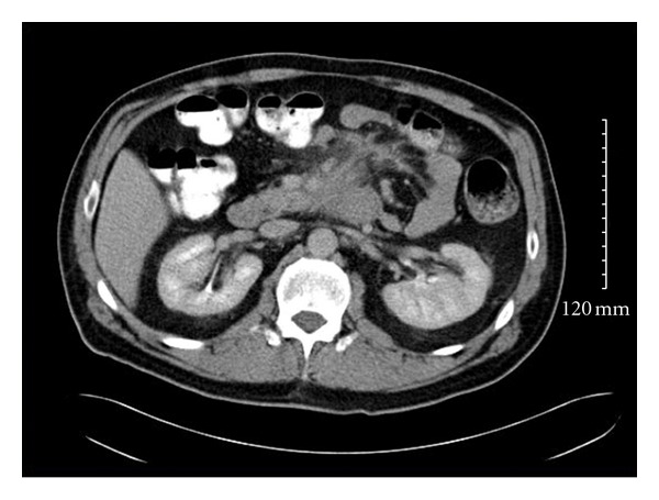
Myeloid Sarcoma (MS), a rare extra hematopoietic carcinoma composed of blast cells, is located primarily in extramedullary sites such as skin, soft tissue, lymph nodes, and bone. MS usually presents in the setting of coexisting acute myeloid leukemia (AML) and myeloproliferative disorders. Gastrointestinal involvement (GI) is extremely rare from nonspecific abdominal symptoms to obstruction. Eight cases of myeloid sarcoma involving the duodenum including the current case have been reported, overall mean age being 40 years (range 17-71) and M : F ratio 7 : 1. The prognosis of patients with de novo MS cases has been reported to be better than those who have a coexisting leukemia. MS is a rare extramedullary tumor, which should be considered in the differential diagnosis of a soft tissue mass involving the duodenum, especially if there is a coexisting hematological disorder. De novo cases often progress to AML, and current therapy involves Daunorubicin- and Cytarabine-based chemotherapy. The wide cytogenetic and molecular heterogeneity of MS implies a potential role for more targeted MS therapies, which may offer a curative strategy.
Figures
Similar articles
-
Myeloid Sarcoma of the Paranasal Sinuses in a Patient with Acute Myeloid Leukemia.Tohoku J Exp Med. 2018 Oct;246(2):141-146. doi: 10.1620/tjem.246.141. Tohoku J Exp Med. 2018. PMID: 30369515
-
Myeloid sarcoma of the periprostatic tissue and prostate: Case report and review of literature.Urol Ann. 2016 Jul-Sep;8(3):348-54. doi: 10.4103/0974-7796.184890. Urol Ann. 2016. PMID: 27453659 Free PMC article.
-
Extramedullary Acute Leukemia-Still an Unforeseen Presentation.Hematol Rep. 2022 Apr 18;14(2):143-148. doi: 10.3390/hematolrep14020021. Hematol Rep. 2022. PMID: 35466185 Free PMC article.
-
Myeloid sarcoma in children - diagnostic and therapeutic difficulties.Contemp Oncol (Pozn). 2016;20(6):444-448. doi: 10.5114/wo.2016.65602. Epub 2017 Jan 12. Contemp Oncol (Pozn). 2016. PMID: 28239280 Free PMC article. Review.
-
Diagnostic Approaches in Myeloid Sarcoma.Curr Issues Mol Biol. 2025 Feb 10;47(2):111. doi: 10.3390/cimb47020111. Curr Issues Mol Biol. 2025. PMID: 39996833 Free PMC article. Review.
Cited by
-
Gastrointestinal Myeloid Sarcoma a Case Presentation and Review of the Literature.Mediterr J Hematol Infect Dis. 2021 Nov 1;13(1):e2021067. doi: 10.4084/MJHID.2021.067. eCollection 2021. Mediterr J Hematol Infect Dis. 2021. PMID: 34804441 Free PMC article. Review.
-
Gastric myeloid sarcoma without acute myeloblastic leukemia.World J Gastroenterol. 2015 Feb 21;21(7):2242-8. doi: 10.3748/wjg.v21.i7.2242. World J Gastroenterol. 2015. PMID: 25717265 Free PMC article.
-
Myeloid sarcoma derived from the gastrointestinal tract: A case report and review of the literature.Oncol Lett. 2016 Jun;11(6):4155-4159. doi: 10.3892/ol.2016.4517. Epub 2016 May 4. Oncol Lett. 2016. PMID: 27313759 Free PMC article.
-
Myeloid sarcoma presenting with leukemoid reaction in a child treated for acute lymphoblastic leukemia.Case Rep Hematol. 2014;2014:757625. doi: 10.1155/2014/757625. Epub 2014 Sep 3. Case Rep Hematol. 2014. PMID: 25276445 Free PMC article.
-
A myeloid sarcoma involving the small intestine, kidneys, mesentery, and mesenteric lymph nodes: A case report and literature review.Medicine (Baltimore). 2017 Oct;96(42):e7934. doi: 10.1097/MD.0000000000007934. Medicine (Baltimore). 2017. PMID: 29049187 Free PMC article. Review.
References
-
- Pileri SA, Ascani S, Cox MC, et al. Myeloid sarcoma: clinico-pathologic, phenotypic and cytogenetic analysis of 92 adult patients. Leukemia. 2007;21(2):340–350. - PubMed
-
- Choi EK, Ha HK, Park SH, et al. Granulocytic sarcoma of bowel: CT findings. Radiology. 2007;243(3):752–759. - PubMed
-
- Burns A. Observation of Surgical Anatomy, Head and Neck. Edinburgh, Scotland: Thomas Royce; 1811.
-
- King A. A case of chloroma. Monthly Journal of the Medical Society. 1853;17:p. 97.
-
- Rappaport H. Atlas of Tumor Pathology, Section III, Fascicle 8. Armed Forces Institute of Pathology. Washington, DC, USA: 1966. Tumours of the hematopoietic system; pp. 241–243.
LinkOut - more resources
Full Text Sources
Research Materials
