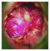
Hemangioma of the tympanic membrane: a case and a review of the literature
- PMID: 23243539
- PMCID: PMC3518088
- DOI: 10.1155/2012/402630
Hemangioma of the tympanic membrane: a case and a review of the literature
Abstract
Hemangiomas of the external auditory canal, involving the posterior bony canal and the adjacent tympanic membrane, although rare, are considered a specific disease entity of the human external auditory canal. Hemangiomas of the tympanic membrane and/or external auditory canal are rare entities; there are 16 previous case reports in the literature. It is a benign vascular tumor. It generally occurs in males in the sixth decade of life. Total surgical excision with or without tympanic membrane grafting appears to be effective in the removal of this benign neoplasm. The authors present a case and a review of the literature discussing diagnostic and surgical approaches.
Figures


Similar articles
-
Hemangioma of the external auditory canal.Am J Otol. 1983 Oct;5(2):125-6. Am J Otol. 1983. PMID: 6650669
-
Cavernous Hemangioma of the External Auditory Canal Involving the Tympanic Membrane: Case Report and Literature Review.Ear Nose Throat J. 2025 Jan 1:1455613241312072. doi: 10.1177/01455613241312072. Online ahead of print. Ear Nose Throat J. 2025. PMID: 39743548
-
Capillary Hemangioma of the Tympanic Membrane and External Auditory Canal.J Craniofac Surg. 2017 May;28(3):e231-e232. doi: 10.1097/SCS.0000000000003437. J Craniofac Surg. 2017. PMID: 28468198
-
Cavernous hemangioma of the tympanic membrane and external ear canal.Am J Otolaryngol. 2007 May-Jun;28(3):180-3. doi: 10.1016/j.amjoto.2006.03.012. Am J Otolaryngol. 2007. PMID: 17499135 Review.
-
Bilateral spontaneous symptomatic temporomandibular joint herniation into the external auditory canal: A case report and literature review.Auris Nasus Larynx. 2018 Apr;45(2):346-350. doi: 10.1016/j.anl.2017.03.011. Epub 2017 Apr 14. Auris Nasus Larynx. 2018. PMID: 28416346 Review.
Cited by
-
Mixed hemangioma of the external auditory canal and the tympanic membrane in a young woman: A case report.Clin Case Rep. 2022 Feb 18;10(2):e05452. doi: 10.1002/ccr3.5452. eCollection 2022 Feb. Clin Case Rep. 2022. PMID: 35223015 Free PMC article.
References
-
- Freedman SI, Barton S, Goodhill V. Cavernous angiomas of the tympanic membrane. Archives of Otolaryngology. 1972;96(2):158–160. - PubMed
-
- Balkany TJ, Meyers AD, Wong ML. Capillary hemangioma of the tympanic membrane. Archives of Otolaryngology. 1978;104(5):296–297. - PubMed
-
- Jackson CG, Levine CS, McKennan KX. Recurrent hemangioma of the external auditory canal. American Journal of Otology. 1990;11(2):117–118. - PubMed
-
- Andrade JM, Gehris CW, Breitenecker R. Cavernous hemangioma of the tympanic membrane. A case report. American Journal of Otology. 1983;4(3):198–199. - PubMed
-
- Kemink JL, Graham MD, McClatchey KD. Hemangioma of the external auditory canal. American Journal of Otology. 1983;5(2):125–126. - PubMed
LinkOut - more resources
Full Text Sources

