. 2012 Dec 1;3(12):3087-104.
doi: 10.1364/BOE.3.003087. Epub 2012 Nov 1.

A 3D glass optrode array for optical neural stimulation

Affiliations

A 3D glass optrode array for optical neural stimulation

T V F Abaya et al. Biomed Opt Express. .

Abstract

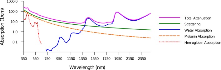
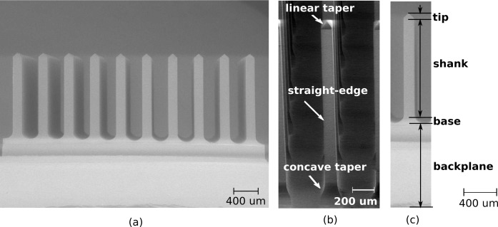
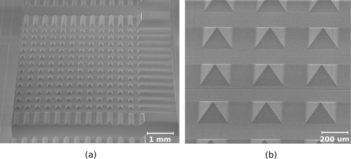
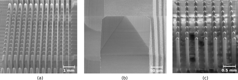
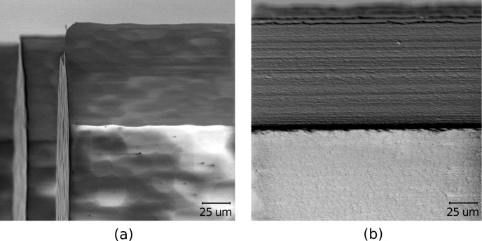
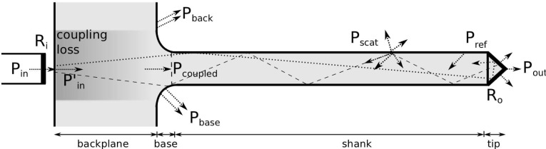
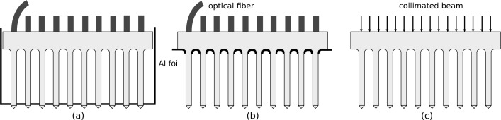
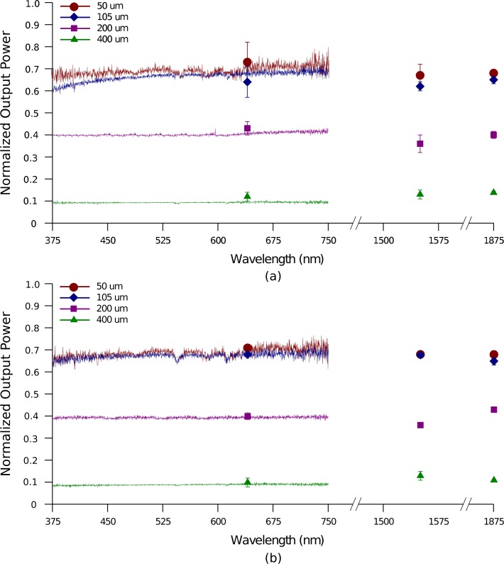
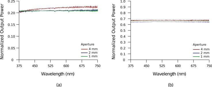
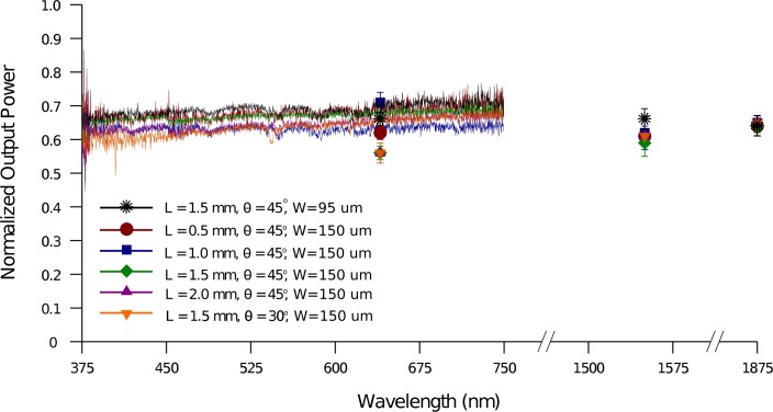
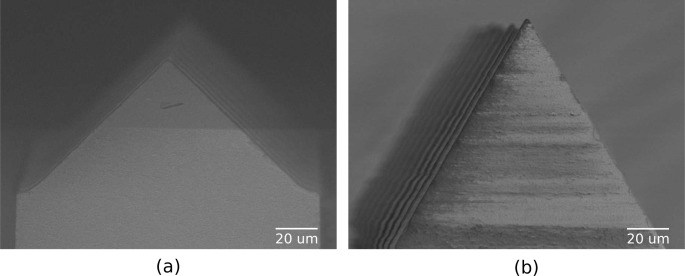
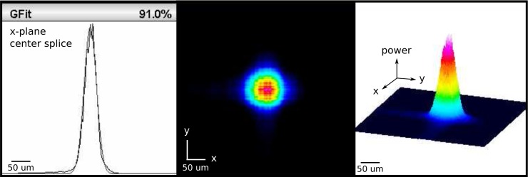
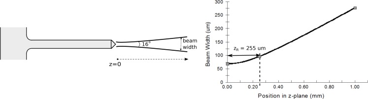
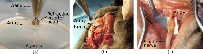
This paper presents optical characterization of a first-generation SiO(2) optrode array as a set of penetrating waveguides for both optogenetic and infrared (IR) neural stimulation. Fused silica and quartz discs of 3-mm thickness and 50-mm diameter were micromachined to yield 10 × 10 arrays of up to 2-mm long optrodes at a 400-μm pitch; array size, length and spacing may be varied along with the width and tip angle. Light delivery and loss mechanisms through these glass optrodes were characterized. Light in-coupling techniques include using optical fibers and collimated beams. Losses involve Fresnel reflection, coupling, scattering and total internal reflection in the tips. Transmission efficiency was constant in the visible and near-IR range, with the highest value measured as 71% using a 50-μm multi-mode in-coupling fiber butt-coupled to the backplane of the device. Transmittance and output beam profiles of optrodes with different geometries was investigated. Length and tip angle do not affect the amount of output power, but optrode width and tip angle influence the beam size and divergence independently. Finally, array insertion in tissue was performed to demonstrate its robustness for optical access in deep tissue.

Keywords: (170.3660) Light propagation in tissues; (170.3890) Medical optics instrumentation; (220.4610) Optical fabrication; (230.7380) Waveguides, channeled.


Figures

Fig. 1
Fig. 1
Tissue attenuation spectrum. Light transport of wavelengths in the visible range is more strongly affected by scattering, while absorption is dominant in the infrared. Penetration depth (i.e., depth where intensity falls to 1/e of surface value) is limited to about 1 mm.
Fig. 2
Fig. 2
SEM image of a 3D optrode array made from glass. (a) 10×10 rows of 1.5-mm long and 150-μm wide optrodes. (b) Profile of optrode geometry. (c) Definition of optrode sections along path of light propagation: 1-mm backplane, base extending 100 μm into straight-edge shank and 120-μm long linearly tapered tip.
Fig. 3
Fig. 3
Array after bevel dicing (a) to form pyramidal tips (b).
Fig. 4
Fig. 4
Shank formation. Array after column dicing (a) has optrodes with pyramidal tips atop rectangular shanks (b). Array after etching (c) has thinner optrodes with the same shape as before.
Fig. 5
Fig. 5
Side-by-side comparison of same glass surface after dicing and HF wet etching (a), and subsequently after annealing (b) to reduce surface roughness. RMS surface roughness after annealing is measured as 22 nm by AFM.
Fig. 6
Fig. 6
Loss mechanisms within the glass optrode include Fresnel reflectance (Ri/o), coupling, backreflection and scattering.
Fig. 7
Fig. 7
Experimental Setup. (a) Determining output power and beam profile from optrode tips using in-coupling fibers. (b) Determining output power from optrode shanks and tips using in-coupling fibers to estimate shank losses. (c) Measuring transmission through optrode tips and array backplane using a collimated beam.
Fig. 8
Fig. 8
Transmission of a broadband light source and several discrete wavelengths through the optrodes (150-μm wide, 1.5-mm long shanks and 45° tip taper). In-coupling fibers of different core sizes with 0.22 NA were used. Optrode output from only the tips (a) and from both shanks and tips (b) was measured relative to power from fiber.
Fig. 9
Fig. 9
Transmission of a broadband light source through optrodes (150-μm wide, 1.5-mm long shanks and 45° tip taper). A 4-mm wide collimated beam was used as input and restricted with apertures of different diameters. Light from optrode tips (a) and through backplane (b) were measured relative to the beam power through the aperture.
Fig. 10
Fig. 10
Transmission of a broadband light source and several discrete wavelengths through the optrodes of varying length L, tip taper angle θ and width W. 50-μm core in-coupling fiber with 0.22 NA was used. Output from optrode tips were measured relative to power from fiber.
Fig. 11
Fig. 11
Optrode tips with 45° (a) and 30° (b) taper angle with respect to the the progation direction (i.e., vertical axis). Shank width is 150 μm.
Fig. 12
Fig. 12
Beam profile from 150-μm wide optrode with 45° tip taper angle using a 105-μm in-coupling fiber. Power is relative to peak.
Fig. 13
Fig. 13
Changes in beam width with propagation distance from 150-μm wide optrodes with 45° tapered tips; λ =1550 nm was coupled to a 105-μm in-coupling fiber of 0.22 NA.
Fig. 14
Fig. 14
A pneumatic wand inserter was used to fully implant the optrode arrays into 2% agarose (a), cat brain (b) and cat sciatic nerve (c). Arrays were intially rested on top of the tissue with the tips facing down; optrodes smoothly penetrated tissue. Optrodes are 150-μm wide and 1.5-mm long with 45° tips

References

    1. Deisseroth K., “Optogenetics,” Nat. Methods 8, 26–29 (2011).10.1038/nmeth.f.324 - DOI - PMC - PubMed
    1. Nagel G., Szellas T., Huhn W., Kateriya S., Adeishvili N., Berthold P., Ollig D., Hegemann P., Bamberg E., “Channelrhodopsin-2, a directly light-gated cation-selective membrane channel,” Proc. Natl. Acad. Sci. U. S. A. 100, 13940–13945 (2003).10.1073/pnas.1936192100 - DOI - PMC - PubMed
    1. Zhang F., Wang L.-P., Brauner M., Liewald J. F., Kay K., Watzke N., Wood P. G., Bamberg E., Nagel G., Gottschalk A., Deisseroth K., “Multimodal fast optical interrogation of neural circuitry,” Nature 446, 633–639 (2007).10.1038/nature05744 - DOI - PubMed
    1. Zhang F., Prigge M., Beyrire F., Tsunoda S. P., Mattis J., Yizhar O., Hegemann P., Deisseroth K., “Red-shifted optogenetic excitation: a tool for fast neural control derived from volvox carteri,” Nat. Neurosci. 11, 631–633 (2008).10.1038/nn.2120 - DOI - PMC - PubMed
    1. Lin J. Y., Lin M. Z., Steinbach P., Tsien R. Y., “Characterization of engineered channelrhodopsin variants with improved properties and kinetics,” Biophys. J. 96, 1803–1814 (2009).10.1016/j.bpj.2008.11.034 - DOI - PMC - PubMed

LinkOut - more resources