

A liquid optical phantom with tissue-like heterogeneities for confocal microscopy
- PMID: 23243566
- PMCID: PMC3521309
- DOI: 10.1364/BOE.3.003153
A liquid optical phantom with tissue-like heterogeneities for confocal microscopy
Abstract
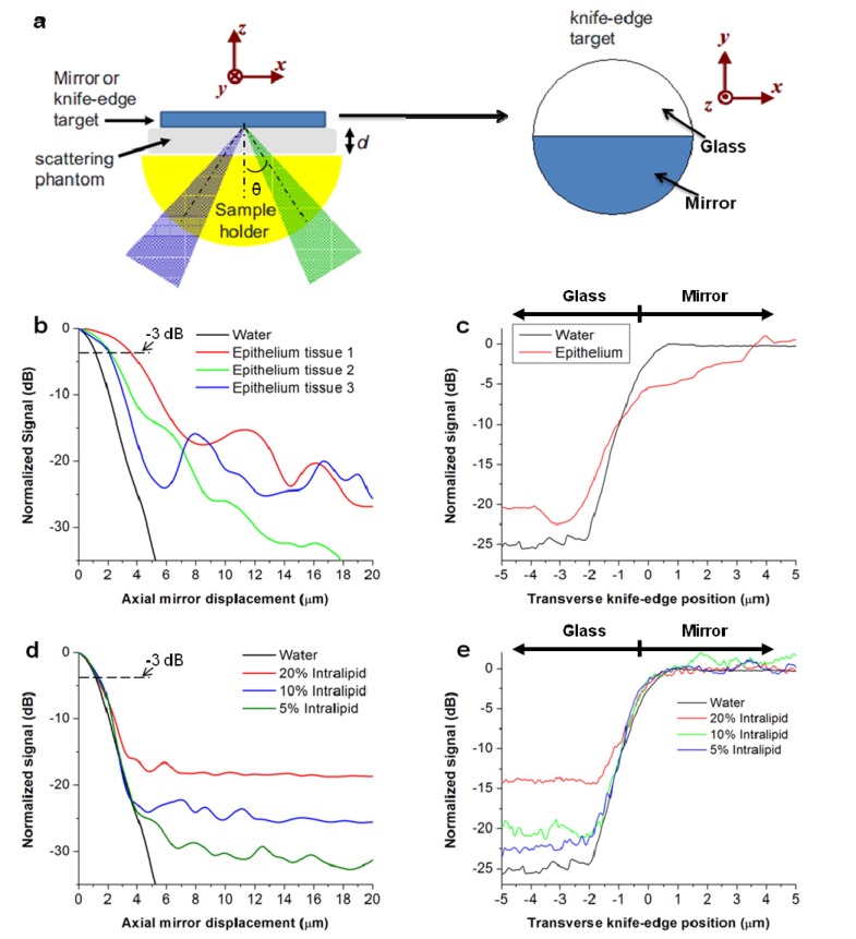
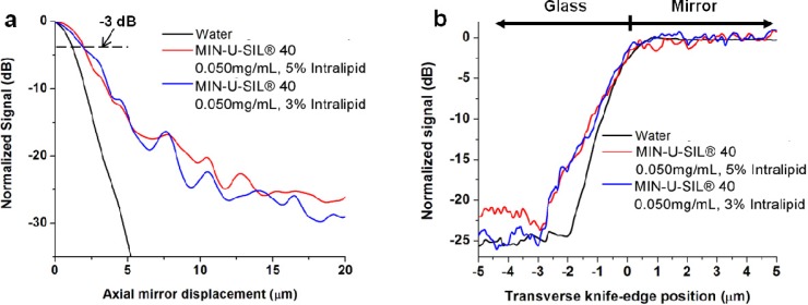
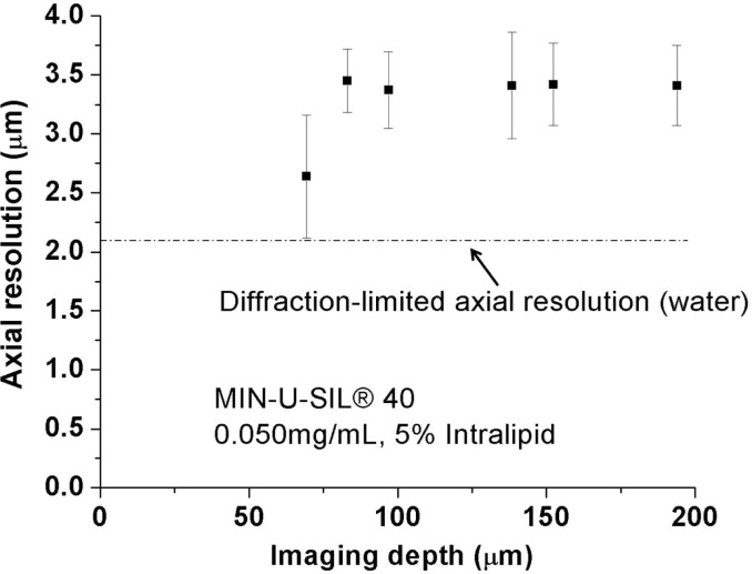
Phantoms play an important role in the development, standardization, and calibration of biomedical imaging devices in laboratory and clinical settings, serving as standards to assess the performance of such devices. Here we present the design of a liquid optical phantom to facilitate the assessment of optical-sectioning microscopes that are being developed to enable point-of-care pathology. This phantom, composed of silica microbeads in an Intralipid base, is specifically designed to characterize a reflectance-based dual-axis confocal (DAC) microscope for skin imaging. The phantom mimics the scattering properties of normal human epithelial tissue in terms of an effective scattering coefficient and a depth-dependent degradation in spatial resolution due to beam steering caused by tissue micro-architectural heterogeneities.
Keywords: (170.1790) Confocal microscopy; (170.3880) Medical and biological imaging; (170.5810) Scanning microscopy; (170.6900) Three-dimensional microscopy; (170.7050) Turbid media.
Figures

Similar articles
-
Bessel-beam illumination in dual-axis confocal microscopy mitigates resolution degradation caused by refractive heterogeneities.J Biophotonics. 2017 Jan;10(1):68-74. doi: 10.1002/jbio.201600196. Epub 2016 Sep 26. J Biophotonics. 2017. PMID: 27667127 Free PMC article.
-
Modulated-alignment dual-axis (MAD) confocal microscopy for deep optical sectioning in tissues.Biomed Opt Express. 2014 Apr 30;5(6):1709-20. doi: 10.1364/BOE.5.001709. eCollection 2014 Jun 1. Biomed Opt Express. 2014. PMID: 24940534 Free PMC article.
-
Miniature in vivo MEMS-based line-scanned dual-axis confocal microscope for point-of-care pathology.Biomed Opt Express. 2016 Jan 5;7(2):251-63. doi: 10.1364/BOE.7.000251. eCollection 2016 Feb 1. Biomed Opt Express. 2016. PMID: 26977337 Free PMC article.
-
Optical sectioning microscopy.Nat Methods. 2005 Dec;2(12):920-31. doi: 10.1038/nmeth815. Nat Methods. 2005. PMID: 16299477 Review.
-
Role of In Vivo Reflectance Confocal Microscopy in the Analysis of Melanocytic Lesions.Acta Dermatovenerol Croat. 2018 Apr;26(1):64-67. Acta Dermatovenerol Croat. 2018. PMID: 29782304 Review.
Cited by
-
A scattering phantom for observing long range order with two-dimensional angle-resolved Low-Coherence Interferometry.Biomed Opt Express. 2013 Aug 26;4(9):1742-8. doi: 10.1364/BOE.4.001742. eCollection 2013. Biomed Opt Express. 2013. PMID: 24049694 Free PMC article.
-
Fractal propagation method enables realistic optical microscopy simulations in biological tissues.Optica. 2016;3(8):861-869. doi: 10.1364/OPTICA.3.000861. Optica. 2016. PMID: 28983499 Free PMC article.
-
Optical-sectioning microscopy of protoporphyrin IX fluorescence in human gliomas: standardization and quantitative comparison with histology.J Biomed Opt. 2017 Apr 1;22(4):46005. doi: 10.1117/1.JBO.22.4.046005. J Biomed Opt. 2017. PMID: 28418534 Free PMC article.
-
Low-cost fabrication of optical tissue phantoms for use in biomedical imaging.Heliyon. 2020 Mar 21;6(3):e03602. doi: 10.1016/j.heliyon.2020.e03602. eCollection 2020 Mar. Heliyon. 2020. PMID: 32258463 Free PMC article.
-
Optimizing the performance of dual-axis confocal microscopes via Monte-Carlo scattering simulations and diffraction theory.J Biomed Opt. 2013 Jun;18(6):066006. doi: 10.1117/1.JBO.18.6.066006. J Biomed Opt. 2013. PMID: 23733022 Free PMC article.
References
-
- Nordstrom R., “Phantoms as Standards in Optical Measurements,” Proc. SPIE 7906, 79060H–, 79060H-5. (2011).10.1117/12.876374 - DOI
Grants and funding
LinkOut - more resources
Full Text Sources