. 2012 Nov 1;1(8):1297-1304.
doi: 10.4161/onci.21659.

Analysis of Vδ1 T cells in clinical grade melanoma-infiltrating lymphocytes

Affiliations

Analysis of Vδ1 T cells in clinical grade melanoma-infiltrating lymphocytes

Marco Donia et al. Oncoimmunology. .

Abstract

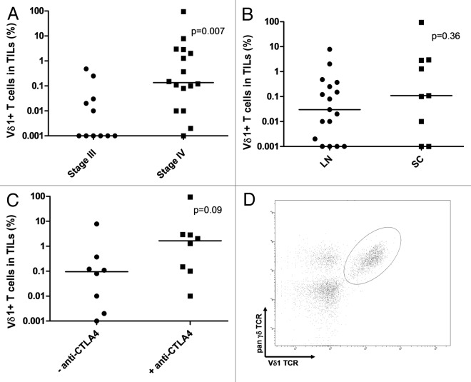
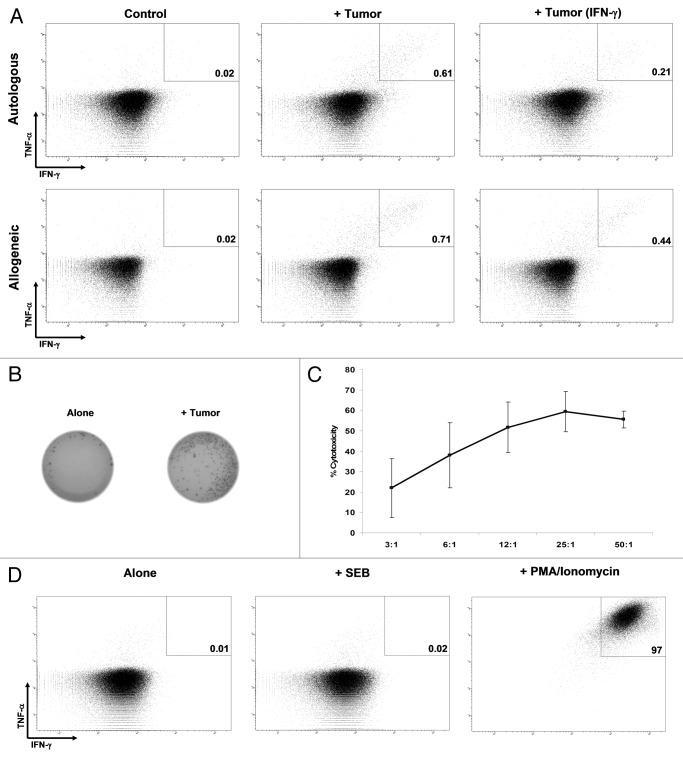
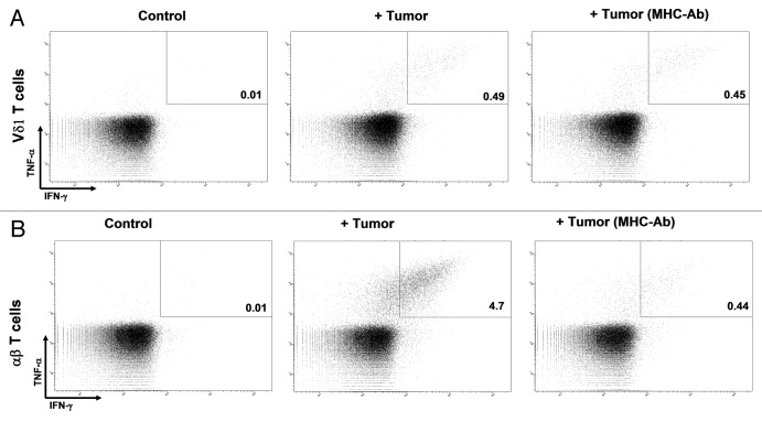
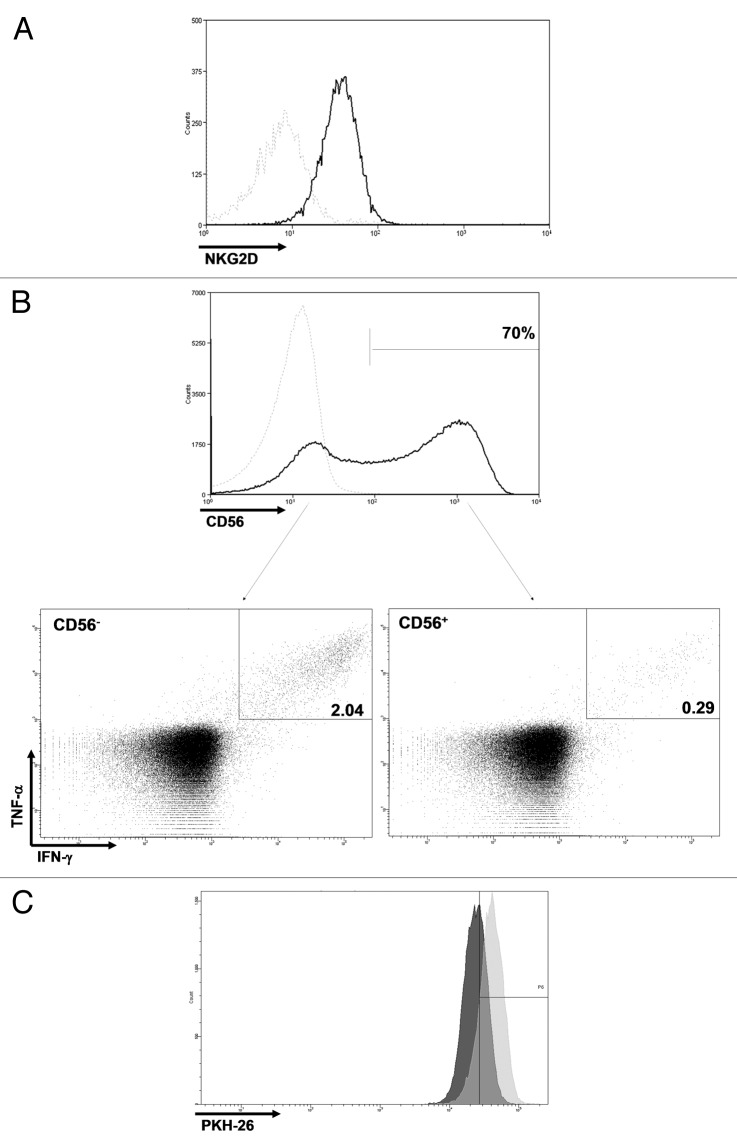
γδ T cells, including Vδ1 and Vδ2 T cells, can recognize tumor-associated ligands neglected by conventional αβ T cells in a MHC-independent manner. Little is known regarding the anticancer potential and the possibility to isolate and expand Vδ1 T cells to therapeutically relevant numbers. In this study, we have detected low frequencies of Vδ1 T cells among tumor-infiltrating lymphocyte (TIL) products for adoptive cell transfer generated from melanoma metastases. An increased frequency of Vδ1 T cells was found among the cell products from patients with an advanced disease stage. Vδ1 T cells displayed in vitro antitumor activities and sufficient proliferative potential to generate over 1 × 10(9) cells using current protocols for T cell transfer. Infusion of Vδ1 T cells together with high numbers of αβ TILs in a clinical trial was safe and well tolerated. These data suggest that Vδ1 T cells should be further scrutinized as a potentially useful tool for the treatment of patients with metastatic melanoma.


Figures

None
Figure 1. Vδ1 T cells in clinical grade tumor-infiltrating lymphocytes (TILs). (A–C) Frequency of detected Vδ1 T cells in clinical grade TIL products grouped for AJCC disease stage (A), biopsy origin (B) and prior treatment with anti-CTLA4 antibodies (only patients with stage IV disease are shown) (C). (D) FACS plot from a representative TIL product (from patient 17). An electonic gate was set on the CD3+CD4-CD8- live cell population. LN, lymph node metastatis; SC, subcutaneous metastasis.
None
Figure 2. Antitumor activity. (A) Responses of Vδ1 T cells (from patient 11, > 90% of Vδ1 T cells) evaluated with the production of tumor necrosis factor α (TNFα) and interferon γ (IFNγ) when unstimulated (control), or stimulated with autologous or an HLA-A-unmatched allogeneic melanoma cells. Both constitutive responses and responses upon stimulation of cancer cells with 100 IU/mL IFNγ for 72 h (+ IFNγ) are shown. All the plots are gated on Vδ1+ T cells. (B) IFNγ ELISPOT. Unstimulated tumor-infiltrating lymphocytes (TILs,control) or TILs stimulated with autologous tumor cells are shown. (C) Percentage of cytotoxicity at different effector:target ratios of unfractionated TILs vs. one HLA-A-unmatched allogeneic melanoma cell line. (D) Production of TNFα and IFNγ by unstimulated (control) TILs or TILs unspecifically stimulated with the Staphylococcal enterotoxin B (SEB) or PMA/Ionomycin.
None
Figure 4. MHC involvement in target recognition. (A and B) Tumor-infiltrating lymphocytes (TILs) were stimulated with autologous tumor cells preincubated with isotype control or a combination of MHC Class I and II-blocking antibodies. While MHC Class I and Class II-blocking antibodies significantly affected tumor cell recognition by αβ T cells (B), they did not modify tumor cell recognition by Vδ1 T cells (A).
None
Figure 3. Phenotypic and proliferative characteristics. (A) Vδ1 T cells expressed NKG2D. (B) CD56 was expressed by a large fraction of Vδ1 T cells, and cells with in vitro anticancer activity were enriched in the CD56- compartment. Dotted light gray line: isotype control. (C) From day 8 to day 10 of the rapid expansion protocol (REP), undivided cells were 85% of αβ T cells vs. 45% of Vδ1 T cells. Light gray, αβ T cells; dark gray, Vδ1 T cells.

References

    1. Hayday AC. [gamma][delta] cells: a right time and a right place for a conserved third way of protection. Annu Rev Immunol. 2000;18:975–1026. doi: 10.1146/annurev.immunol.18.1.975. - DOI - PubMed
    1. Kabelitz D, Wesch D, He W. Perspectives of gammadelta T cells in tumor immunology. Cancer Res. 2007;67:5–8. doi: 10.1158/0008-5472.CAN-06-3069. - DOI - PubMed
    1. Shimura E, Hozumi N, Kanagawa O, Metzger D, Chambon P, Radtke F, et al. Epidermal gammadelta T cells sense precancerous cellular dysregulation and initiate immune responses. Int Immunol. 2010;22:329–40. doi: 10.1093/intimm/dxq014. - DOI - PubMed
    1. Groh V, Rhinehart R, Secrist H, Bauer S, Grabstein KH, Spies T. Broad tumor-associated expression and recognition by tumor-derived gamma delta T cells of MICA and MICB. Proc Natl Acad Sci U S A. 1999;96:6879–84. doi: 10.1073/pnas.96.12.6879. - DOI - PMC - PubMed
    1. Groh V, Steinle A, Bauer S, Spies T. Recognition of stress-induced MHC molecules by intestinal epithelial gammadelta T cells. Science. 1998;279:1737–40. doi: 10.1126/science.279.5357.1737. - DOI - PubMed

Publication types