

Phosphorylation of hepatic AMP-activated protein kinase and liver kinase B1 is increased after a single oral dose of green tea extract to mice
- PMID: 23244544
- PMCID: PMC3590820
- DOI: 10.1016/j.nutres.2012.10.005
Phosphorylation of hepatic AMP-activated protein kinase and liver kinase B1 is increased after a single oral dose of green tea extract to mice
Abstract
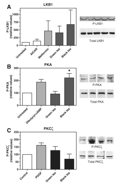
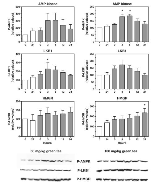
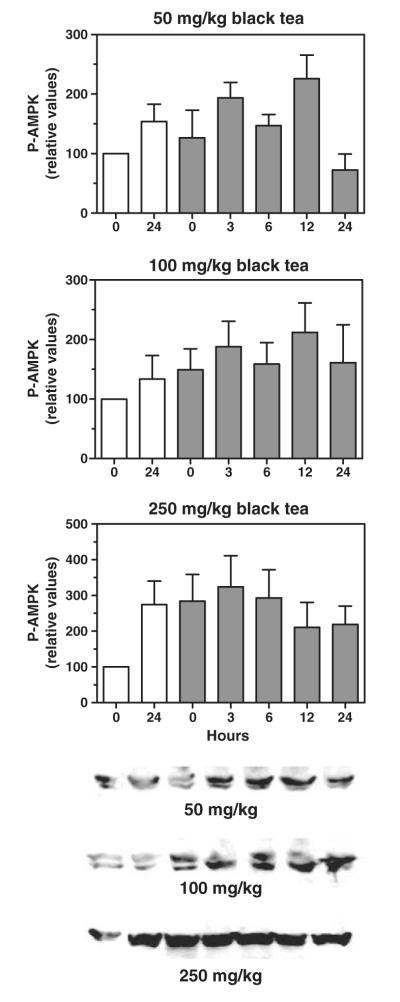
We have previously shown that green and black tea extracts increase the phosphorylation of AMP-activated protein kinase (AMPK) and HMG-CoA reductase in rat hepatoma cells in culture, concomitant with a decrease in cholesterol synthesis. In the present study, we evaluated the ability of a single oral dose of green or black tea extract to promote the phosphorylation of AMPK, liver kinase B1 (LKB1, an AMPK-kinase), and HMG-CoA reductase in mouse liver. Green tea extract administered by gavage at 50 and 100 mg/kg caused a 2- to 3-fold increase in hepatic AMPK phosphorylation at 3 and 6 hours after dosing and a 1.5- to 2-fold increase in LKB1 phosphorylation at these same time points. The phosphorylation of HMG-CoA reductase at these and later time points was not significantly increased. Black tea administered by gavage at up to 250 mg/kg was ineffective in increasing hepatic AMPK phosphorylation. Both green and black tea extracts increased LKB1 phosphorylation in hepatoma cells in culture at 15 μg/mL, and black tea also increased the phosphorylation of protein kinase A in hepatoma cells. These results suggest that compounds in both tea extracts activate AMPK by activating its upstream kinase, LKB1, and that black tea may do so by first activating protein kinase A, a known kinase for LKB1. Only green tea, at 50 and 100 mg/kg, was able to activate AMPK and LKB1 in mouse liver after oral dosing, suggesting that the polymerized catechins present in black tea do not reach the liver in sufficient concentration to affect AMPK activity.
Copyright © 2012 Elsevier Inc. All rights reserved.
Figures
References
-
- Davies MJ, Judd JT, Baer DJ, Clevidence BA, Paul DR, Edwards AJ, et al. Black tea consumption reduces total and LDL cholesterol in mildly hypercholesterolemic adults. J Nutr. 2003;133:3298S–302S. - PubMed
-
- Maron DJ, Lu GP, Cai NS, Wu ZG, Li YH, Chen H, et al. Cholesterol-lowering effect of a theaflavin-enriched green tea extract: a randomized controlled trial. Arch Intern Med. 2003;163:1448–53. - PubMed
-
- Bursill CA, Abbey M, Roach PD. A green tea extract lowers plasma cholesterol by inhibiting cholesterol synthesis and upregulating the LDL receptor in the cholesterol-fed rabbit. Atherosclerosis. 2007;193:86–93. - PubMed
-
- Kim A, Chiu A, Barone MK, Avino D, Wang F, Coleman CI, et al. Green tea catechins decrease total and low-density lipoprotein cholesterol: a systematic review and meta-analysis. J Am Diet Assoc. 2011;111:1720–9. - PubMed
Publication types
MeSH terms
Substances
Grants and funding
LinkOut - more resources
Full Text Sources
Medical
