Randomized Controlled Trial
. 2013 May 1;73(9):887-94.
doi: 10.1016/j.biopsych.2012.10.022. Epub 2012 Dec 14.

Neural and behavioral effects of a novel mu opioid receptor antagonist in binge-eating obese people

Affiliations
Randomized Controlled Trial

Neural and behavioral effects of a novel mu opioid receptor antagonist in binge-eating obese people

Victoria C Cambridge et al. Biol Psychiatry. .

Abstract

Background: Binge eating is associated with obesity and has been conceptualized as "food addiction." However, this view has received only inconsistent support in humans, and limited evidence relates key neurocircuitry to the disorder. Moreover, relatively few studies have used pharmacologic functional magnetic resonance imaging to probe the underlying basis of altered eating behaviors.

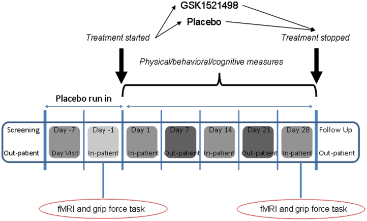
Methods: In a double-blind, placebo-controlled, parallel group study, we explored the effects of a potent mu-opioid receptor antagonist, GSK1521498, in obese individuals with moderate binge eating. Subjects were tested during a baseline placebo run-in period and retested after 28-days of drug (n = 21) or placebo (n = 21) treatment. Using functional magnetic resonance imaging and behavioral measures, we determined the drug's effects on brain responses to food images and, separately, on motivation to expend energy to view comparable images.

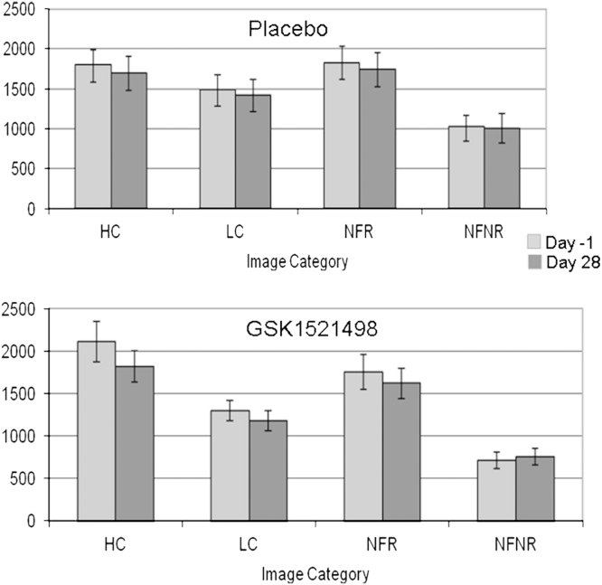
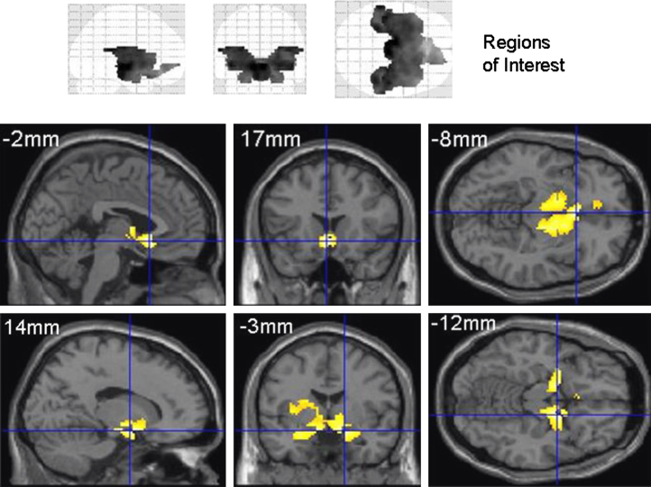
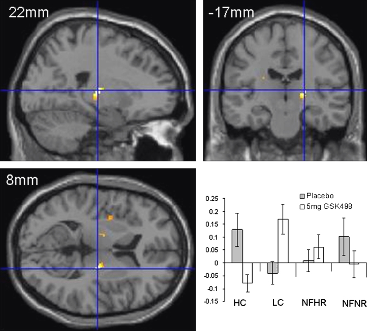
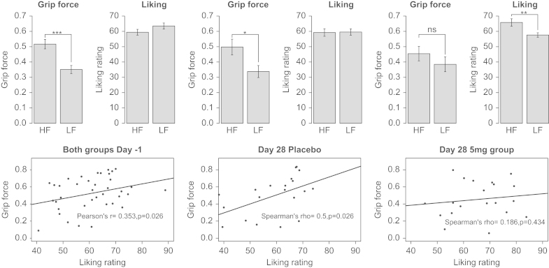
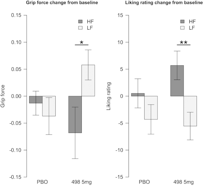
Results: Compared with placebo, GSK1521498 was associated with a significant reduction in pallidum/putamen responses to pictures of high-calorie food and a reduction in motivation to view images of high-calorie food. Intriguingly, although motivational responding was reduced, subjective liking for the same images actually increased following drug treatment.

Conclusions: Stimulus-specific putamen/pallidal responses in obese people with binge eating are sensitive to altered mu-opioid function. This neuromodulation was accompanied by reductions in motivational responding, as measured by grip force, although subjective liking responses to the same stimuli actually increased. As well as providing evidence for a link between the opioid system and food-related behavior in binge-eating obese individuals, these results support a dissociation across measures of motivation and liking associated with food-related stimuli in these individuals.

Trial registration: ClinicalTrials.gov NCT01195792.


Figures

Figure 1
Figure 1
Study design. Following screening and a 7-day single-blind, placebo run-in, a baseline assessment was performed during a 2-day stay on the clinical research unit. This included the functional magnetic resonance imaging (fMRI) and grip force tasks that are the focus of this article. Thereafter, participants were randomised to receive oral GSK1521498 (5 mg) or placebo for 28 days. Further inpatient assessments were performed on Day 14 and Day 28. fMRI and grip force task were repeated on Day 28. Several other physical, cognitive, and eating measures were performed over the course of the visits (see Ziauddeen et al. for details).
Figure 2
Figure 2
Functional magnetic resonance imaging task: liking ratings. Subjective liking of food images was assessed (see Methods and Materials) using duration of button press and is represented here in milliseconds. Average values for each of the four image categories are shown before (Day –1) and after (Day 28) drug and placebo treatment. HC, high-calorie images; LC, low calorie images; NFR, nonfood rewarding images; NFNR, nonfood, nonrewarding images.
Figure 3
Figure 3
Functional magnetic resonance imaging results at Day –1. The upper panel depicts the regions of interest used on orthogonal maximum intensity projections (“glass brains” viewed from the right (left projection), from behind (middle) and from above (right). The lower panels show the significant (p<.05, uncorrected for display) areas of activation when contrasting viewing of high-calorie to low-calorie food images at Day –1. This contrast used the placebo and GSK1521498 treatment groups combined and shows activity across key regions of reward circuitry. Full details of the activation foci are presented in Table 1. Areas of significant activation are rendered onto a standard template image in Montreal Neurological Institute space with sections chosen at the coordinates most appropriate to display the key activations. This activation map was used as a mask to constrain subsequent analysis exploring condition-specific effects of GSK1521498.
Figure 4
Figure 4
Functional magnetic resonance imaging (fMRI) results Day 28: drug-by-stimulus-by-reward interaction. fMRI results at Day 28 showing drug by stimulus type (food vs. nonfood) by reward type (high calorie/high reward vs. low calorie/low reward). The pallidum/putamen region demonstrating this interaction (p<.05, uncorrected for multiple comparisons for display) is shown superimposed onto orthogonal sections of a structural MRI in standard Montreal Neurological Institute space. In the lower right panel are plotted the parameter estimates for each of the stimulus types after placebo and GSK1521498 treatment. HC, high-calorie images; LC, low calorie images; NFHR, nonfood high reward images; NFNR, nonfood, nonrewarding images.
Figure 5
Figure 5
Grip force task: effort expended and liking ratings. The top panel shows the grip force exerted and the corresponding liking ratings at baseline in both groups and separately for placebo and the GSK1521498 group at the end of treatment. It can be seen that the difference between force exerted for high-fat (HF) versus low-fat (LF) food images is no longer significant after drug treatment, even though the subjective liking for these images is higher. The bottom panel displays the correlations between the exerted force and liking ratings for the high-fat food images, again at baseline for both groups and separately at Day 28. The correlation between these two measures is seen at baseline and in the placebo group at the end of treatment but is lost in the drug group. *p < .05; **p < .01; ***p < .001.
Figure 6
Figure 6
Change from baseline in grip force and liking ratings. *p < .05; **p < .01. HF, high fat; LF, low fat; PBO, placebo.

Similar articles

Cited by

References

    1. Berridge K.C. Food reward: Brain substrates of wanting and liking. Neurosci Biobehav Rev. 1996;20:1–25. - PubMed
    1. Robbins T.W., Everitt B.J. A role for mesencephalic dopamine in activation: Commentary on Berridge (2006) Psychopharmacology (Berl) 2007;191:433–437. - PubMed
    1. de Wit S., Dickinson A. Associative theories of goal-directed behaviour: A case for animal-human translational models. Psychol Res. 2009;73:463–476. - PMC - PubMed
    1. Everitt B.J., Belin D., Economidou D., Pelloux Y., Dalley J.W., Robbins T.W. Review. Neural mechanisms underlying the vulnerability to develop compulsive drug-seeking habits and addiction. Philos Trans R Soc Lond B Biol Sci. 2008;363:3125–3135. - PMC - PubMed
    1. Everitt B.J., Robbins T.W. Neural systems of reinforcement for drug addiction: From actions to habits to compulsion. Nat Neurosci. 2005;8:1481–1489. - PubMed

Publication types

MeSH terms

Substances

Associated data