. 2013 Jan 8;21(1):154-166.
doi: 10.1016/j.str.2012.11.004. Epub 2012 Dec 13.

Sculpting of DNA at abasic sites by DNA glycosylase homolog mag2

Affiliations

Sculpting of DNA at abasic sites by DNA glycosylase homolog mag2

Bjørn Dalhus et al. Structure. .

Abstract

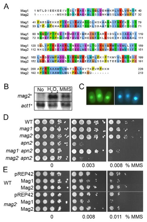
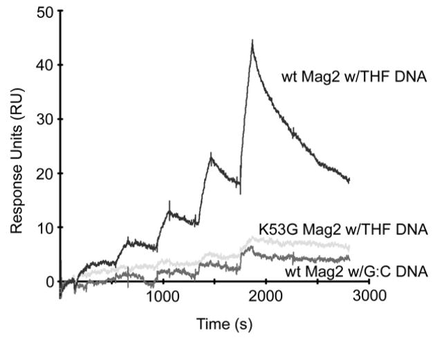
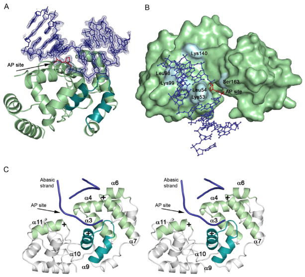
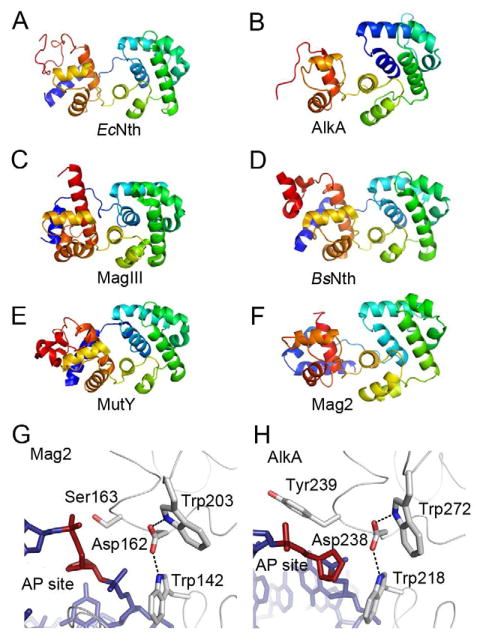
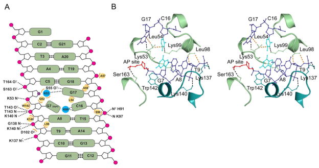
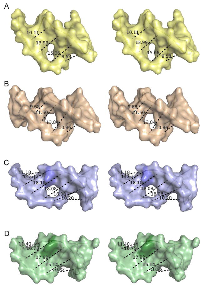
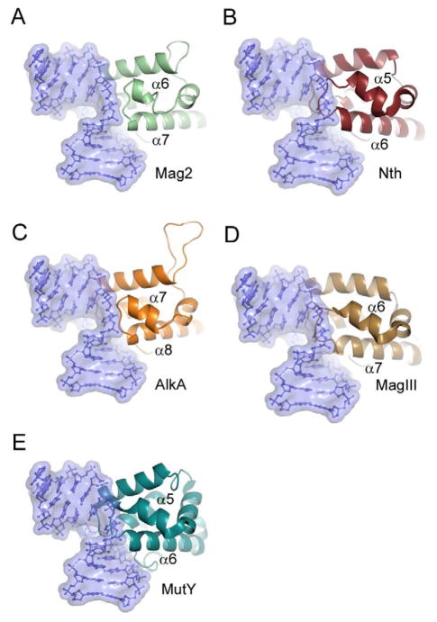
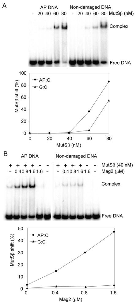
Modifications and loss of bases are frequent types of DNA lesions, often handled by the base excision repair (BER) pathway. BER is initiated by DNA glycosylases, generating abasic (AP) sites that are subsequently cleaved by AP endonucleases, which further pass on nicked DNA to downstream DNA polymerases and ligases. The coordinated handover of cytotoxic intermediates between different BER enzymes is most likely facilitated by the DNA conformation. Here, we present the atomic structure of Schizosaccharomyces pombe Mag2 in complex with DNA to reveal an unexpected structural basis for nonenzymatic AP site recognition with an unflipped AP site. Two surface-exposed loops intercalate and widen the DNA minor groove to generate a DNA conformation previously only found in the mismatch repair MutS-DNA complex. Consequently, the molecular role of Mag2 appears to be AP site recognition and protection, while possibly facilitating damage signaling by structurally sculpting the DNA substrate.


Figures

Figure 1
Figure 1. Characterization of the mag2 Gene Function
(A) Multiple-sequence alignment of S. pombe Mag1 and Mag2. Panel made using Jalview (Waterhouse et al., 2009) (B) Northern blot analysis of mag2 gene expression. Total RNA was isolated from untreated control (No) and from wildtype cells exposed to H2O2 or MMS and the filter was hybridized with a 642 bp mag2 probe. A β-a ctin (act1) probe was used as control. (C) Fluorescence microscopy of S. pombe cells expressing fusions of Mag2 to GFP. Yeast cells were transformed by DNA constructs expressing Mag2-GFPc (left panel) and stained with Hoechst 33342 as a nuclear marker (right panel). (D) MMS sensitivity of S. pombe BER mutant strains. S. pombe wild type (WT), mag1, mag2, apn2, mag1 apn2 and mag2 apn2 mutant cells were spread on solid media containing increasing doses of MMS and cell survival was evaluated relative to plates without MMS. (E) Overexpression of Mag1, but not Mag2 is toxic for the fission yeast cells. S. pombe wild type (WT) and mag2 mutant strains overexpressing Mag1 or Mag2 from pREP42 were spread on minimal media containing increasing doses of MMS and cell survival was evaluated relative to plates without MMS. Cells with empty vector (pREP42) were used as control. See also Figure S1.
Figure 2
Figure 2. SPR Analyses of Mag2 wt and K53G Mutant Binding to Abasic DNA
Increasing amounts (25, 50, 100, 200 and 400 nM) of Mag2 wt (dark grey) and K53G mutant (light gray) were injected on chip containing the abasic site analogue THF, or WT Mag2 on non-damaged DNA (intermediate grey).
Figure 3
Figure 3. Overall Structure and DNA Conformation in the S. pombe Mag2-DNA Complex
(A) Ribbon display of Mag2 in complex with abasic DNA (blue DNA with simulated annealing composite omit map) showing the distortion of the bound DNA. The canonical helix-hairpin-helix (HhH) motif is highlighted (cyan) and the unflipped AP site analogue is coloured red. (B) Surface representation of Mag2 with DNA in ball-and-stick. (C) Stereo-view of the various helices and loops involved in DNA bending, unwinding and strand separation. Helices α10 and α11 both have their positive dipole toward the negatively charged phosphate backbone, and pull and stretch the DNA strand containing the abasic site. Helices α6 and α7 form a “push-and-pull” pair as they have opposite charged dipoles close to the undamaged strand. The loop between helix α3 and α4 include residues Lys53 and Leu54, both of which intercalate with the DNA. Helix α7 contains Leu98 and Lys99, whereas the last interacting loop between helix α9 and α10 is part of the helix-hairpin-helix motif.
Figure 4
Figure 4. Protein Folding of Selected DNA Glycosylases and Trp-Asp-Trp Triad in S. pombe Mag2 and E. coli AlkA
(A)–(F) Protein folding of (A) E. coli endonuclease III (Nth) (Thayer et al., 1995), (B) E. coli 3mA DNA glycosylase II (AlkA) (Hollis et al., 2000a), (C) H. pylori 3mA DNA glycosylase MagIII (Eichman et al., 2003), (D) B. stearothermophilus endonuclease III (Nth) (Fromme and Verdine, 2003), (E) B. stearothermophilus adenine DNA glycosylase (MutY) (Fromme et al., 2004), and (F) S. pombe Mag2. Color spectrum goes from N-terminal blue to C-terminal red. Only the C-terminal α-helical part of AlkA and the N-terminal part of B. stearothermophilus Nth are shown. (G) and (H) Close-up view of the AP site binding region in (G) S. pombe Mag2 and (H) E. coli AlkA (Hollis et al., 2000a) with stick models of the DNA (blue with abasic site in red). Both structures contain a Trp-Asp-Trp motif. The bulky side chain of Tyr239 in AlkA (H) pushes on the 5′ phosphate of the AP site as part of the flipping mechanism, while the less bulky Ser163 in Mag2 (G) allow the accommodation of a non-flipped AP site.
Figure 5
Figure 5. Protein-DNA Contacts in the S. pombe Mag2-DNA Complex
(A) Diagram showing all contacts between Mag2 and the abasic DNA. Hydrogen bond/ionic interactions < 3.35Å are shown with dashed lines, wherein symbol N denotes main chain amide. Steric interactions < 3.75Å are shown as orange arcs with the respective amino acids as yellow circles. Leu54 is wedged in between Cyt16 and the orphan Gua17, and together with Lys99 dictate the spatial orientation of Cyt16. Gua7 has the unusual syn conformation. The abasic site analogue is shown as a violet circle. (B) Stereo view of the intercalating loops in Mag2, showing key interactions between the protein and the abasic DNA. Only the central part of the bound DNA (blue) with the abasic site analogue (AP site, red) and the distorted base pair Gua7:Cyt16 (cyan) is shown. Amino acid side chains involved in DNA-bending, strand separation and widening of the DNA minor groove are shown in ball-and-stick. Lys53 occupies the empty space close to the abasic site in the DNA-ladder, splitting the two DNA strands. The side chain of Leu54 is wedged in between the C16 and G17 bases. Gln52 (partly hidden behind nucleotides G7 and A8) and Lys99 protrude into the DNA helix, forcing G7 to adopt a syn-conformation, abolishing normal Watson-Crick base pairing with C16. Leu98 contacts the DNA backbone near the ribose of Thy9, pushing the DNA toward Lys140 and Lys137 in the helix-hairpin-helix motif. Trp142 form a platform onto which the 5′ phosphate of G7 can rest. Finally, Ser163 form a hydrogen bond to the 5′ phosphate of the AP site. See also Figure S3.
Figure 6
Figure 6. Comparison of Minor Groove Widening in Selected Protein-DNA Complexes
(A) Stereo view of DNA in the structure of E. coli 3mA DNA glycosylase AlkA (Hollis et al., 2000a). (B) Stereo view of DNA in the structure of human AP endonuclease APE1 (Mol et al., 2000b). (C) Stereo view of DNA in the structure of S. pombe Mag2. (D) Stereo view of DNA in the structure of E. Coli MutS bound to DNA harbouring an A:A mismatch (Natrajan et al., 2003). The displayed distances correspond to the C4′-C4′ distance between nucleotide n and (n + 2). The protruding Cyt16 in the DNA bound to Mag2 and the corresponding Ade in the MutS-DNA complex are coloured in a darker shade. The holes in the middle of the DNA show the position of the abasic sites. The DNA from the AlkA and MutS complexes has been reduced to 11 base pairs for the sake of clarity. See also Figures S2 and S3.
Figure 7
Figure 7. Close-up View of the Wedging Loop Between Helix α6 and α7 in Mag2 and Corresponding Loops in Selected 3mA Glycosylases, AP Endonuclease and MutY, Showing Steric Conflicts With the Mag2 DNA Conformation
(A) S. pombe Mag2, (B) E. coli endonuclease III (Nth) (Thayer et al., 1995), (C) E. coli 3mA DNA glycosylase II (AlkA) (Hollis et al., 2000a), (D) H. pylori 3mA DNA glycosylase MagIII (Eichman et al., 2003) and (E) B. stearothermophilus adenine DNA glycosylase MutY (Fromme et al., 2004). DNA from Mag2 is shown in blue ball-and-stick representation within a semi-transparent surface boundary in all five panels. The different proteins have been superimposed onto the Cα-trace of Mag2, with an r.m.s.d of less than 3.3 Å for a selection of 53 – 103 atoms in a core domain comprising residues 40 – 170 in Mag2.
Figure 8
Figure 8. Electrophoretic Mobility Shift Assay With Mag2 and Human MutSβ
(A) Duplex DNA containing a single AP site or non-damaged DNA (5 nM) was incubated with increasing concentrations of MutSβ (20–80 nM) in a buffer containing 20 mM HEPES, pH 7.5, 2 mM MgCl2, 100 mM NaCl, 5% glycerol, 0.1 mg/ml BSA and 1 mM DTT. Formation of a MutSβ-DNA complex was observed, both for AP DNA and undamaged DNA. (B) Specific binding of MutSβ to AP DNA was stimulated >10 fold by increasing the concentration of Mag2 (0.4–1.6 μM). The amounts of free DNA and DNA in complex with MutSβ were analysed on 10% polyacrylamide gels, visualised using PhosphoImager and quantified using ImageQuant. These data represent a typical result of three independent experiments. See also Figure S4.

Similar articles

Cited by

References

    1. Adhikary S, Eichman BF. Analysis of substrate specificity of Schizosaccharomyces pombe Mag1 alkylpurine DNA glycosylase. EMBO Rep. 2011;12:1286–1292. - PMC - PubMed
    1. Alseth I, Osman F, Korvald H, Tsaneva I, Whitby MC, Seeberg E, Bjoras M. Biochemical characterization and DNA repair pathway interactions of Mag1-mediated base excision repair in Schizosaccharomyces pombe. Nucleic Acids Res. 2005;33:1123–1131. - PMC - PubMed
    1. Berti PJ, McCann JA. Toward a detailed understanding of base excision repair enzymes: transition state and mechanistic analyses of N-glycoside hydrolysis and N-glycoside transfer. Chem Rev. 2006;106:506–555. - PubMed
    1. Boiteux S, Guillet M. Abasic sites in DNA: repair and biological consequences in Saccharomyces cerevisiae. DNA Repair (Amst) 2004;3:1–12. - PubMed
    1. Bruner SD, Norman DP, Verdine GL. Structural basis for recognition and repair of the endogenous mutagen 8-oxoguanine in DNA. Nature. 2000;403:859–866. - PubMed

Publication types

MeSH terms

LinkOut - more resources