. 2013 Jan 8;21(1):20-31.
doi: 10.1016/j.str.2012.11.005. Epub 2012 Dec 13.

Y-family polymerase conformation is a major determinant of fidelity and translesion specificity

Affiliations

Y-family polymerase conformation is a major determinant of fidelity and translesion specificity

Ryan C Wilson et al. Structure. .

Abstract

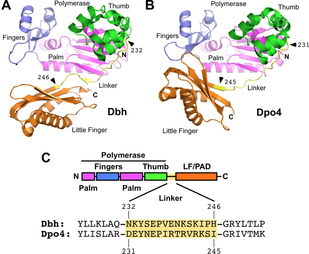
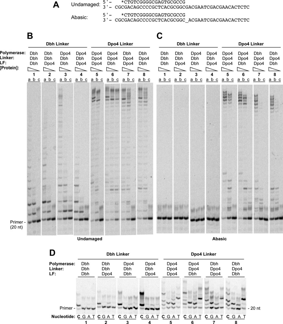
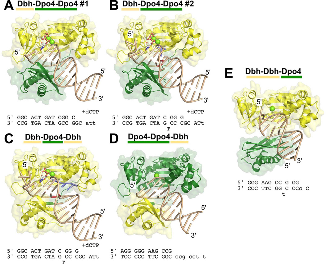
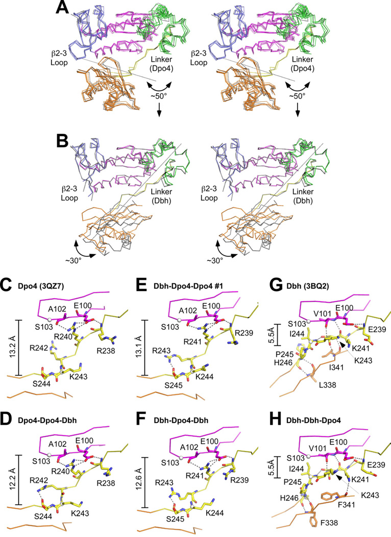
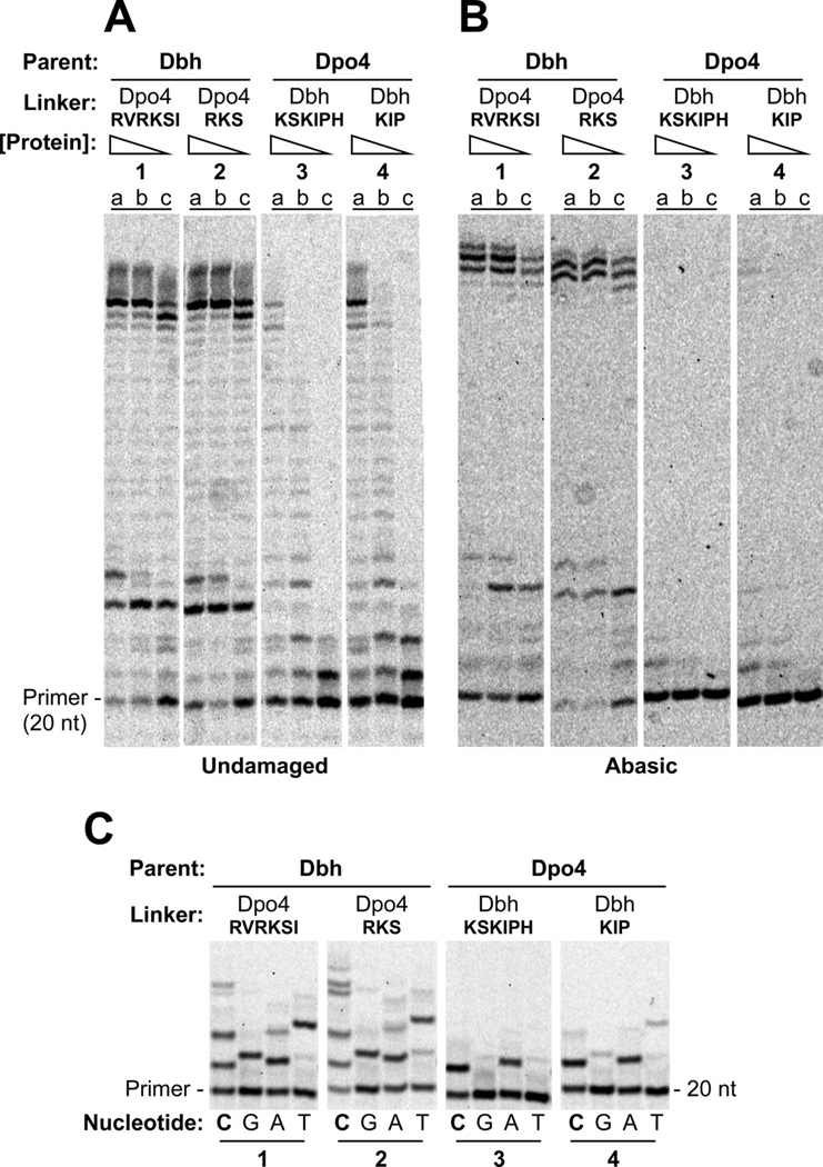
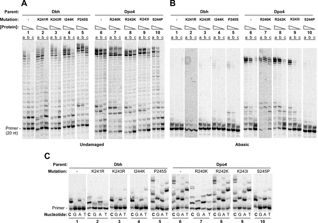
Y-family polymerases help cells tolerate DNA damage by performing translesion synthesis opposite damaged DNA bases, yet they also have a high intrinsic error rate. We constructed chimeras of two closely related Y-family polymerases that display distinctly different activity profiles and found that the polypeptide linker that tethers the catalytic polymerase domain to the C-terminal DNA-binding domain is a major determinant of overall polymerase activity, nucleotide incorporation fidelity, and abasic site-bypass ability. Exchanging just 3 out of the 15 linker residues is sufficient to interconvert the polymerase activities tested. Crystal structures of four chimeras show that the conformation of the protein correlates with the identity of the interdomain linker sequence. Thus, residues that are more than 15 Å away from the active site are able to influence many aspects of polymerase activity by altering the relative orientations of the catalytic and DNA-binding domains.


Conflict of interest statement

CONFLICT OF INTEREST

The authors declare no conflict of interest.

Figures

Figure 1
Figure 1. Parental Y-family Polymerases Used to Construct Chimeric Enzymes
(A) Structure of Dbh showing overall protein conformation and junctions (marked with arrowheads) before and after the linkers that were exchanged in chimeric polymerases. Note the short β-strand in the linker that contacts both the palm and the LF/PAD domains. Made using coordinates from PDB code 3BQ1 (Wilson and Pata, 2008), a ternary complex that contained incoming dNTP and primer-template DNA with an extrahelical nucleotide in the template strand three nucleotides to the 3’ side of the templating base (substrates not shown). Colored by domain: blue (fingers), magenta (palm), green (thumb), yellow (linker), and orange (LF/PAD). (B) Structure of Dpo4. Note the contact between the LF/PAD and fingers domains. Domains are colored as in A and junctions are marked with arrowheads. Made using coordinates from PDB code 3QZ7 (Wu et al., 2011). (C) Diagram showing the linear organization of the polymerase domains and the protein sequences of Dbh and Dpo4 in the region of the linker. Domains are colored as in A.
Figure 2
Figure 2. Single and Multiple Nucleotide Incorporation by Chimeric Polymerases on Undamaged and Abasic Site DNA
(A) Primer-template sequences showing undamaged template, top, and template containing an abasic site (denoted by _), bottom. Primers were labeled at the 5’ end during synthesis with 6-FAM (denoted by *). (B) Polymerase assays on undamaged primer-template DNA containing (a) 160 nM, (b) 40 nM, or (c) 10 nM protein with 40 nM DNA and 1 mM each dATP, dCTP, dGTP and dTTP. Reactions were incubated at 60°C for 5 min. (C) Polymerase assays on primer-template DNA containing abasic site in the template strand immediately adjacent to the terminal basepair. Protein and substrate concentrations were the same as in B. Reactions were incubated at 60°C for 10 min. (D) The undamaged primer-template DNA shown in A was used as the substrate in reactions that separately contained 1mM dCTP (lanes C), dGTP (lanes G), dATP (lanes A), or dTTP (lanes T) with 1µM enzyme, and 40nM primer-template DNA. Reactions were incubated at 37°C for 5 min. See Figure S2 for quantitation.
Figure 3
Figure 3. Structural Overview of Chimeric Polymerases
The structures of five complexes between chimeric polymerases and DNA are shown: (A) Dbh-Dpo4-Dpo4, complex #1, (B) Dbh-Dpo4-Dpo4, complex #2, (C) Dbh-Dpo4-Dbh, (D) Dpo4-Dpo4-Dbh, and (E) Dbh-Dbh-Dpo4. The protein is shown with a molecular surface representation and is colored by the parental sequence: Dpo4 (green), Dbh (yellow). DNA primer-template sequences and nucleotide (if any) that were included during co-crystallization are shown below each structure; sequences shown in lower case were not visible in the electron density maps. DNA is shown in a ladder representation. Nucleotides shown in red (in B and C) were designed to be unpaired; nucleotides shown in blue (in B and C) were added to the primer-terminus during co-crystallization (see Supplemental Material, Figure S3 D and E). Calcium ions at the active site are shown as chartruse spheres; incoming nucleotides are shown in stick representation, colored by atom: carbon (white), oxygen (red), nitrogen (blue), phosphate (yellow). See Figures S3 and S4 for additional details.
Figure 4
Figure 4. Chimeric polymerase conformation depends on the identity of the linker sequence
(A) Stereo diagram of the Dpo4-linker-containing chimeric polymerases (Figure 3A–D) superimposed on Dpo4 (PDB code 2AGQ (Vaisman et al., 2005))). Individual proteins superimpose with RMSDs ranging from 0.80 to 1.82 Å over 340 CA atoms. Chimeric proteins are colored as in Figure 1A; Dpo4 is colored gray. Arrow and dotted line show the magnitude and axis of rotation that would be needed to align the LF/PAD of these proteins onto the LF/PAD of Dbh. (B) Stereo diagram of the Dbh-linker-containing chimera, Dbh-Dbh-Dpo4 (Figure 3E), superimposed on Dbh (PDB code 3BQ2 (Wilson and Pata, 2008)). RMSD 2.97 Å over 337 CA atoms. Chimeric proteins are colored as in Figure 1A; Dbh is colored gray. Arrow and dotted line show the magnitude and axis of rotation that would be needed to align the LF/PAD of Dbh-Dbh-Dpo4 with the LF/PAD of Dbh. (C–H) Close-up views of the linker (yellow) and nearby sequences in the palm (magenta) and LF/PAD (orange) domains of (C) Dpo4 (PDB code 3QZ7), (D) Dpo4-Dpo4-Dbh, (E) Dbh-Dpo4-Dpo4 #1, (F) Dbh-Dpo4-Dbh, (G) Dbh (PDB code 3BQ2), and (H) Dbh-Dbh-Dpo4. Residues discussed in the text are show in stick representation. Dotted lines indicate hydrogen bonds.
Figure 5
Figure 5. Nucleotide Incorporation on Undamaged and Abasic Site DNA by Chimeric Polymerases with 3 or 6 Linker Residues Exchanged
(A) Polymerase assays on undamaged primer-template DNA containing (a) 160 nM, (b) 40 nM, or (c) 10 nM protein with 40 nM DNA and 1 mM each dATP, dCTP, dGTP and dTTP. Reactions were incubated at 60°C for 5 min. DNA sequences shown in Figure 2A. (B) Polymerase assays on primer-template DNA containing abasic site in the template strand immediately adjacent to the terminal basepair. Protein and substrate concentrations were the same as in A. Reactions were incubated at 60°C for 10 min. (C) The undamaged primer-template DNA used in A was used as the substrate in reactions that separately contained 1mM dCTP (lanes C), dGTP (lanes G), dATP (lanes A), or dTTP (lanes T) with 1µM enzyme, and 40nM primer-template DNA. Reactions were incubated at 37°C for 5 min. See Figure S2 for quantitation.
Figure 6
Figure 6. Single and Multiple Nucleotide Incorporation by Dbh and Dpo4 with Individual Point Mutations in the Linker
Primer-template sequences same as shown in Figure 2A. (A) Polymerase assays on undamaged primer-template DNA containing (a) 160 nM, (b) 40 nM, or (c) 10 nM protein with 40 nM DNA and 1 mM each dATP, dCTP, dGTP and dTTP. Reactions were incubated at 60°C for 5 min. (B) Polymerase assays on primer-template DNA containing abasic site in the template strand immediately adjacent to the terminal basepair. Protein and substrate concentrations were the same as in A. Reactions were incubated at 60°C for 10 min. (C) The undamaged primer-template DNA was used as the substrate in reactions that separately contained 1mM dCTP (lanes C), dGTP (lanes G), dATP (lanes A), or dTTP (lanes T) with 1µM enzyme, and 40nM primer-template DNA. Reactions were incubated at 37°C for 5 min. See Figure S2 for quantitation.

Similar articles

Cited by

References

    1. Adams PD, Afonine PV, Bunkoczi G, Chen VB, Davis IW, Echols N, Headd JJ, Hung LW, Kapral GJ, Grosse-Kunstleve RW, et al. PHENIX: a comprehensive Python-based system for macromolecular structure solution. Acta crystallographica. Section D. Biological crystallography. 2010;66:213–221. - PMC - PubMed
    1. Alt A, Lammens K, Chiocchini C, Lammens A, Pieck JC, Kuch D, Hopfner KP, Carell T. Bypass of DNA lesions generated during anticancer treatment with cisplatin by DNA polymerase eta. Science. 2007;318:967–970. - PubMed
    1. Beard WA, Wilson SH. Structural insights into the origins of DNA polymerase fidelity. Structure. 2003;11:489–496. - PubMed
    1. Boudsocq F, Kokoska RJ, Plosky BS, Vaisman A, Ling H, Kunkel TA, Yang W, Woodgate R. Investigating the role of the little finger domain of Y-family DNA polymerases in low fidelity synthesis and translesion replication. J Biol Chem. 2004;279:32932–32940. - PubMed
    1. DeLucia AM, Chaudhuri S, Potapova O, Grindley ND, Joyce CM. The properties of steric gate mutants reveal different constraints within the active sites of Y-family and A-family DNA polymerases. The Journal of biological chemistry. 2006;281:27286–27291. - PubMed

Publication types

MeSH terms