

Langerhans cell histiocytosis of the orbit: five clinicopathologic cases and review of the literature
- PMID: 23246282
- PMCID: PMC4100607
- DOI: 10.1016/j.survophthal.2012.09.004
Langerhans cell histiocytosis of the orbit: five clinicopathologic cases and review of the literature
Abstract
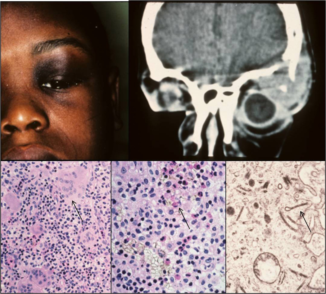
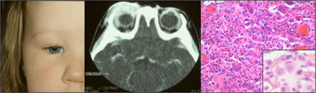
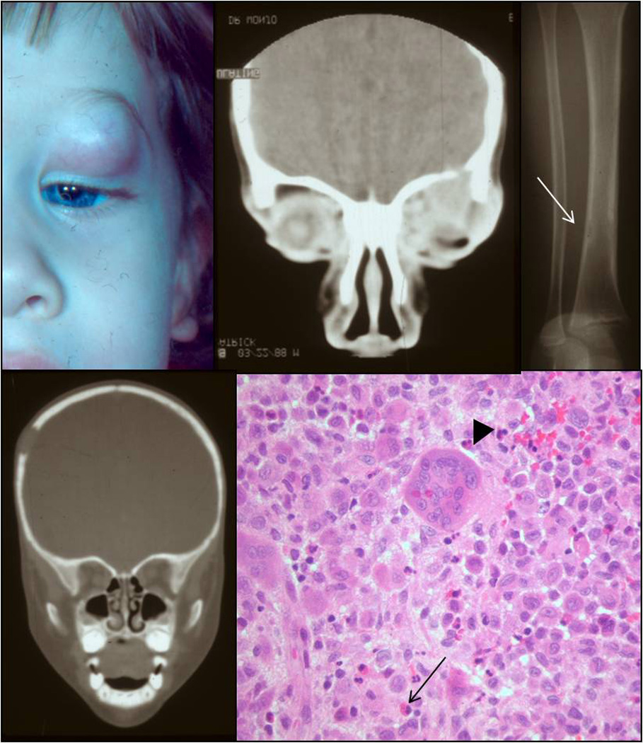
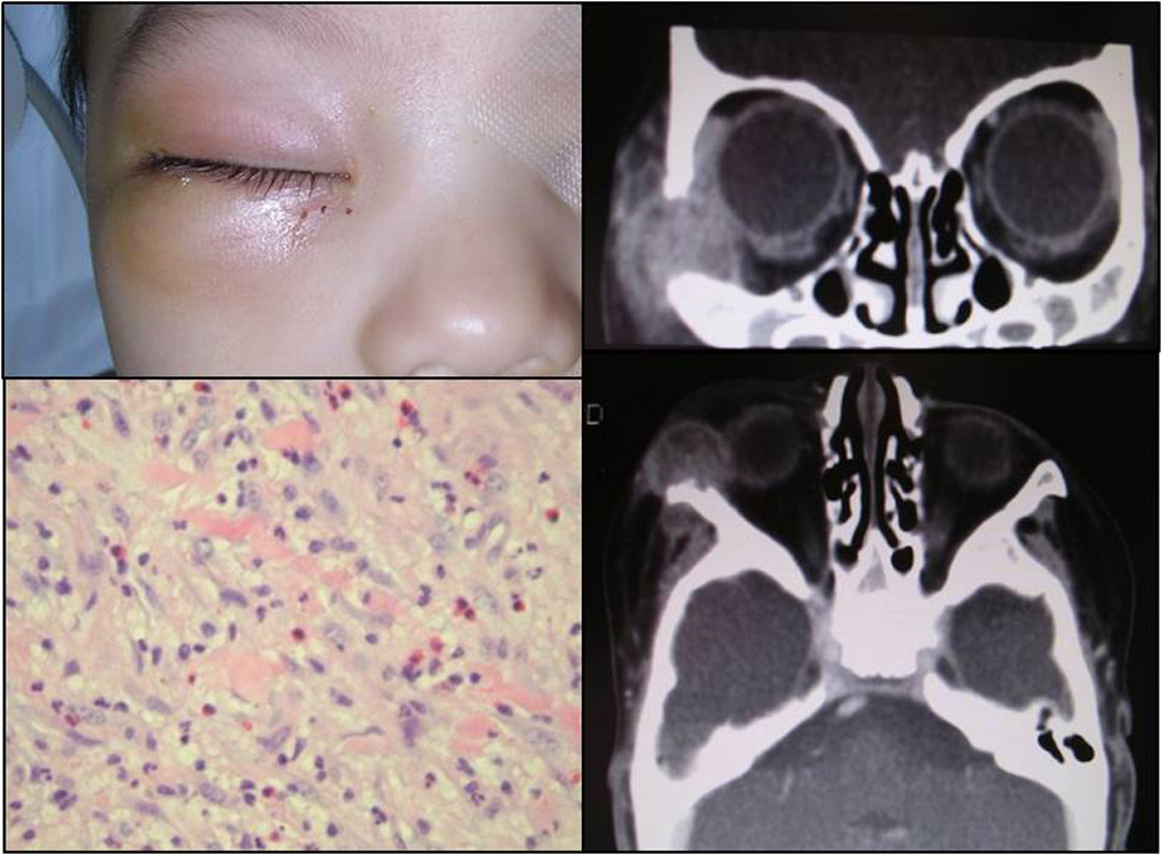
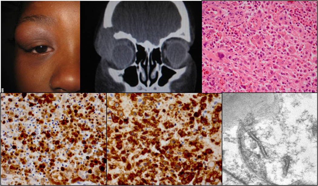
Langerhans cell histiocytosis (LCH) is a proliferation of Langerhans cells intermixed with inflammatory cells, in particular eosinophils, that may manifest as unisystem (unifocal or multifocal) or multisystem disease. Orbital involvement typically manifests as a solitary lesion that carries a favorable prognosis. We describe the clinical and histologic spectrum of LCH of the orbit in our five cases. One patient exhibited multifocal unisystem disease; the other four had a localized process. Typical histologic features included numerous histiocytes with varying degrees of giant cell formation and scattered eosinophilic granulocytes. The presence of Langerhans cells was confirmed by CD1a and S100 immunohistochemistry. Transmission electron microscopy demonstrated characteristic intracytoplasmic Birbeck granules. We review the different ophthalmic manifestations of LCH and treatment strategies. As LCH may solely involve the orbit, treatment is based on the degree of organ involvement. LCH should included in the differential diagnosis in tumors of the ocular adnexae, especially in young children.
Copyright © 2013 Elsevier Inc. All rights reserved.
Figures
References
-
- Margo CE, Goldman DR. Langerhans cell histiocytosis. Surv Ophthalmol. 2008;53(4):332–358. - PubMed
-
- Nezelof C, Basset F, Rousseau MF. Histiocytosis X histogenetic arguments for a Langerhans cell origin. Biomedicine. 1973;18(5):365–371. - PubMed
-
- Willman CL, Busque L, Griffith BB, et al. Langerhans'-cell histiocytosis (histiocytosis X)--a clonal proliferative disease. N Engl J Med. 1994;331(3):154–160. - PubMed
-
- Yu RC, Chu C, Buluwela L, Chu AC. Clonal proliferation of Langerhans cells in Langerhans cell histiocytosis. Lancet. 1994;343(8900):767–768. - PubMed
Publication types
MeSH terms
Substances
Grants and funding
LinkOut - more resources
Full Text Sources
Other Literature Sources
Research Materials
