. 2013 Jan 7;23(1):70-5.
doi: 10.1016/j.cub.2012.11.031. Epub 2012 Dec 13.

Rare codons regulate KRas oncogenesis

Affiliations

Rare codons regulate KRas oncogenesis

Benjamin L Lampson et al. Curr Biol. .

Abstract

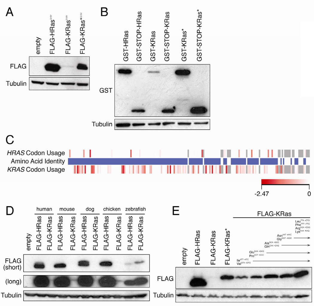
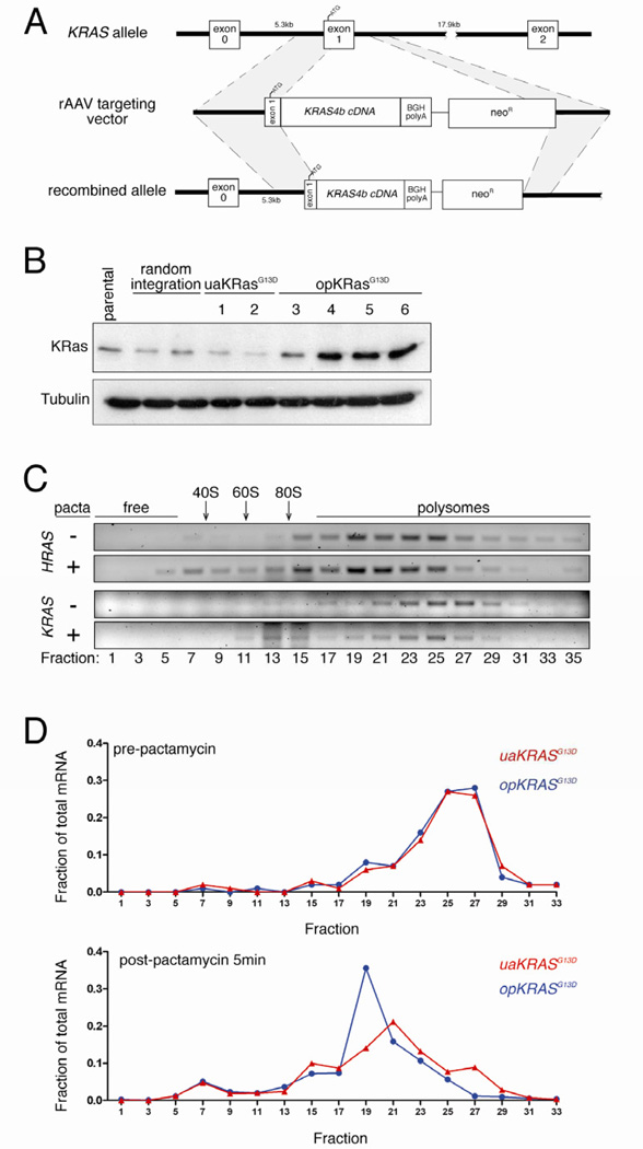
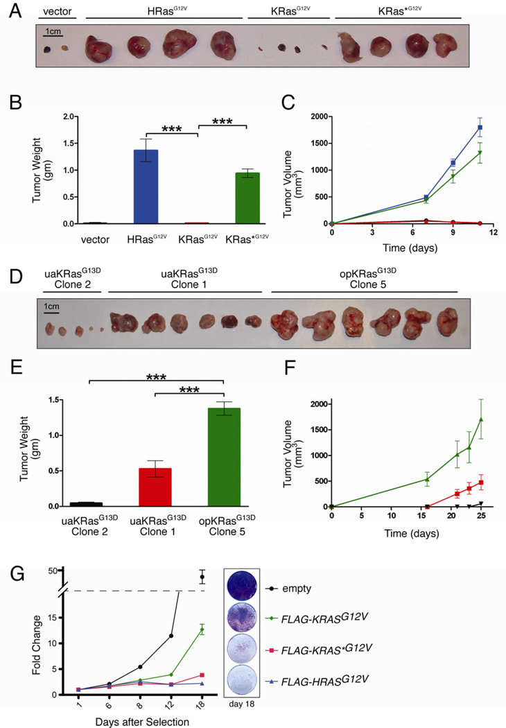
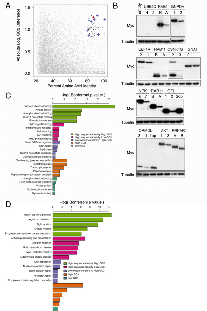
Oncogenic mutations in the small Ras GTPases KRas, HRas, and NRas render the proteins constitutively GTP bound and active, a state that promotes cancer. Ras proteins share ~85% amino acid identity, are activated by and signal through the same proteins, and can exhibit functional redundancy. Nevertheless, manipulating expression or activation of each isoform yields different cellular responses and tumorigenic phenotypes, even when different ras genes are expressed from the same locus. We now report a novel regulatory mechanism hardwired into the very sequence of RAS genes that underlies how such similar proteins impact tumorigenesis differently. Specifically, despite their high sequence similarity, KRAS is poorly translated compared to HRAS due to enrichment in genomically underrepresented or rare codons. Converting rare to common codons increases KRas expression and tumorigenicity to mirror that of HRas. Furthermore, in a genome-wide survey, similar gene pairs with opposing codon bias were identified that not only manifest dichotomous protein expression but also are enriched in key signaling protein classes and pathways. Thus, synonymous nucleotide differences affecting codon usage account for differences between HRas and KRas expression and function and may represent a broader regulation strategy in cell signaling.


Figures

Figure 1
Figure 1. Translation of KRAS is limited by rare codons
(A) Immunoblot of lysates isolated from human HEK-HT cells stably infected with the retrovirus pBabepuro encoding the indicated N-terminal FLAG-epitope tagged oncogenic (G12V) human Ras proteins (FLAG-RasG12V) with an αFLAG or αtubulin antibody. One of three experiments. (B) Immunoblot of lystates isolated from HEK-HT cells transiently transfected with the plasmid pCIneo encoding GST cDNA, a STOP codon where indicated, and in frame the indicated FLAG-Ras cDNAs, with an αGST or αtubulin antibody. One of three experiments. (C) Amino acid identity of HRas and KRas is shown in the middle bar. Blue: identical amino acids. White: non-identical amino acids. Relative codon usage of HRAS versus KRAS is shown in the upper and lower bars, respectively. Shade of red box: relative rarity of the codon. Grey box: gaps in alignment or non-identical amino acids. (D) Immunoblot of lysates isolated from human HEK-HT cells stably infected with the retrovirus pBabepuro encoding the indicated FLAG-Ras proteins isolated from the indicated species with an αFLAG or αtubulin antibody. One of two experiments. (E) Immunoblot of lysates isolated from human HEK-HT cells stably infected with the retrovirus pBabepuro encoding FLAG-HRas, FLAG-KRas or FLAG-KRas* (in which the indicated rare codons were progressively converted to the indicated common codons) with an αFLAG or αtubulin antibody. One of two experiments. Detailed methodologies and regent descriptions are provided in Supplemental Experimental Procedures. See also Figure S1.
Figure 2
Figure 2. Rare codon bias limits endogenous KRAS translation
(A) Targeting strategy to knock into exon 1 the endogenous KRAS gene in human HCT116 colon cancer cells (ATCC) oncogenic KRASG13D cDNA in which 130 rare codons were either optimized to common codons (opKRASG13D) or left unaltered (uaKRASG13D) by AAV-based recombination [16]. (B) Immunoblot of lysates isolated from parental HCT116 cell line, stable clones with non-homologous vector integration, clones with homologous integration of uaKRASG13D, and clones with homologous integration of opKRASG13D, with an αKRas or αtubulin antibody. One of one to two experiments. (C) Semi-quantitative RT-PCR using primers specific for HRAS and the non-targeted KRAS mRNA of the indicated sedimentation fractions (relative positions of ribosome-free, ribosome subunits 40S, 60S and 80S and polysome fractions) isolated from HCT116 opKRASG13D clone 5 pre- (-) and post- (+) pactamycin (pacta) treatment to halt translation initiation. (D) Quantitative RT-PCR using primers specific for the KRASG13D knock-in mRNA of the indicated sedimentation fractions isolated from HCT116 opKRASG13D clone 5 and uaKRASG13D clone 1 pre- and post-pactamycin treatment to halt translation initiation. Detailed methodologies and regent descriptions are provided in Supplemental Experimental Procedures. See also Figure S2.
Figure 3
Figure 3. Rare codons limit oncogenic KRas-driven tumorigenesis
(A) Photograph and (B) mean weight ± SEM of tumors at endpoint, as well as (C) mean size ± SEM tumors over time derived from HEK-HT cells stably expressing: ● empty vector, ■ HRasG12V, ▲ KRasG12V, and ▼ KRas*G12V, n=4. (D) Photograph and (E) mean weight ± SEM of tumors at endpoint, as well as (F) mean size ± SEM tumors over time derived from HCT116 clones expressing ▲ opKRasG13D (clone 5), ■ uaKRasG13D (clone 1), or ▼ uaKRasG13D (clone 2), n=6. One of two experiments. ***P<0.001. (G) Change in IMR90 cell number over time following acute expression of the indicated FLAG-RasG12V constructs. Left: Fold change in crystal violet absorbance at 590nm at the indicated time points after cell plating is normalized to day 1 for each cell line. Relative cell number for IMR90 cells expressing any of the oncogenic FLAG-Ras constructs was significantly lower than empty vector control cells on days 8, 12, and 18 (P<0.01). Additionally, relative cell numbers for both empty vector and FLAG-KRASG12V expressing cells were significantly different from each other and FLAG-KRAS* G12V and FLAG-HRASG12V cells on days 12 and 18 (P<0.0001). Right: Crystal violet staining of intact cells at day 18, representative of four replicates. One of one to two experiments. Detailed methodologies and regent descriptions are provided in Supplemental Experimental Procedures. See also Figure S3.
Figure 4
Figure 4. Gene pairs with divergent codon bias demonstrate correlating differences in expression and cluster in unique signaling protein classes
(A) Percent amino acid identity versus log difference in CDS GC3 content of individual protein pairs identified by BLAST alignment (grey points). Black points: gene pairs with >80% identity and >1.8-fold difference in GC3 content. Blue cross: gene pair tested for protein expression. Red cross: HRAS-KRAS gene pair. (B) Immunoblot of lysates isolated from human 293 cells (ATCC) stably infected with the retrovirus pBabepuro encoding the indicated N-terminal FLAG-epitope tagged cDNAs corresponding to human genes pairs of high amino acid sequence identity that have a common (first) versus a rare (second) codon bias, or the gene pair enriched in rare codons after rare codons were optizimized (op) to common codons, with an αFLAG or αtubulin antibody. One of one to two experiments. Histogram comparing p values of (C) gene ontology categories or (D) KEGG signaling pathways enriched in lists of gene pairs with high amino acid sequence identity and high GC3 difference (green), high identity and low GC3 difference (pink), low identity and high GC3 difference (purple), or lists of genes with high GC3 content (brown) or low GC3 content (aquamarine). Detailed methodologies and regent descriptions are provided in Supplemental Experimental Procedures. See also Tables S1–3.

Comment in

References

    1. Pylayeva-Gupta Y, Grabocka E, Bar-Sagi D. RAS oncogenes: weaving a tumorigenic web. Nature Rev. Cancer. 2011;11:761–774. - PMC - PubMed
    1. Colicelli J. Human RAS superfamily proteins and related GTPases. Sci STKE. 2004;2004:RE13. - PMC - PubMed
    1. Quilliam LA, Rebhun JF, Castro AF. A growing family of guanine nucleotide exchange factors is responsible for activation of Ras-family GTPases. Prog. Nucleic Acid Res. Mol. Biol. 2002;71:391–444. - PubMed
    1. Rodriguez-Viciana P, Sabatier C, McCormick F. Signaling specificity by Ras family GTPases is determined by the full spectrum of effectors they regulate. Mol. Cell. Biol. 2004;24:4943–4954. - PMC - PubMed
    1. Potenza N, Vecchione C, Notte A, De Rienzo A, Rosica A, Bauer L, Affuso A, De Felice M, Russo T, Poulet R, et al. Replacement of K-Ras with H-Ras supports normal embryonic development despite inducing cardiovascular pathology in adult mice. EMBO Rep. 2005;6:432–437. - PMC - PubMed

Publication types