Comparative Study
. 2013 Apr;10(4):575-82.
doi: 10.1016/j.hrthm.2012.12.017. Epub 2012 Dec 12.

Pro- and antiarrhythmic effects of ATP-sensitive potassium current activation on reentry during early afterdepolarization-mediated arrhythmias

Affiliations
Comparative Study

Pro- and antiarrhythmic effects of ATP-sensitive potassium current activation on reentry during early afterdepolarization-mediated arrhythmias

Marvin G Chang et al. Heart Rhythm. 2013 Apr.

Abstract

Background: Under conditions promoting early afterdepolarizations (EADs), ventricular tissue can become bi-excitable, that is, capable of wave propagation mediated by either the Na current (INa) or the L-type calcium current (ICa,L), raising the possibility that ICa,L-mediated reentry may contribute to polymorphic ventricular tachycardia (PVT) and torsades de pointes. ATP-sensitive K current (IKATP) activation suppresses EADs, but the effects on ICa,L-mediated reentry are unknown.

Objective: To investigate the effects of IKATP activation on ICa,L-mediated reentry.

Methods: We performed optical voltage mapping in cultured neonatal rat ventricular myocyte monolayers exposed to BayK8644 and isoproterenol. The effects of pharmacologically activating IKATP with pinacidil were analyzed.

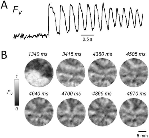
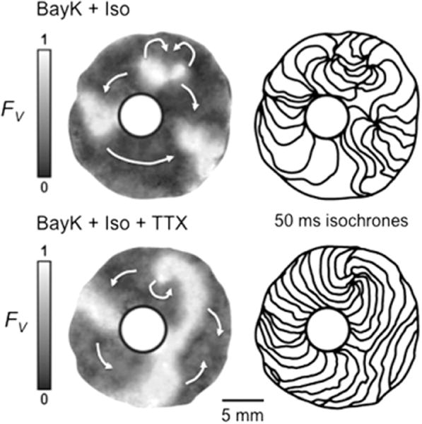
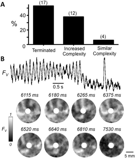
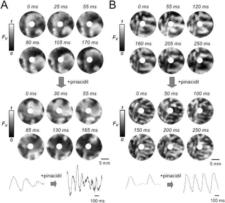
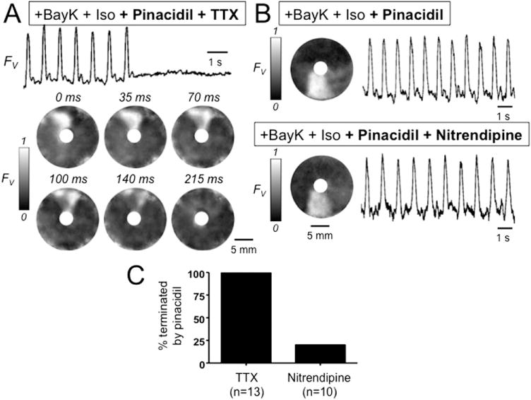
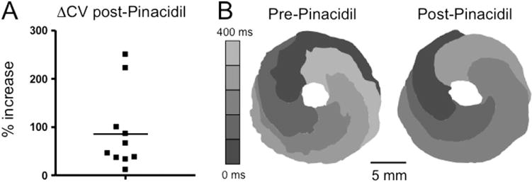
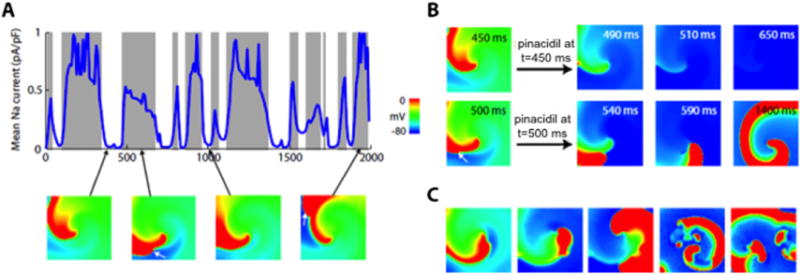
Results: In 13 monolayers with anatomic ICa,L-mediated reentry around a central obstacle, pinacidil (50 μM) converted ICa,L-mediated reentry to INa-mediated reentry. In 33 monolayers with functional ICa,L-mediated reentry (spiral waves), pinacidil terminated reentry in 17, converted reentry into more complex INa-mediated reentry resembling fibrillation in 12, and had no effect in 4. In simulated 2-dimensional bi-excitable tissue in which ICa,L- and INa-mediated wave fronts coexisted, slow IKATP activation (over minutes) reliably terminated rotors but rapid IKATP activation (over seconds) often converted ICa,L-mediated reentry to INa-mediated reentry resembling fibrillation.

Conclusions: IKATP activation can have proarrhythmic effects on EAD-mediated arrhythmias if ICa,L-mediated reentry is present.


Figures

Figure 1
Figure 1
Early afterdepolarization (EAD)-related arrhythmias induced by BayK4688 + isoproterenol in neonatal rat ventricular myocyte monolayers. A: Optical voltage trace (FV) illustrating a burst of EAD-mediated triggered activity following a paced beat. Note the incomplete repolarization between beats. B: Snapshots of voltage fluorescence (FV) at the times indicated following the paced beat at 1340 ms, revealing a complex pattern of wave fronts due to a mixture of focal activity and reentry. FV is shown on a gray scale, with 0 = maximally repolarized (<−70 mV) and 1 = maximally depolarized (>0 mV), on the basis of our previously reported patch electrode recording estimates.
Figure 2
Figure 2
Early afterdepolarization-related reentry around a central obstacle in a neonatal rat ventricular myocyte monolayer. A: Snapshot (left) and trace (right) of voltage fluorescence (FV), illustrating that reentry around the central obstacle was unaffected by tetrodotoxin (TTX) (50 μM). B: In contrast, nitrendipine (5 μM) terminated reentry. C: Bar graph summarizing incidence of termination of reentry by tetrodotoxin (TTX) vs nitrendipine in 23 monolayers. FV gray scale as described in Figure 1 legend. BayK + Iso = BayK4688 + isoproterenol.
Figure 3
Figure 3
Effect of tetrodotoxin (TTX) on early afterdepolarization (EAD)-related reentry around a central obstacle in a neonatal rat ventricular myocyte monolayer. Top panels: Sustained reentry was driven by a figure-eight rotor (curved arrows at 1 o’clock). At right, isochrone maps (50 ms between isochrones) show slow propagation (closely spaced isochrones) clockwise from 9 to 6 o’clock, consistent with L-type calcium current (ICa,L)-mediated propagation, but much faster (widely spaced isochrones) from 6 to 9 o’clock, consistent with a significant contribution of Na current (INa)-mediated propagation. Bottom panels: After TTX (50 μM), a single rotor with the same cycle length (~500 ms) continues, but conduction velocity is now uniformly slow, consistent with ICa,L-mediated rotor and wave propagation throughout the tissue. FV gray scale as described in Figure 1 legend. BayK + Iso = BayK4688 + isoproterenol.
Figure 4
Figure 4
Effect of the ATP-sensitive K channel agonist pinacidil in neonatal rat ventricular myocyte (NRVM) monolayers with complex sustained early afterdepolarization (EAD)-mediated arrhythmias. A: Histogram illustrating the outcomes of pinacidil (100 μM) treatment on complex EAD-mediated arrhythmias induced by BayK4688 + isoproterenol in 33 NRVM monolayers with a central obstacle. B: Representative optical trace (top) and snapshots (below) of voltage fluorescence (FV) at the times indicated, illustrating termination of a complex EAD-mediated arrhythmia by pinacidil. In this example, the arrhythmia was driven primarily by a functional rotor (spiral wave) at 1 o’clock. FV gray scale as described in Figure 1 legend.
Figure 5
Figure 5
Effect of the ATP-sensitive K channel agonist pinacidil in neonatal rat ventricular myocyte monolayers with complex sustained early afterdepolarization-mediated arrhythmias. A: Snapshots of voltage fluorescence (FV) at the times indicated in a monolayer with a central obstacle in which BayK4688 + isoproterenol induced an arrhythmia driven primarily by functional rotors (spiral waves) at 6 and 8 o’clock (left panels). Pinacidil (100 μM, right panels) accelerated but did not significantly affect the complexity of the arrhythmia. B: Voltage snapshots from another monolayer with a central obstacle, in which pinacidil (right panels) increased the complexity of the BayK4688 + isoproterenol-induced arrhythmia, as evident from the greater number of wave fronts. FV gray scale as described in Figure 1 legend.
Figure 6
Figure 6
Effect of pinacidil on early afterdepolarization-mediated reentry around a central obstacle in a neonatal rat ventricular myocyte monolayer. A: Optical trace (above) and snapshots (below) of voltage fluorescence (FV) in a monolayer in which BayK4688 + isoproterenol induced sustained reentry around a central obstacle. After pinacidil treatment, the addition of tetrodotoxin (TTX) (50 μM) terminated reentry. B: Same, but with nitrendipine (5 μM) added instead of TTX, showing that reentry persists. C: Bar graph summarizing the incidence of termination of reentry by TTX vs nitrendipine in 23 monolayers exposed to BayK4688 + isoproterenol + pinacidil. FV gray scale as described in Figure 1 legend. BayK + Iso = BayK4688 + isoproterenol.
Figure 7
Figure 7
Effect of pinacidil on conduction velocity of early afterdepolarization-mediated reentry around a central obstacle in neonatal rat ventricular myocyte monolayers. A: The percent increase in conduction velocity after pinacidil in 10 monolayers (solid squares) in which BayK4688 + isoproterenol induced sustained reentry around a central obstacle. Mean increase is indicated by the horizontal bar. B: Isochrome map during reentry in a representative monolayer, pre-, and post-pinacidil treatment. Increased spacing between 80-ms isochrome lines after pinacidil reflects increased conduction velocity. FV gray scale as described in Figure 1 legend.
Figure 8
Figure 8
Effect of ATP-sensitive K current (IKATP) activation on L-type calcium current (ICa,L)-mediated reentry in simulated 2-dimensional heterogeneous cardiac tissue (300 × 300 myocytes). A: Outcomes of rapidly activating IKATP at various time points during reentry driven by an ICa,L-mediated rotor in the center of the tissue. Gray zones indicate that ICa,L-mediated reentry converted to Na current (INa)-mediated reentry after IKATP activation; white zones indicate that reentry terminated. The blue line shows INa amplitude averaged over all cells in the tissue over time. IKATP activation terminated reentry only if no significant INa-mediated wave fronts were present. Voltage snapshots (below) show the ICa,L-mediated rotor in the center of the tissue, with INa-mediated wave fronts intermittently present along the spiral arm (2nd and 4th panels; white arrows). B: Voltage snapshots showing termination of reentry by IKATP activation at t = 450 ms (top row) but conversion to INa-mediated reentry at t = 500 ms (bottom row). C: When the number of KATP channels was randomly varied from myocyte to myocyte (using a Gaussian distribution with mean 3.8 channels/μm2 and standard deviation 2.0), IKATP activation caused the stable ICa,L-mediated rotor to convert to an INa-mediated rotor, which then broke up into multiwavelet VF. FV scale as described in Figure 1 legend.

Comment in

Similar articles

Cited by

References

    1. Chang MG, Sato D, de Lange E, et al. Bi-stable wave propagation and early afterdepolarization-mediated cardiac arrhythmias. Heart Rhythm. 2012;9:115–122. - PMC - PubMed
    1. Fish FA, Prakash C, Roden DM. Suppression of repolarization-related arrhythmias in vitro and in vivo by low-dose potassium channel activators. Circ Res. 1990;82:1362–1369. - PubMed
    1. Spinelli W, Sorota S, Siegal M, et al. Antiarrhythmic actions of the ATP-regulated K current activated by pinacidil. Circ Res. 1991;68:1127–1137. - PubMed
    1. Carlsson L, Abrahamsson C, Drews L, et al. Antiarrhythmic effects of potassium channel openers in rhythm abnormalities related to delayed repolarization. Circulation. 1992;85:1491–1500. - PubMed
    1. Sato T, Hata Y, Yamamoto M, Morita H, et al. Early afterdepolarization abolished by potassium channel opener in a patient with idiopathic long QT syndrome. J Cardiovasc Electrophysiol. 1995;6:279–282. - PubMed

Publication types

MeSH terms