Comparative Study
. 2013 Apr;10(4):592-9.
doi: 10.1016/j.hrthm.2012.12.016. Epub 2012 Dec 12.

Inhibition of CaMKII phosphorylation of RyR2 prevents inducible ventricular arrhythmias in mice with Duchenne muscular dystrophy

Affiliations
Comparative Study

Inhibition of CaMKII phosphorylation of RyR2 prevents inducible ventricular arrhythmias in mice with Duchenne muscular dystrophy

Sameer Ather et al. Heart Rhythm. 2013 Apr.

Abstract

Background: Ventricular tachycardia (VT) is the second most common cause of death in patients with Duchenne muscular dystrophy (DMD). Recent studies have implicated enhanced sarcoplasmic reticulum (SR) Ca(2+) leak via type 2 ryanodine receptor (RyR2) as a cause of VT in the mdx mouse model of DMD. However, the signaling mechanisms underlying induction of SR Ca(2+) leak and VT are poorly understood.

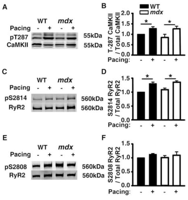
Objective: To test whether enhanced Ca(2+)/calmodulin-dependent protein kinase II (CaMKII) phosphorylation of RyR2 underlies SR Ca(2+) leak and induction of VT in mdx mice.

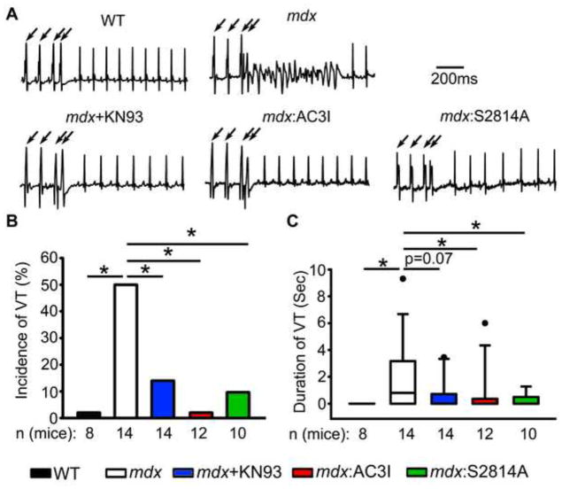
Methods: Programmed electrical stimulation was performed on anesthetized mice and confocal imaging of Ca(2+) release events in isolated ventricular myocytes.

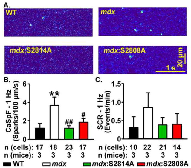
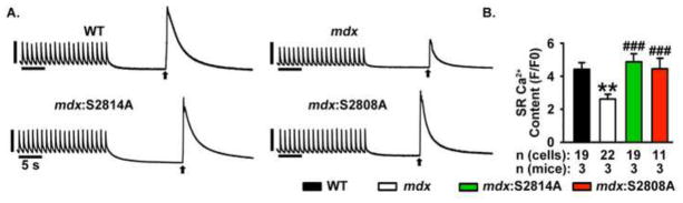
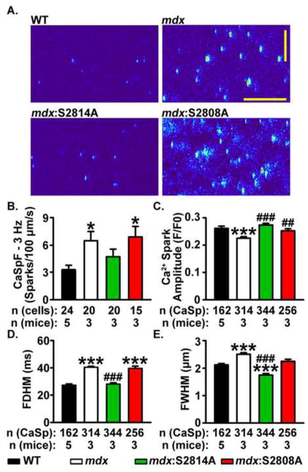
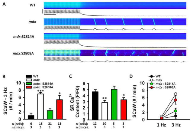
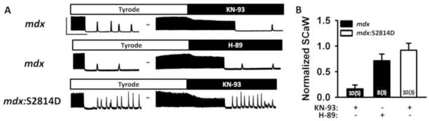
Results: Programmed electrical stimulation revealed inducible VT in mdx mice, which was inhibited by CaMKII inhibition or mutation S2814A in RyR2. Myocytes from mdx mice exhibited more Ca(2+) sparks and Ca(2+) waves compared with wild-type mice, in particular at faster pacing rates. Arrhythmogenic Ca(2+) waves were inhibited by CaMKII but not by protein kinase A inhibition. Moreover, mutation S2814A but not S2808A in RyR2 suppressed spontaneous Ca(2+) waves in myocytes from mdx mice.

Conclusions: CaMKII blockade and genetic inhibition of RyR2-S2814 phosphorylation prevent VT induction in a mouse model of DMD. In ventricular myocytes from mdx mice, spontaneous Ca(2+) sparks and Ca(2+) waves can be suppressed by CaMKII inhibition or mutation S2814A in RyR2. Thus, the inhibition of CaMKII-induced SR Ca(2+) leak might be a new strategy to prevent arrhythmias in patients with DMD without heart failure.


Conflict of interest statement

Conflicts of Interest: None of the authors have a conflict of interest.

Figures

Figure 1
Figure 1. Genetic inhibition of CaMKII phosphorylation of RyR2 prevents induction of ventricular tachycardia (VT) in mdx mice
(A) Representative surface ECG tracings revealing ventricular tachycardia (VT) in an mdx mouse following intracardiac pacing (indicated by arrows). (B) Bar graph showing quantification of the incidence of pacing-induced sustained VT in WT and mdx mice. (C) Boxplot showing quantification of the duration of pacing induced sustained VT in WT and mdx mice. Pharmacologic (KN93) or genetic inhibition of CaMKII (AC3I peptide), as well as mutation S2814A on RyR2, prevented VT induction in mdx mice (B, C). *p<0.05 vs. WT. #p<0.05 vs. mdx.
Figure 2
Figure 2. Pacing-induced CaMKII activation and increased S2814 phosphorylation on RyR2 in WT and mdx mice
Representative Western blots and bar graphs with quantification of averaged data showing (A, B) Increased CaMKII autophosphorylation levels at T287 following intracardiac pacing in both WT and mdx mice. (C, D) Increased RyR2 phosphorylation levels at S2814 following intracardiac pacing in both WT and mdx mice. (E, F) Unaltered RyR2 phosphorylation levels at S2808 following pacing. Each bar represents averaged data from 4–5 mice. * p<0.05.
Figure 3
Figure 3. Mutations S2814A and S2808A in RyR2 reduce Ca2+ spark frequency (CaSpF) leak in mdx mice at 1-Hz
(A) Representative line scans showing spontaneous Ca2+ sparks in ventricular myocytes from wildtype (WT), mdx, mdx:S2814A and mdx:S2808A cardiomyocytes following a 20-s 1 Hz pacing train. Bar graph showing (B) CaSpF and (C) frequency of spontaneous Ca2+ waves (SCaW), during the 1-minute pause. **p<0.01 vs. WT. #p<0.05, ##p<0.01 vs. mdx.
Figure 4
Figure 4. Reduced SR Ca2+ stores in mdx ventricular myocytes at 1-Hz
(A) Representative [Ca2+]i recordings from ventricular myocytes during 1-Hz pacing train, followed by caffeine-evoked SR Ca2+ dump, and (B) SR Ca2+ content, in ventricular myocytes from WT, mdx, and mdx:S2814A mice. Numbers in bar graphs represent number of cardiomyocytes obtained from 3–5 mice in each group. * p<0.01, ### P<0.001.
Figure 5
Figure 5. Mutation S2814A but not S2808A in RyR2 normalizes Ca2+ spark frequency (CaSpF) and mass in mdx mice at 3 Hz
(A) Representative line scans showing spontaneous Ca2+ sparks in ventricular myocytes from wildtype (WT), mdx, mdx:S2814A and mdx:S2808A cardiomyocytes following a 20-s 3 Hz pacing train. Bar graph showing (B) CaSpF, (C) Spark amplitude, (D) Full diameter half maximum (FDHM), and (E) Full width half maximum (FWHM), during the 1-minute pause. *p<0.05, ***p<0.001 vs. WT. ##p<0.01, ###p<0.001 vs. mdx.
Figure 6
Figure 6. Mutation S2814A but not S2808A in RyR2 reduces spontaneous Ca2+ waves (SCaW) in mdx mice at 3 Hz
(A) Representative line scan images and [Ca2+]i tracings showing spontaneous Ca2+ waves in ventricular myocytes from mdx mice following a 20-s 3-Hz pacing train. Scale bars: 2 F/F0 (vertical) and 5 seconds (horizontal). (B) Bar graph showing incidence of SCaWs following pacing train. (C) Bar graph showing SR Ca2+ load at 3 Hz. (D) Line graph showing the increase in SCaW frequency when pacing rate was increased from 1-Hz to 3-Hz. The proportional increase in SCaWs was greater in mdx and mdx:S2808A mice, compared to WT and mdx:S2814A mice. *p<0.05, **p<0.01, ***p<0.001 vs. WT. ###p<0.001 vs. mdx.
Figure 7
Figure 7. CaMKII inhibition suppresses SCaW in mdx ventricular myocytes at 3-Hz
(A) Representative tracings showing spontaneous Ca2+ waves (SCaW) following 3-Hz pacing. CaMKII inhibitor KN-93 (1 μM) or PKA inhibitor H-89 (1 μM) were added subsequently, followed by another observation period for SCaWs. Scale bars: 2 F/F0 (vertical) and 20 seconds (horizontal). (B) Bar graph showing SCaW frequency after inhibitor administration normalized to SCaW frequency in Tyrode.

References

    1. Groh WJ, Zipes DL. Neurological disorders and cardiovascular disease. In: Bonow RO, Mann DL, Zipes DL, Libby P, editors. Braunwald’s heart disease. A textbook of cardiovascular medicine. Philadephia: Saunders; 2011. pp. 1916–1919.
    1. Jung C, Martins AS, Niggli E, Shirokova N. Dystrophic cardiomyopathy: amplification of cellular damage by Ca2+ signalling and reactive oxygen species-generating pathways. Cardiovascular research. 2008;77:766–773. - PubMed
    1. Sarma S, Li N, van Oort RJ, Reynolds C, Skapura DG, Wehrens XH. Genetic inhibition of PKA phosphorylation of RyR2 prevents dystrophic cardiomyopathy. Proc Natl Acad Sci U S A. 2010;107:13165–13170. - PMC - PubMed
    1. Fauconnier J, Thireau J, Reiken S, et al. Leaky RyR2 trigger ventricular arrhythmias in Duchenne muscular dystrophy. Proc Natl Acad Sci U S A. 2010;107:1559–1564. - PMC - PubMed
    1. Dobrev D, Wehrens XH. Calmodulin kinase II, sarcoplasmic reticulum Ca2+ leak, and atrial fibrillation. Trends in cardiovascular medicine. 2010;20:30–34. - PMC - PubMed

Publication types

MeSH terms

Substances