. 2013 Mar;21(3):554-60.
doi: 10.1038/mt.2012.259. Epub 2012 Dec 18.

A molecular imaging system based on both transcriptional and genomic amplification to detect prostate cancer cells in vivo

Affiliations

A molecular imaging system based on both transcriptional and genomic amplification to detect prostate cancer cells in vivo

Frédéric Pouliot et al. Mol Ther. 2013 Mar.

Abstract

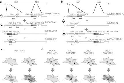
An imaging modality that can accurately discern prostate cancer (PCa) foci would be useful to detect PCa early or guide treatment. We have engineered numerous adenoviral vectors (Ads) to carry out reporter gene-based imaging using specific promoters to express a potent transcriptional activator, which in turn activates the reporter gene in PCa. This two-step transcriptional amplification (TSTA) method can boost promoters' activity, while maintaining cell specificity. Here, we examined a dual TSTA (DTSTA) approach, which utilizes TSTA not only to express the imaging reporter, but also to direct viral genome replication of a conditionally replicating Ad (CRAd) to further augment the expression levels of the reporter gene by genomic amplification supported in trans by coadministered CRAd. In vitro studies showed up to 50-fold increase of the reporter genome by DTSTA. Compared with TSTA reporter alone, DTSTA application exhibited a 25-fold increase in imaging signal in PCa xenografts. DTSTA approach is also beneficial for a combination of two TSTA Ads with distinct promoters, although amplification is observed only when TSTA-CRAd can replicate. Consequently, the DTSTA approach is a hybrid method of transcriptional and genomic augmentation that can provide higher level reporter gene expression potentially with a lower dose of viral administration.


Figures

Figure 1
Figure 1
The experimental design and concept of the Dual-TSTA study. The Dual-TSTA (DTSTA) system was examined in tissue culture with two different experimental designs. (a,b) Show the list and combination of adenovirus vectors used in each experiment; (c,d) Cartoon of expected gene expression following infection. (a) PSA-positive (androgen receptor positive) prostate cancer cell lines were infected with the combination of AdPSA-TSTA-FL and either of TSTA-CRAd, AdPSA-VP16 or AdCMV-GFP, the latter two being control, balancing vectors. Both TSTA are regulated under the modified PSA promoter PSE-BC. (c) The expression of firefly luciferase (FL) reporter gene will be amplified further when coinfected with TSTA-CRAd compared to single infection. (b) The combined infection with two distinct promoters, the PSE-BC–based TSTA-CRAd, and MUC1 promoter-based reporter vector (AdMUC1-FL or AdMUC1-TSTA-FL). A nonreplicating AdPSA-TSTA-TK, expressing HSV-TK, was used as the control balancing Ad. (d) The schema depicts the expected gene expression in different cells, with respect to MUC1 and PSA expression status. In MUC1 and AR cells, reporter gene expression should be negligible. In AR+ cells, the presence of TSTA-CRAd will amplify the reporter signal. Highest FL expression is expected in MUC1+ and AR+ cells, as both transcriptional and genomic amplification mechanism will be at play. Ad, adenoviral vector; AR, androgen receptor; CMV, cytomegalovirus; CRAd, conditionally replicating Ad; GFP, green fluorescent protein; MUC1, mucin 1; PSA, prostate-specific antigen; TSTA, two-step transcriptional amplification.
Figure 2
Figure 2
Coinfection of TSTA-CRAd with AdPSA-TSTA-FL further amplifies reporter gene expression by genome copy amplification in prostate cancer cells. (a,b) LNCaP and (c,d) CWR22Rv1 PCa cells were infected with the AdPSA-TSTA-FL virus (MOI = 0.5) and either with the TSTA-CRAd or controls (AdCMV-GFP or AdPSA-VP16 viruses) at MOI = 3. (a,c) Show the FL activity, assessed on day 2, 4, 6, and 8 post-infection. Each point on the graph represents the average of duplicates ± SE of FL activity per µg protein. (b,d) Cell cytotoxicity of the coinfection experiments described above, determined at day 2, 4, 6 or 8 after adenoviral infections. LDH cell cytotoxicity was normalized to the level on day 2 after viral infection. (e) Amplification of the AdPSA-TSTA-FL genome by TSTA-CRAd. Real-time PCR was performed on total DNA extracted from LNCaP coinfected either with AdCMV-GFP, AdPSA-VP16 or TSTA-CRAd. The copy number of AdPSA-TSTA-FL genomes increases with TSTA-CRAd addition but not in controls (AdCMV-GFP or AdPSA-VP16 viruses), suggesting the viral replication of AdPSA-TSTA-FL supported by TSTA-CRAd. Values are reported as fold-FL gene copy number normalized to that of day 2 ± SE of FL activity per µg protein from duplicates. Ad, adenoviral vector; CMV, cytomegalovirus; CRAd, conditionally replicating Ad; FL, firefly luciferase; GFP, green fluorescent protein; LDH, lactate dehydrogenase; MOI, multiplicity of infection; PCa, prostate cancer; PSA, prostate-specific antigen; TSTA, two-step transcriptional amplification.
Figure 3
Figure 3
The highly amplified activity of DTSTA in vivo in the xenograft LAPC-9 PCa model. (a) LAPC-9-RL (overexpressing the Renilla luciferase) tumors were grown as xenografts in SCID/Beige mice. After randomization for tumor size, mice were injected with AdPSA-TSTA-FL in combination with AdCMV-GFP (left panels) or with TSTA-CRAd (right panels). Mice were then imaged in vivo at different timepoints using a CCD-camera to detect bioluminescence emitted by FL following luciferin intraperitoneal injection. (b) In vivo bioluminescence quantification at 7 days after infection for each mouse. In average, animals with CRAd showed total flux of 4.4 ± 1.3 × 10⁷ photons per second (p/s) compared with control animals (+CMV-GFP): 31.0 ± 8.3 × 10⁵ p/s. These are statistically significant (P < 0.05) based on the Student's t-test with equal variance. (c) Ex-vivo firefly-to-Renilla luciferase activity ratio for each mouse tumor extract. On average, ratios of group with control and with CRAd were 1.9 ± 1.3 × 10⁻⁴ and 15.0 ± 2.8 × 10⁻³, respectively. These are statistically significant (P < 0.05) based on the t-test with equal variance. (d) Ex-vivo real-time PCR using FL gene-specific primers on total DNA extracts for each mouse. In average, group with control and with CRAd were 1.8 ± 1.1 and 3.0 ± 1.1 × 10⁻², respectively (P = 0.05). (e) FL activity increases with time after DTSTA injection in LAPC-9-RL tumors. Mice 101R (AdPSA-TSTA-FL) and 101B (DTSTA) were imaged in vivo at different timepoints using a CCD-camera to detect bioluminescence emitted by FL. The FL activity increases with time after infection in the DTSTA-infected mouse while it is stable for the mouse infected with AdPSA-TSTA-FL alone. Ad, adenoviral vector; CMV, cytomegalovirus; CRAd, conditionally replicating Ad; DTSTA, dual two-step transcriptional amplification; FL, firefly luciferase; GFP, green fluorescent protein; PCa, prostate cancer; PSA, prostate-specific antigen; SCID, severe combined immunodeficiency.
Figure 4
Figure 4
The enhanced expression of combining a cancer-specific (MUC1) and prostate-specific promoters in the DTSTA system. (a) FL activity in LNCaP was examined with two different coinfection; AdMUC1-FL plus AdPSA-TSTA-TK (closed bars) or AdMUC1-FL plus TSTA-CRAd (open bars). (b) LNCaP cells were infected with AdMUC1-TSTA-FL plus AdPSA-TSTA-TK (darker hatched bars) or AdMUC1-TSTA-FL plus TSTA-CRAd (lightly dotted bars). (c) The AR+ prostate cancer line, LAPC-4, (d) untransformed prostatic epithelial BPH1 cell line, (e) MDA-MB-435 breast cancer cell line, (f) HepG2 hepatic carcinoma line, and (g) HeLa cervical carcinoma cell line are coinfected with either AdMUC1-FL plus control, replication-incompetent AdPSA-TSTA-TK (closed bars) or AdMUC1-FL plus TSTA-CRAd (open bars). Infection of all the virus were carried out at MOI = 1. Cells harvested on day 2, 4, and 6 are subjected to luciferase assay. Numbers on top of each graph shows ratios of FL activity of coinfection with or without TSTA-CRAd (e.g., AdMUC1-FL + TSTA-CRAd activity divided by AdMUC1-FL + AdPSA-TSTA-TK activity). Ad, adenoviral vector; AR, androgen receptor; CRAd, conditionally replicating Ad; DTSTA, dual two-step transcriptional amplification; FL, firefly luciferase; MOI, multiplicity of infection; MUC1, mucin 1.

Similar articles

Cited by

References

    1. Min JJ., and, Gambhir SS. Molecular imaging of PET reporter gene expression. Handb Exp Pharmacol. 2008. pp. 277–303. - PubMed
    1. Pouliot F, Johnson M., and, Wu L. Non-invasive molecular imaging of prostate cancer lymph node metastasis. Trends Mol Med. 2009;15:254–262. - PMC - PubMed
    1. Brand AH., and, Perrimon N. Targeted gene expression as a means of altering cell fates and generating dominant phenotypes. Development. 1993;118:401–415. - PubMed
    1. Fischer JA, Giniger E, Maniatis T., and, Ptashne M. GAL4 activates transcription in Drosophila. Nature. 1988;332:853–856. - PubMed
    1. Block A, Milasinovic D, Mueller J, Schaefer P, Schaefer H., and, Greten H. Amplified Muc1-specific gene expression in colon cancer cells utilizing a binary system in adenoviral vectors. Anticancer Res. 2002;22 6A:3285–3292. - PubMed

Publication types

Substances