Review
. 2013 Jan;14(1):62-75.
doi: 10.1038/nrg3345.

Chromatin and the genome integrity network

Affiliations
Review

Chromatin and the genome integrity network

Manolis Papamichos-Chronakis et al. Nat Rev Genet. 2013 Jan.

Abstract

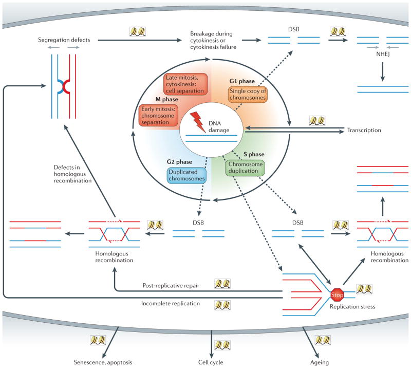
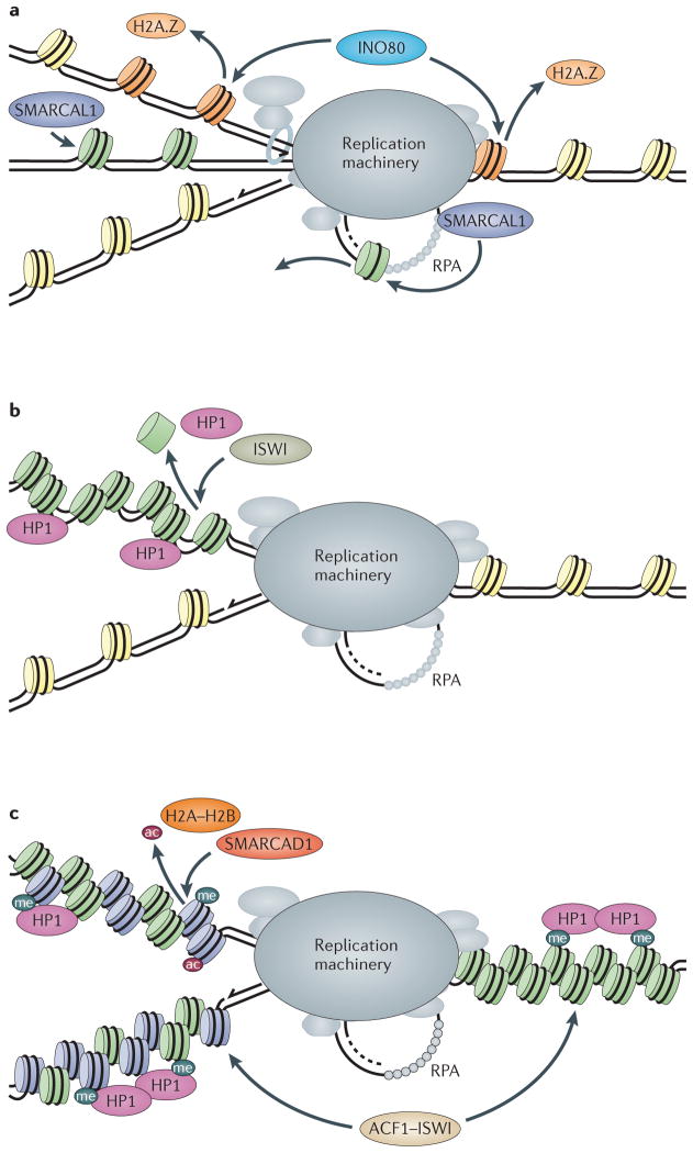
The maintenance of genome integrity is essential for organism survival and for the inheritance of traits to offspring. Genomic instability is caused by DNA damage, aberrant DNA replication or uncoordinated cell division, which can lead to chromosomal aberrations and gene mutations. Recently, chromatin regulators that shape the epigenetic landscape have emerged as potential gatekeepers and signalling coordinators for the maintenance of genome integrity. Here, we review chromatin functions during the two major pathways that control genome integrity: namely, repair of DNA damage and DNA replication. We also discuss recent evidence that suggests a novel role for chromatin-remodelling factors in chromosome segregation and in the prevention of aneuploidy.


Conflict of interest statement

Competing interests statement

The authors declare no competing financial interests.

Figures

Figure 1
Figure 1. The role of the genome stability network in cell homeostasis
DNA damage (shown by the lightning bolt) can create double-strand breaks (DSBs) that are repaired by the appropriate pathway, depending on the cell cycle phase. DNA DSBs in G1 phase are preferably repaired by non-homologous end joining (NHEJ), whereas DSBs in S phase or in G2 phase are mainly repaired by homologous recombination. During S phase, replication forks that encounter DNA damage or that undergo other types of replication stress may induce DNA DSBs and/or lead to the formation of aberrant chromatin structures between chromosomes. If they are not resolved, these aberrant structures, which are derived from replication defects or incomplete homologous recombination, can lead to chromosome segregation failure in mitosis or to chromosomal breakage during cytokinesis. To ensure that the most appropriate response is activated, crosstalk between the genome stability pathways is essential. In addition, the genome stability network successfully connects the repair process with other pathways that regulate cell homeostasis. Chromatin has a major role in the different genome stability pathways (as depicted by the nucleosomes in boxes).
Figure 2
Figure 2. Two primary pathways for double-strand break repair
Following its formation, a double-strand break (DSB) is initially bound by the yeast Mre11–Rad50–Xrs2 (MRX) or human MRE11–RAD50–NBS1 (MRN) complex. If a DSB is formed in the G1 phase of the cell cycle, the DSB is preferentially repaired by the non-homologous end-joining (NHEJ) pathway (left panel). In this case, binding of the Ku heterodimer (Ku70–Ku80) prevents extensive nucleolytic processing of the DSB and promotes subsequent ligation of the DSB by the Dnl4–Lif1–Nej1 complex in budding yeast and the LIG4–XRCC4 factors in mammals. If a DSB is formed in the S or G2 cell cycle phase, the homologous recombination repair pathway predominates (right panel). Key steps for successful repair by homologous recombination include: nucleolytic processing of the 5′ ends of the dsDNA ends into 3′ single-stranded tails by the initial action of the yeast MRX complex and Sae2 enzyme (known as RBBP8 or CTIP in mammals); extensive processing by either the exodeoxyribonuclease 1 (Exo1) or the Dna2–Sgs1 DNA-end-processing enzymes (the human Bloom’s syndrome protein (BLM) helicase is the homologue of yeast Sgs1); assembly of a recombination protein A (RPA)–ssDNA filament that is subsequently converted into a Rad51–ssDNA filament; completion of a successful homology search and formation of heteroduplex DNA; DNA synthesis that uses the 3′ end of the broken DNA, resolution of the heteroduplex or a double-sided Holliday junction; and, finally, ligation of the ssDNA nicks and termination of the process. Note that formation of the RPA–ssDNA filament is also essential for activation of the DNA damage cell cycle checkpoint. For a recent Review, see REF. . Protein or complex names given in brackets in the figure are the human homologues.
Figure 3
Figure 3. Chromatin dynamics in double-strand break checkpoint response in S. cerevisiae
a | Formation of a double-strand break (DSB; shown by lightning bolt), initial chromatin alterations and checkpoint activation. Recognition of the broken ends by the Mre11–Rad50–Xrs2 (MRX) complex initiates DNA end processing. The MRX complex recruits the RSC chromatin-remodelling complex, which restructures chromatin at the DSB ends; checkpoint kinases Mec1 and Tel1 (‘Mec1/Tec1’ in the figure) phosphorylate the histone variant H2A.X, leading to the NuA4 complex and SWR complex (SWR-C) recruitment. SWR-C incorporates H2A.Z, which can lead to relocalization of the DSB to the nuclear periphery; NuA4 is self-activated by acetylating its subunit Yng2 (depicted as Yng2ac in the figure) a nd targets histones H2A.Z and H4 for acetylation (abbreviated to ‘ac’ in the figure). Rad9 recruitment is stabilized by its interactions with H2A.X phosphorylated at serine 139 (H2A.XS139ph; known as γH2A.X and abbreviated to ‘γ’ in the figure) and H3 trimethylated at 79 (H3K79me3; abbreviated to ‘me’ in the figure), leading to checkpoint activation. b | The DSB is processed by the Sgs1–Dna2 and exodeoxyribonuclease 1 (Exo1) pathways and is assisted by the chromatin-remodelling complex Fun30, which appears to counteract the inhibitory effect of Rad9 on DNA resection. Initial steps for removing epigenetic marks are also shown: histone deacetylase 1 (Hda1) deacetylates H2A.Z, and Rpd3–Sin3 deacetylates H4 and Yng2. Inactivation of NuA4 occurs by targeted degradation of Yng2 by the 26S proteasome (the degraded Yng2 is represented by the jagged oval). INO80 recruitment leads to eviction of H2A.Z, facilitating checkpoint termination and adaptation. Restoration of chromatin structure is mediated in part by Asf1, which deposits new histones and serine/threonine protein phosphatase 4 catalytic subunit (Pph3) dephosphorylates H2A.X.
Figure 4
Figure 4. Chromatin dynamics in checkpoint signalling in mammalian cells
a | DSB formation and initial histone modifications. Ataxia telangiectasia mutated (ATM) and ataxia telangiectasia and RAD3-related (ATR) checkpoint kinases (‘ATM/ATR’ in the figure) phosphorylate the histone variant H2A.X at serine 139 (H2A.XS139ph; termed γH2A.X and depicted as ‘γ’ in the figure) and promote nucleosome eviction. ATM recruits GCN5, which catalyses H3 acetylation (the red ‘ac’ in the figure). Recruitment of MOF catalyses acetylation of histone H4 at lysine 16 (H4K16; the black ‘ac’ in the figure), leading to chromatin decompaction; poly(ADP-ribose) polymerase 1 (PARP1) action leads to PARylation of proteins and histones (‘PAR’). The RING finger heterodimer RNF20–RNF40 ubiquitylates histone H2BK120, which also promotes decompaction (‘ub1’). b | Chromatin decondensation induced by the histone modifications catalysed in panel a leads to subsequent recruitment of WSTF, which dephosphorylates H2A.X at tyrosine 142 (H2A.XY142; phY→ph), leading to mediator of DNA damage checkpoint 1 (MDC1) recruitment by γH2A.X. The SWI/SNF complex is recruited by histone H3 acetylation, and its chromatin-remodelling activity facilitates further spreading of γH2A.X and stabilization of MDC1 on chromatin. MDC1 in turn recruits the KAT5–p400 enzyme, which is activated by trimethylation of H3 at lysine 9 (H3K9; not shown), leading to further histone acetylation, and also recruits RNF8 and RNF168 (‘RNF8/RNF168’ in the figure), which ubiquitylate H2A and/or other proteins (‘ub2’). Ubiquitylation by RNF8 and RNF168 recruits chromodomain helicase DNA-binding protein 4 (CHD4). Histone PARylation (and/or modification by other proteins) leads to recruitment of the nucleosome-remodelling and histone deacetylase (NuRD) complex. Histone H4 is further modified by MMSET-mediated methylation (H4K20me2 or H4K20me3; depicted as ‘me’ in the figure). The combination of ubiquitylation by RNF8 and RNF168 with either H4K20me or PAR-ylation recruits tumour suppressor p53-binding protein 1 (53BP1) or ALC1, respectively.
Figure 5
Figure 5. Chromatin-remodelling activities at the replication fork
a | INO80 may remove H2A.Z–H2B dimers (‘H2A.Z’ in the figure) from nucleosomes (H2A.Z nucleosomes depicted in orange) ahead of the fork or from newly formed nucleosomes on replicated DNA (orange nucleosomes). SMARCA-like protein 1 (SMARCAL1) catalyses fork regression and Holliday junction migration and may remove nucleosomes from regressed dsDNA or from ssDNA. b | The ISWI complex may protect newly replicated DNA from unwarranted heterochromatinization (green nucleosomes) and aberrantly recruited heterochromatin protein 1 (HP1) by repositioning nucleosomes behind the fork. c | SMARCA-containing DEAD/H box 1 (SMARCAD1) may protect heterochromatin (depicted as HP1-associated nucleosomes) during replication by deacetylating newly synthesized histones (purple nucleosomes), promoting histone H3 trimethylated at lysine 9 (H3K9me3; ‘me’ in the figure) and exchanging H2A–H2B histone dimers. Human ACF1–ISWI complex promotes replication of late-replicating heterochromatin regions, which act either in front or behind the fork. RPA, replication protein A.

References

    1. Ciccia A, Elledge SJ. The DNA damage response: making it safe to play with knives. Mol Cell. 2010;40:179–204. - PMC - PubMed
    1. Branzei D, Foiani M. Maintaining genome stability at the replication fork. Nature Rev Mol Cell Biol. 2010;11:208–219. - PubMed
    1. Gordon DJ, Resio B, Pellman D. Causes and consequences of aneuploidy in cancer. Nature Rev Genet. 2012;13:189–203. - PubMed
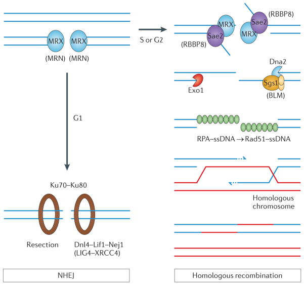
    1. Symington LS, Gautier J. Double-strand break end resection and repair pathway choice. Annu Rev Genet. 2011;45:247–271. - PubMed
    1. Green CM, Almouzni G. When repair meets chromatin. EMBO Rep. 2002;3:28–33. - PMC - PubMed