. 2013 Feb;4(1):9-27.
doi: 10.1007/s13244-012-0207-7. Epub 2012 Dec 18.

Mimics in chest disease: interstitial opacities

Affiliations

Mimics in chest disease: interstitial opacities

Anastasia Oikonomou et al. Insights Imaging. 2013 Feb.

Abstract

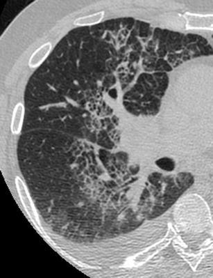
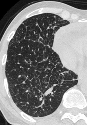
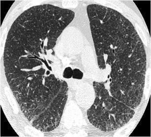
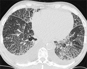
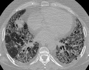
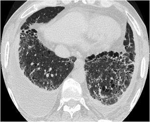
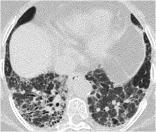
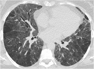
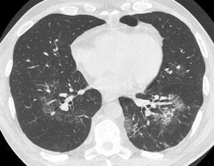
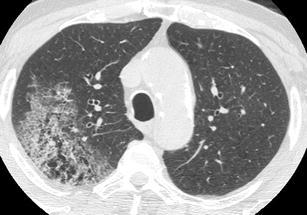
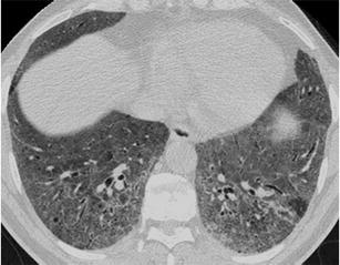
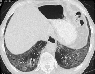
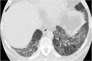
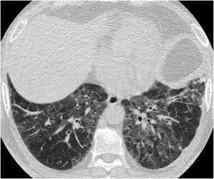
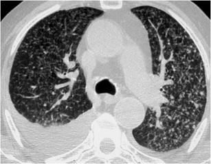
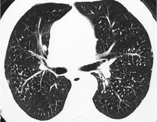
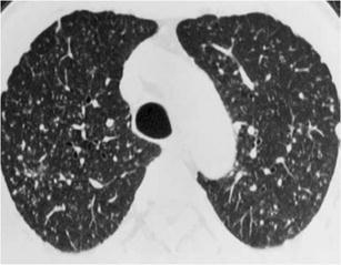
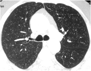
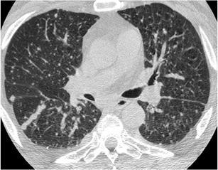
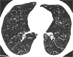
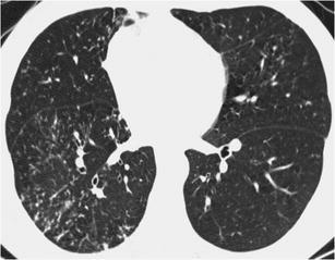
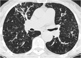
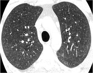
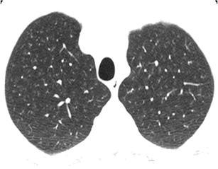
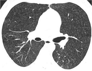
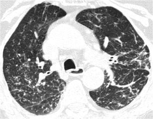
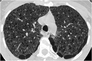
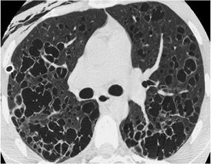
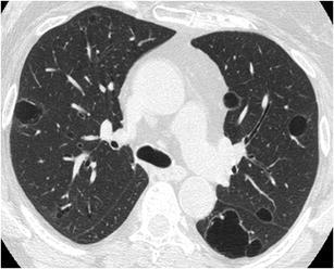
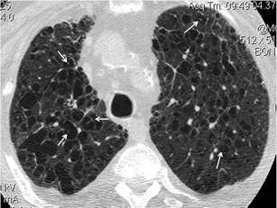
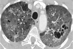
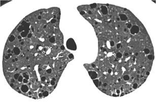
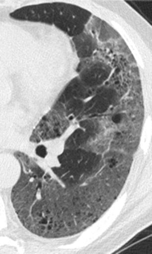
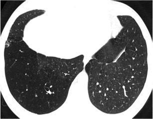
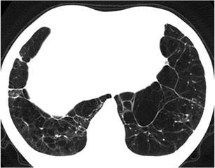
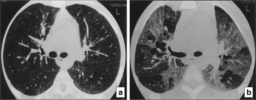
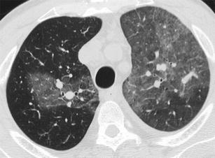
Septal, reticular, nodular, reticulonodular, ground-glass, crazy paving, cystic, ground-glass with reticular, cystic with ground-glass, decreased and mosaic attenuation pattern characterise interstitial lung diseases on high-resolution computed tomography (HRCT). Occasionally different entities mimic each other, either because they share identical HRCT findings or because of superimposition of patterns. Idiopathic pulmonary fibrosis (IPF), fibrosis associated with connective tissue disease, asbestosis, end-stage sarcoidosis or chronic hypersensitivity pneumonitis (HP) may present with lower zone, subpleural reticular pattern associated with honeycombing. Lymphangiomyomatosis may be indistinguishable from histiocytosis or extensive emphysema. Both pulmonary oedema and lymphangitic carcinomatosis may be characterised by septal pattern resulting from thickened interlobular septa. Ill-defined centrilobular nodular pattern may be identically present in HP and respiratory bronchiolitis-associated with interstitial lung disease (RBILD). Sarcoidosis may mimic miliary tuberculosis or haematogenous metastases presenting with miliary pattern, while endobronchial spread of tuberculosis may be indistinguishable from panbronchiolitis, both presenting with tree-in-bud pattern. Atypical infection presenting with ground-glass mimics haemorrhage. Ground-glass pattern with minimal reticulation is seen in desquamative interstitial pneumonia (DIP), RBILD and non-specific interstitial pneumonia (NSIP). Obliterative bronchiolitis and panlobular emphysema may present with decreased attenuation pattern, while obliterative bronchiolitis, chronic pulmonary embolism and HP may manifest with mosaic attenuation pattern. Various mimics in interstitial lung diseases exist. Differential diagnosis is narrowed based on integration of predominant HRCT pattern and clinical history. Teaching Points • To learn about the different HRCT patterns, which are related to interstitial lung diseases. • To be familiar with the more "classical" entities presenting with each HRCT pattern. • To discuss possible overlap of different HRCT patterns and the more common mimics in each case. • To learn about some clues that help differentiate the various diagnostic mimics on HRCT.


Figures

Fig. 1
Fig. 1
Hydrostatic pulmonary oedema secondary to endocarditis and rupture of a leaflet of the mitral valve. HRCT scan of the right lung shows a “septal pattern” characterised by thickened smoothly interlobular septae in the right parahilar area. Right pleural effusion is also seen
Fig. 2
Fig. 2
Lymphangitic carcinomatosis. HRCT of the right lung shows a “septal pattern” characterised by diffuse nodular thickening of the interlobular septae and the right major fissure
Fig. 3
Fig. 3
Sarcoidosis. HRCT at the level of the upper lobes shows “septal pattern” characterised by mild thickening of the interlobular septae predominantly in the peripheral and subpleural posterior areas of the lungs. A calcified right paratracheal lymphnode is also noted
Fig. 4
Fig. 4
Niemann-Pick. HRCT at the level of the lower lobes shows intense and homogenous thickening of the interlobular septae diffusely in the lungs. Superimposed areas of ground-glass opacity are also noted. Note also bilateral pleural effusions
Fig. 5
Fig. 5
Idiopathic pulmonary fibrosis. HRCT at the level of the lower lobes shows a subpleural “reticular pattern” characterised by thickened interlobular septae, thickened intralobular interstitium, traction bronchiolectasis and bronchiectasis and minimal honeycombing
Fig. 6
Fig. 6
Non-specific interstitial pneumonia. HRCT at the level of the lower lobes exhibits a “reticular pattern” characterised by the presence of thickened peribronchovascular interstitium and traction bronchiolectasis and bronchiectasis
Fig. 7
Fig. 7
Asbestosis. HRCT scan at the level of the lower lobes shows “reticular pattern” characterised by the presence of subpleural traction bronchiolectasis and bronchiectasis, honeycombing and thickening of the peribronchovascular interstitium and interlobular septae. The presence of calcified subpleural and diaphragmatic pleural plaques (white and black arrows) gives a definitive clue for the diagnosis of asbestosis
Fig. 8
Fig. 8
Chronic hypersensitivity pneumonitis. HRCT at the level of the lower lobes demonstrates a coarse “reticular pattern” with traction bronchiolectasis and bronchiectasis, thickening of the intralobular and peribronchovascular interstitium and ground-glass opacity. The presence of lobular areas of decreased attenuation offers a “key” for the diagnosis of chronic hypersensitivity pneumonitis
Fig. 9
Fig. 9
Rheumatoid arthritis associated pulmonary fibrosis. HRCT at the level of the lower lobes exhibits a coarse “reticular pattern” consisted of traction bronchiolectasis and bronchiectasis, thickening of the intralobular and peribronchovascular interstitium, ground-glass opacity and honeycombing
Fig. 10
Fig. 10
Desquamative interstitial pneumonia (DIP). HRCT at the level of the lower lobes demonstrates a “reticular pattern” consisted of subpleural traction bronchiolectasis and bronchiectasis, thickening of the peribronchovascular interstitium, ground-glass opacity and minimal honeycombing. Bilateral pleural effusions—which are not a finding of DIP—are attributed to cardiac failure
Fig. 11
Fig. 11
End-stage sarcoidosis. HRCT at the level of the lower lobes demonstrates a coarse “reticular pattern” with architectural distortion, subpleural traction bronchiolectasis and bronchiectasis, thickening of the peribronchovascular interstitium and honeycombing
Fig. 12
Fig. 12
Subacute hypersensitivity pneumonitis. HRCT shows geographic areas of “ground-glass pattern” with a few spared secondary lobules and no associated findings of fibrosis
Fig. 13
Fig. 13
Respiratory bronchiolitis associated interstitial lung disease. HRCT at the level of the upper lobes shows an almost diffuse “ground-glass pattern” with small centrilobular radiolucent areas representing concomitant centrilobular emphysema
Fig. 14
Fig. 14
Pneumocystis pneumonia. HRCT at the level of the lower lobes demonstrates geographic areas of “ground-glass pattern” with no associated findings of pulmonary fibrosis or lung cysts
Fig. 15
Fig. 15
Alveolar proteinosis. HRCT at the level of the lower and middle lobes demonstrates a “crazy paving pattern” characterised by geographic areas of involved pulmonary lobules with ground-glass opacity surrounded by thickened interlobular septae
Fig. 16
Fig. 16
Sarcoidosis. HRCT at the level of the lower lobes demonstrates a “crazy paving pattern” at the subpleural areas of the lungs associated with smoothly and nodular thickened interlobular septae. Note the “beaded” appearance of the major fissures, especially on the left
Fig. 17
Fig. 17
Alveolar haemorrhage in a patient with history of haemoptysis 2 days before. HRCT at the level of the lower and middle lobes exhibits small geographic areas of “crazy paving pattern” (pulmonary lobules with ground-glass opacity surrounded by thickened interlobular septae) consistent with alveolar haemorrhage in remission
Fig. 18
Fig. 18
Invasive mucinus adenicarcinoma. HRCT at the level of the upper lobes shows a focal area of “crazy paving pattern” in the right upper lobe intermingled with centrilobular emphysema
Fig. 19
Fig. 19
Non-specific interstitial pneumonia. HRCT at the level of the lower lobes exhibits a mixed “ground-glass and reticular pattern” characterised by diffuse ground-glass opacity and traction bronchiolectasis. There is no associated honeycombing
Fig. 20
Fig. 20
Scleroderma. HRCT at the level of the lower lobes demonstrates a mixed “ground-glass and reticular pattern” characterised by geographic areas of ground-glass opacity and traction bronchiolectasis. Note the characteristic 5 mm subpleural sparing, which is characteristic for NSIP—the most common histological type of scleroderma associated pulmonary fibrosis
Fig. 21
Fig. 21
Idiopathic pulmonary fibrosis. HRCT at the level of the lung bases exhibits a mixed “ground-glass and reticular pattern” characterised by geographic areas of ground-glass opacity within which traction bronchiolectasis and bronchiectasis is seen. Honeycombing is minimal, if any
Fig. 22
Fig. 22
Desquamative interstitial pneumonia. HRCT at the level of the lower lobes demonstrates geographic areas of mixed “ground-glass opacity and mild reticulation”. Honeycombing is absent
Fig. 23
Fig. 23
Sarcoidosis. HRCT at the level of the upper lobes exhibits a “nodular without tree-in-bud pattern” characterised by the presence of perilymphatic micronodules around the interlobular septae, fissures, bronchovascular bundles and pleura
Fig. 24
Fig. 24
Silicosis. CT at the level of the upper lobes exhibits a “nodular without tree-in-bud pattern” characterised by well-defined homogeneous centrilobular nodules that have an upper lobe and posterior predominance
Fig. 25
Fig. 25
Coal workers pneumoconiosis. HRCT at the level of the upper lobes exhibits a “nodular without tree-in-bud pattern” characterised by ill-defined centrilobular nodules of slightly variable size that have an upper lobe and posterior predominance
Fig. 26
Fig. 26
Miliary TB. HRCT at the level of the upper lobes exhibits a “miliary nodular pattern” characterised by random micronodules diffusely and symmetrically distributed within the lungs having approximately the same size
Fig. 27
Fig. 27
Miliary metastatic disease. HRCT at the level of the upper lobes shows a “milary nodular pattern” characterised by random and perilymphatic micronodules diffusely distributed throughout the lungs that have a more variable size compared with Fig. 26
Fig. 28
Fig. 28
Panbronchiolitis. HRCT at the level of the lower and middle lobes exhibits a “nodular with tree-in-bud pattern” diffusely throughout the lungs
Fig. 29
Fig. 29
Tuberculosis. HRCT at the level of the lower and middle lobes exhibits a “nodular with tree-in-bud pattern” predominantly in the right lower lobe, consistent with endobronchial spread of tuberculosis
Fig. 30
Fig. 30
Atypical mycobacteria infection in an elderly woman. HRCT at the level of the middle lobe, lingula and the superior segments of the lower lobes reveals a “nodular with tree-in-bud pattern” associated with mild cylindrical bronchiectasis in middle lobe and lingula and mucus plugging
Fig. 31
Fig. 31
Hypersensitivity pneumonitis. HRCT at the level of the upper lobes reveals an “ill-defined centrilobular nodular pattern” characterised by micronodules of ground-glass opacity that are diffusely distributed characteristically in the centre of the pulmonary lobules
Fig. 32
Fig. 32
RBILD. HRCT at the level of the upper lobes exhibits an “ill-defined centrilobular nodular pattern” characterised by micronodules of ground-glass opacity that are diffusely distributed characteristically in the centre of the pulmonary lobules. In this case the history of smoking favours the diagnosis of respiratory bronchiolitis interstitial lung disease
Fig. 33
Fig. 33
COP. HRCT at the level of the upper lobes exhibits an “ill-defined centrilobular nodular pattern” characterised by micronodules of ground-glass opacity that are diffusely distributed characteristically in the centre of the pulmonary lobules. The HRCT findings are almost indistinguishable from those in Fig. 31 or 32
Fig. 34
Fig. 34
Sarcoidosis. HRCT at the level of the upper lobes exhibits a “reticulonodular pattern” characterised by the presence of thickening of the interlobular septae and bronchovascular bundles, perilymphatic and perifissural micronodules and architectural distortion
Fig. 35
Fig. 35
LAM. HRCT at the level of the upper lobes exhibits a “cystic pattern” characterised by the presence of numerous thin walled “true” cysts of variable sized that are located in the lung parenchyma and paraseptally
Fig. 36
Fig. 36
Histiocytosis. HRCT at the level of the upper lobes reveals a “cystic pattern” characterised by numerous thin- and thick-walled “true” cysts with bizarre shapes and variable size. A drainage catheter is noted in the right pleural space in order to treat pneumothorax, which was the presenting symptom of this 27-year-old heavy smoker
Fig. 37
Fig. 37
Lymphocytic interstitial pneumonia. HRCT at the level of the upper lobes reveals a “cystic pattern” characterised by few thin–walled “true” cysts that may have rounded or more lobulated shape. The history of immunosuppression (AIDS, Sjogren, autoimmune disorders, dysproteinemia) favours the specific diagnosis of LIP
Fig. 38
Fig. 38
Centrilobular emphysema. HRCT shows centrilobular areas of radiolucency with no discernible walls in most of the cases (except in those areas where there is thickening of the interlobular septae) and with the presence of a central white dot (at the centre of the radiolucencies) representing the centrilobular artery (white arrows). Centrilobular emphysema mimics occasionally interstitial diseases that present with a “true cystic pattern”
Fig. 39
Fig. 39
Pneumocystis pneumonia in an AIDS patient. HRCT at the level of the upper lobes reveals a mixed “ground-glass and cystic pattern” characterised by the presence of diffuse areas of ground-glass opacity and a few thin-walled multilocular cysts
Fig. 40
Fig. 40
Lymphocytic interstitial pneumonia in a female patient with Sjogren’s disease. HRCT at the level of the upper lobes exhibits a mixed “ground-glass and cystic pattern” characterised by the presence of diffuse areas of ground-glass opacity and numerous thin-walled rounded and lobulated cysts of variable size
Fig. 41
Fig. 41
Desquamative interstitial pneumonia. HRCT at the level of the left lower lobe (magnified view of the left lower lobe) reveals a mixed “ground glass and cystic pattern” characterized by the presence of patchy areas of ground glass opacity and numerous small scattered “lucencies” that may represent either true cysts, bronciolectasis or emphysema
Fig. 42
Fig. 42
Obliterative bronchiolitis. HRCT at the level of the lower lobes exhibits a “decreased attenuation pattern” characterised by patchy “black” areas with paucity of vessels and few scattered bronchiectasis
Fig. 43
Fig. 43
Panlobular emphysema in a α-1 antitrypsin deficiency patient. HRCT at the level of the lower lobes reveals a “decreased attenuation pattern” characterised by the presence of confluent “black pulmonary lobules surrounded by few interlobular septae resembling a “spider’s web”
Fig. 44
Fig. 44
Obliterative bronchiolitis in a patient with cystic fibrosis. HRCT at the level of the carina at (a) inspiration and (b) expiration reveals at expiration a “mosaic attenuation pattern” secondary to air-trapping (b) which is not revealed on inspiration (a)
Fig. 45
Fig. 45
Chronic thromoboembolic pulmonary arterial hypertension. HRCT at the level of the upper lobes exhibits a “mosaic attenuation pattern” with patchy areas of increased attenuation within which large calibre vessels are seen representing the hyperperfused areas compared with the areas of decreased attenuation with small calibre vessels, which represent the hypoperfused areas (from [45])

Similar articles

Cited by

References

    1. Glaspole IN, du Bois RM, Wells AU. The application of high-resolution CT to diagnosis in diffuse parenchymal lung disease. Monaldi Arch Chest Dis. 2001;56:233–239. - PubMed
    1. Elicker B, Pereira CA, Webb R, Leslie KO. High-resolution computed tomography patterns of diffuse interstitial lung disease with clinical and pathological correlation. J Bras Pneumol. 2008;34:715–744. doi: 10.1590/S1806-37132008000900013. - DOI - PubMed
    1. Gotway MB, Reddy GP, Webb WR, Elicker BM, Leung JW. High-resolution CT of the lung: patterns of disease and differential diagnoses. Radiol Clin North Am. 2005;43:513–542. doi: 10.1016/j.rcl.2005.01.010. - DOI - PubMed
    1. Hansell DM, Bankier AA, MacMahon H, McLoud TC, Müller NL, Remy J. Fleischner society: glossary of terms for thoracic imaging. Radiology. 2008;246:697–722. doi: 10.1148/radiol.2462070712. - DOI - PubMed
    1. Storto ML, Kee ST, Golden JA, Webb WR. Hydrostatic pulmonary edema: high-resolution CT findings. AJR Am J Roentgenol. 1995;165:817–820. doi: 10.2214/ajr.165.4.7676973. - DOI - PubMed