

Growth plate alteration precedes cam-type deformity in elite basketball players
- PMID: 23247816
- PMCID: PMC3585998
- DOI: 10.1007/s11999-012-2740-6
Growth plate alteration precedes cam-type deformity in elite basketball players
Erratum in
- Clin Orthop Relat Res. 2013 May;471(5):1737
Abstract
Background: Vigorous sporting activity during the growth years is associated with an increased risk of having a cam-type deformity develop. The underlying cause of this osseous deformity is unclear. One may speculate whether this is caused by reactive bone apposition in the region of the anterosuperior head-neck junction or whether sports activity alters the shape of and growth in the growth plate. If the latter is true, then one would expect athletes to show an abnormal shape of the capital growth plate (specifically, the epiphyseal extension) before and/or after physeal closure.
Questions/purposes: We therefore raised three questions: (1) Do adolescent basketball players show abnormal epiphyseal extension? (2) Does the epiphyseal extension differ before and after physeal closure? (3) Is abnormal epiphyseal extension associated with high alpha angles?
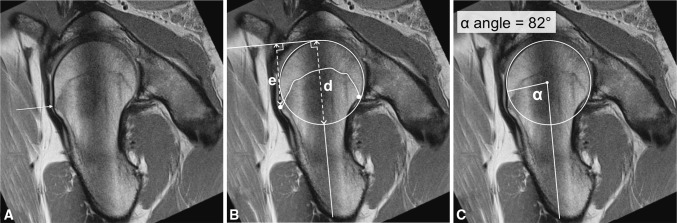
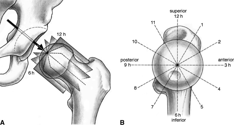
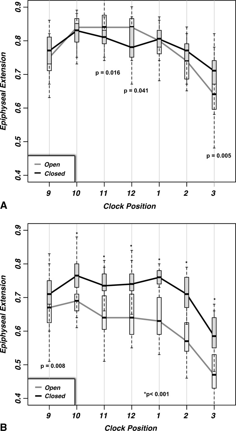
Methods: We performed a case-control comparative analysis of young (age range, 9-22 years) male elite basketball athletes with age-matched nonathletes, substratified by whether they had open or closed physes. We measured epiphyseal extension on radial-sequence MRI cuts throughout the cranial hemisphere from 9 o'clock (posterior) to 3 o'clock (anterior). Epiphyseal extension was correlated to alpha angle measurements at the same points.
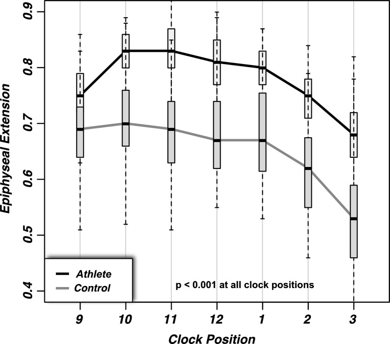
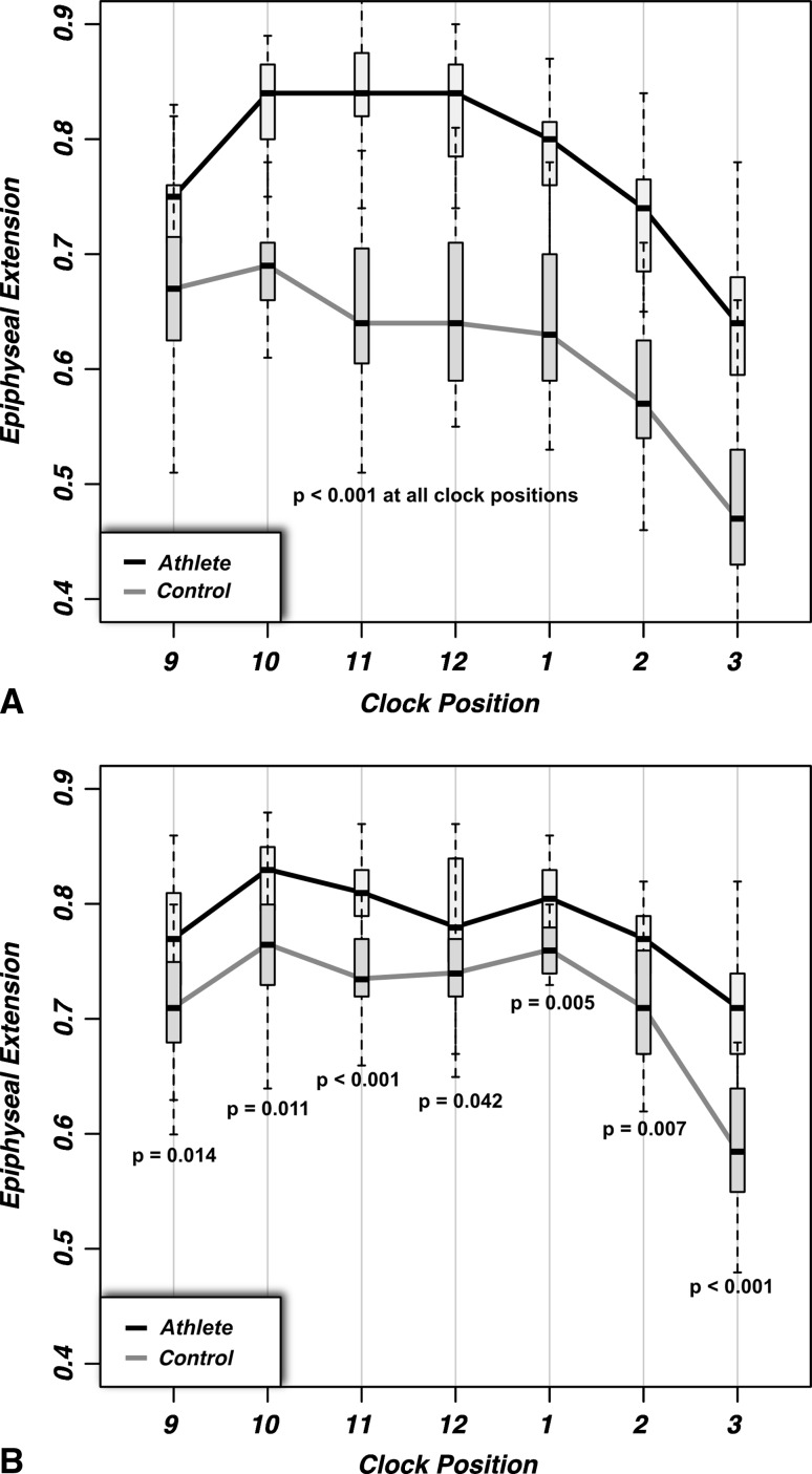
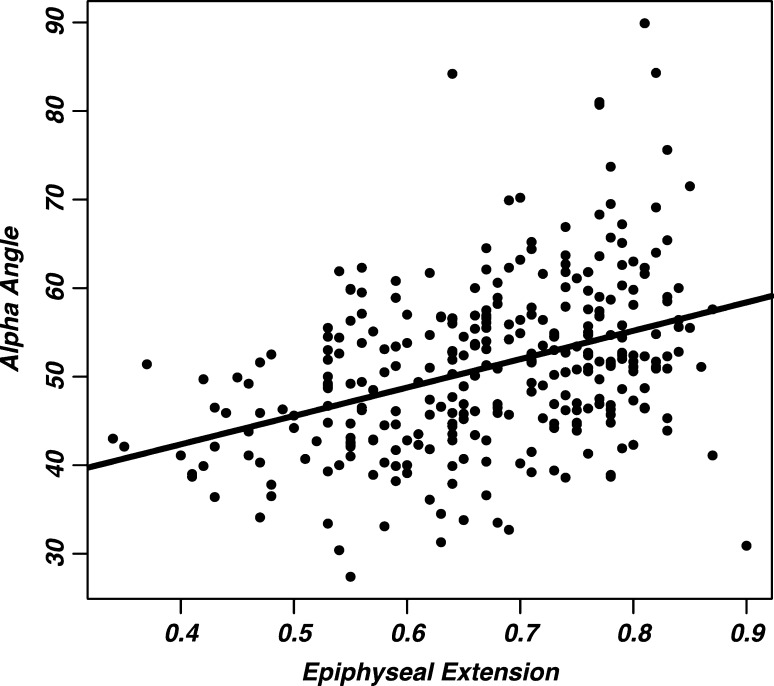
Results: Epiphyseal extension was increased in all positions in the athletes compared with the control group. On average, athletes showed epiphyseal extension of 0.67 to 0.83 versus 0.53 to 0.71 in control subjects. In the control group epiphyseal extension was increased at all measurement points in hips after physeal closure compared with before physeal closure. In contrast, the subgroup of athletes with a closed growth plate only had increased epiphyseal extension at the 3 o'clock position compared with the athletes with an open [corrected] growth plate (0.64-0.70). We observed a correlation between an alpha angle greater than 55° and greater epiphyseal extension in the anterosuperior femoral head quadrant: the corresponding Spearman r values were 0.387 (all hips) and 0.285 (alpha angle>55°) for the aggregate anterosuperior quadrant.
Conclusions: These findings suggest that a cam-type abnormality in athletes is a consequence of an alteration of the growth plate rather than reactive bone formation. High-level sports activity during growth may be a new and distinct risk factor for a cam-type deformity.
Figures
Comment in
-
Editor's spotlight/take 5: Growth plate alteration precedes cam-type deformity in elite basketball players (DOI 10.1007/s11999-012-2740-6). Interview by Seth S. Leopold.Clin Orthop Relat Res. 2013 Apr;471(4):1081-3. doi: 10.1007/s11999-013-2839-4. Epub 2013 Feb 13. Clin Orthop Relat Res. 2013. PMID: 23404420 Free PMC article. No abstract available.
References
Publication types
MeSH terms
LinkOut - more resources
Full Text Sources
Other Literature Sources
Medical
