. 2013 Apr;471(4):1334-42.
doi: 10.1007/s11999-012-2745-1. Epub 2012 Dec 18.

Lateral soft tissue laxity increases but medial laxity does not contract with varus deformity in total knee arthroplasty

Affiliations

Lateral soft tissue laxity increases but medial laxity does not contract with varus deformity in total knee arthroplasty

Shigetoshi Okamoto et al. Clin Orthop Relat Res. 2013 Apr.

Abstract

Background: In TKA, soft tissue balance (the joint gap) depends on the amount of resected bone and soft tissue release. Some studies report preoperative bony deformity correlates with soft tissue balance evaluated intraoperatively and that the medial tissues are contracted with varus deformity. However, these studies did not take into account the amount of resected bone and did not describe whether the soft tissue was tight or loose. Therefore, it remains unclear whether in varus deformity the soft tissues on the medial side are contracted.

Questions/purposes: We compared (1) intraoperative joint gap, (2) amount of resected bone, and (3) intraoperative soft tissue laxity on the lateral and medial sides according to severity of preoperative varus deformity.

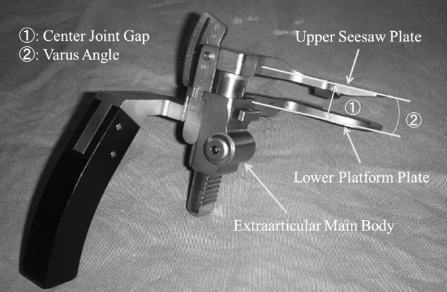
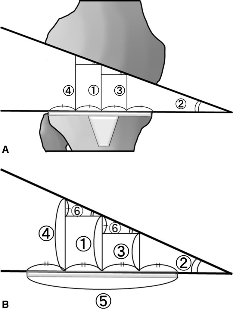
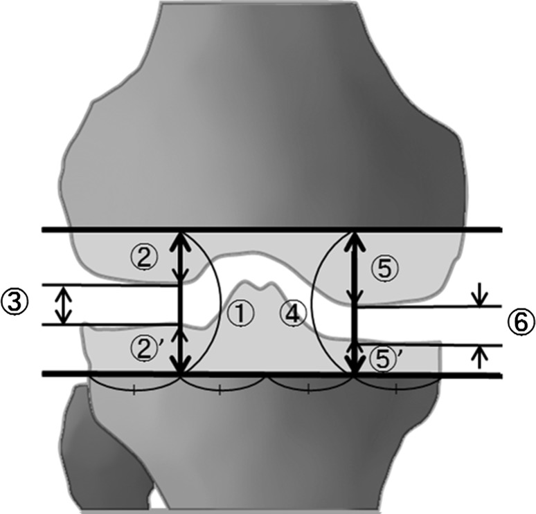
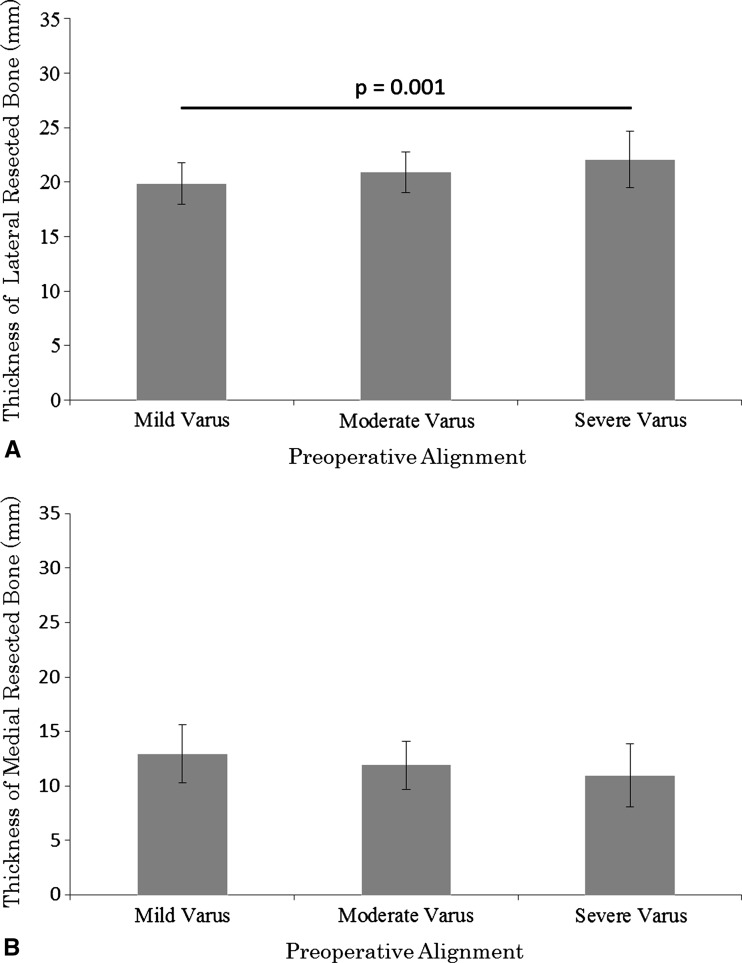
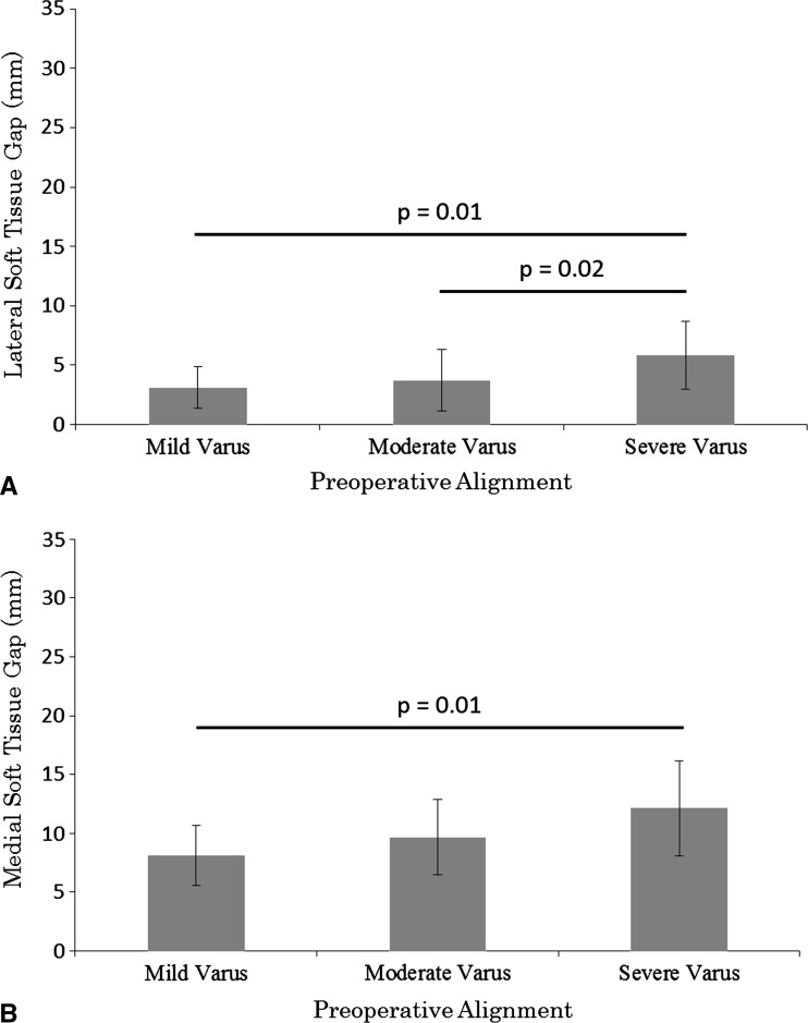
Methods: We retrospectively reviewed 70 patients with osteoarthritis and varus deformities who underwent 90 TKAs. We retrospectively divided the 90 knees into three groups according to degree of preoperative alignment: mild varus group (<10°), moderate varus group (10°-20°), and severe varus group (>20°). To evaluate intraoperative soft tissue tension, we calculated the soft tissue gap by subtracting the thickness of the resected bone from the joint gaps on the medial and lateral sides, respectively. We then explored the relationship between the soft tissue gap and preoperative alignment.

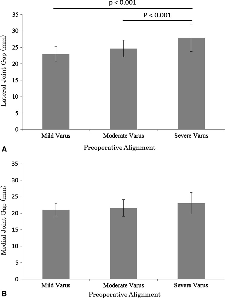
Results: The lateral soft tissue gap was larger in the severe varus group than in the mild and moderate varus groups. The medial soft tissue gap was larger in the severe varus group than in the mild varus group, but there were no differences in the medial joint gaps among the groups.

Conclusions: After the bone is resected, the soft tissue on the lateral side is more lax; however, the soft tissue on the medial side is not shorter with greater preoperative varus deformity.


Figures

Fig. 1
Fig. 1
The tension device consists of three parts: an upper seesaw plate, a lower platform plate, and an extraarticular main body. Two measurements were made: (1) the center joint gap and (2) the angle between the seesaw plate and the platform plate.
Fig. 2A–B
Fig. 2A–B
(A) A diagram illustrates the: (1) center joint gap, (2) varus angle, (3) medial joint gap, and (4) lateral joint gap. (B) A diagram illustrates the following parameters: (1) center joint gap, (2) varus angle, (3) medial joint gap, (4) lateral joint gap, and (5) transverse diameter of the tibial component. Using these measurements, we calculated the medial and lateral joint gaps as follows: (6) = 0.25 × (5) × tan (2), (3) = (1) − (6), and (4) = (1) + (6).
Fig. 3
Fig. 3
The (1) lateral joint gap, (2) + (2)′ thickness of the lateral resected bone, (3) lateral soft tissue gap, (4) medial joint gap, (5) + (5)′ thickness of the medial resected bone, and (6) medial soft tissue gap are shown.
Fig. 4A–B
Fig. 4A–B
The graphs compare the joint gaps among preoperative alignments. Data are shown as mean ± SD. (A) The lateral joint gap was larger in the severe varus group than in the mild and moderate varus groups. (B) There were no differences in the medial joint gaps among groups.
Fig. 5A–B
Fig. 5A–B
The graphs compare the thicknesses of the resected bone with preoperative alignments. The data are shown as mean ± SD. (A) The thickness of the lateral resected bone was larger in the severe varus group than in the mild varus group. (B) There were no differences in the thickness of the medial resected bone among groups.
Fig. 6A–B
Fig. 6A–B
Comparison of the soft tissue gaps among preoperative alignments are shown in these graphs. The data are shown as mean ± SD. (A) The lateral soft tissue gap was larger in the severe varus group than in the mild and moderate varus groups. (B) The medial soft tissue gap was larger in the severe varus group than in the mild varus group.

References

    1. Bellemans J, Vandenneucker H, Vanlauwe J, Victor J. The influence of coronal plane deformity on mediolateral ligament status: an observational study in varus knees. Knee Surg Sports Traumatol Arthrosc. 2010;18:152–156. doi: 10.1007/s00167-009-0903-0. - DOI - PubMed
    1. Berend ME, Small SR, Ritter MA, Buckley CA. The effects of bone resection depth and malalignment on strain in the proximal tibia after total knee arthroplasty. J Arthroplasty. 2010;25:314–318. doi: 10.1016/j.arth.2009.01.021. - DOI - PubMed
    1. Brage ME, Draganich LF, Pottenger LA, Curran JJ. Knee laxity in symptomatic osteoarthritis. Clin Orthop Relat Res. 1994;304:184–189. - PubMed
    1. Brooks P. Seven cuts to the perfect total knee. Orthopedics. 2009;32. pii: orthosupersite.com/view.asp?rID = 42848. DOI: 10.3928/01477447-20090728-27. - PubMed
    1. Fang DM, Ritter MA, Davis KE. Coronal alignment in total knee arthroplasty: just how important is it? J Arthroplasty. 2009;24(6 suppl):39–43. doi: 10.1016/j.arth.2009.04.034. - DOI - PubMed

Publication types