

Case Reports
doi: 10.4103/0973-029X.102522.
Chronic invasive aspergillosis of paranasal sinuses: A case report with review of literature
Affiliations
- PMID: 23248489
- PMCID: PMC3519232
- DOI: 10.4103/0973-029X.102522
Item in Clipboard
Case Reports
Chronic invasive aspergillosis of paranasal sinuses: A case report with review of literature
J Oral Maxillofac Pathol.
2012 Sep.
Abstract
Aspergillosis of the nasal and paranasal sinuses is recognized as being second to candidiasis, among opportunistic fungal infections in immunocompromised patients. However, invasive variant in normal and mildly immunocompromised hosts is a very rare occurrence. We report one such case of aspergillosis involving paranasal sinuses in mildly immunocompromised patient.
Keywords: Aspergillosis; fruiting body; hyphae; methanamine silver stain.
Conflict of interest statement
Figures
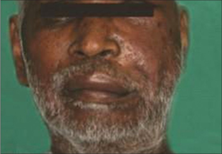
Extra-oral photograph showing swelling on left side of face
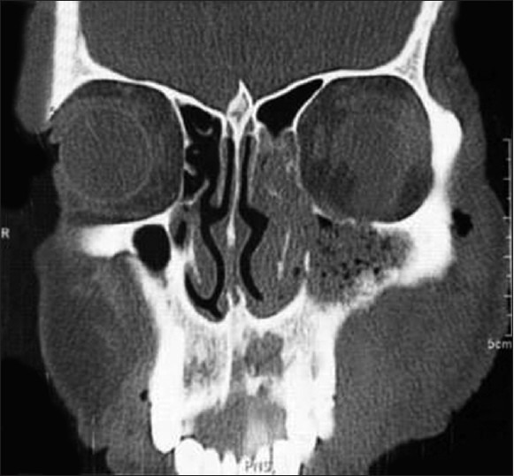
CT PNS showing there is erosion of alveolar cortex of the maxilla in the region of maxillary central incisors, posterolateral wall of the left maxillary sinus and inferior orbital wall (orbital floor) with extension of the soft tissue within the maxillary sinus, contiguous with buccal space and mild extension into orbital cavity
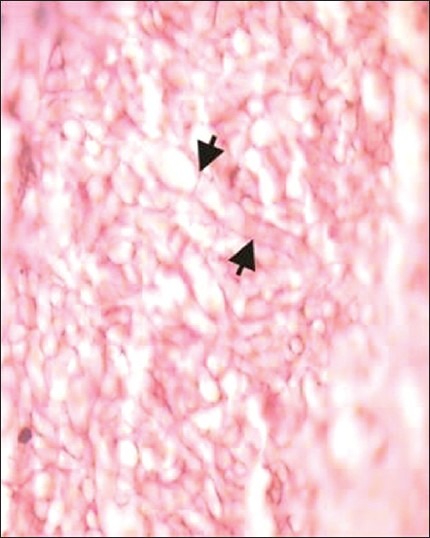
Photomicrograph reveals characteristic septate fungal hyphae of an Aspergillus species (H and E, ×100)
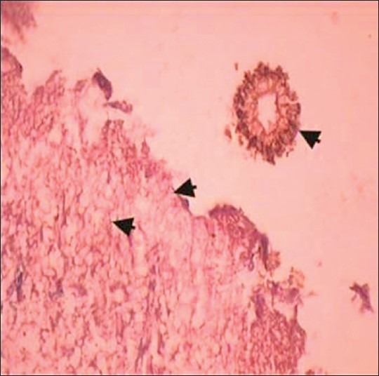
Photomicrograph reveals fungal hyphae and a fruiting body of an Aspergillus species (H and E, ×40)
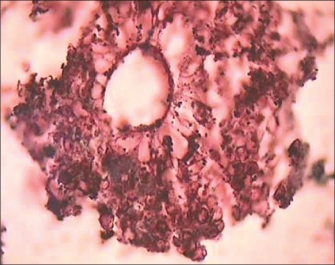
Photomicrograph reveals fruiting body of an Aspergillus species (H and E, ×100)
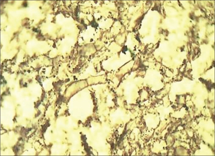
Photomicrograph reveals characteristic septate fungal hyphae of an Aspergillus species (Gnott-Gomori methanamine silver stain, ×100)
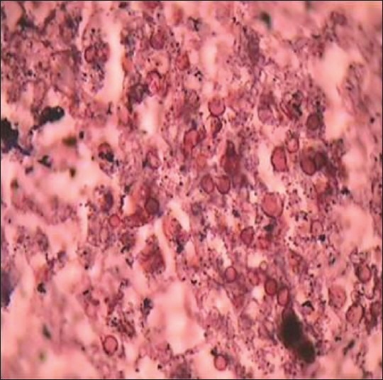
Photomicrograph reveals spores of fungal hyphae of an Aspergillus species (Gnott-Gomori methanamine silver stain, ×100)
References
-
- Sharma OP, Chwogule R. Many faces of pulmonary aspergillosis. Eur Respir J. 1998;12:705–15. - PubMed
-
- Warder FR, Chikes PG, Hudson WR. Aspergillosis of the paranasalsinusis. Arch Otolaryngol. 1975;101:683–5. - PubMed
-
- Sapp JP, Eversole LR, Wysocki GP. Contemporary Oral and Maxillofacial Pathology. 2nd ed. Missouri: Mosby - An Affiliate of Elsevier; 2004. Oral infections; pp. 207–51.
-
- Kwon J, Park KH, Park SI, Jin SY. Aspergillosis of the paranasal sinuses–diagnostic significance of the computed tomography. Yonsei Med J. 1989;30:294–7. - PubMed
-
- Stammberger H, Jakse R, Beaufort F. Aspergillosis of the paranasal sinuses; X-ray diagnosis, histopathology, and clinical aspects. Ann Otol Rhinol Laryngol. 1984;93:251–6. - PubMed
