. 2012 Dec 13:3:201.
doi: 10.3389/fphar.2012.00201. eCollection 2012.

Mechanisms of CFTR Folding at the Endoplasmic Reticulum

Affiliations

Mechanisms of CFTR Folding at the Endoplasmic Reticulum

Soo Jung Kim et al. Front Pharmacol. .

Abstract

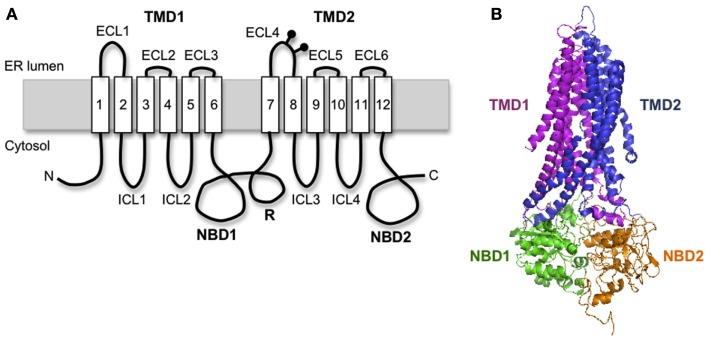
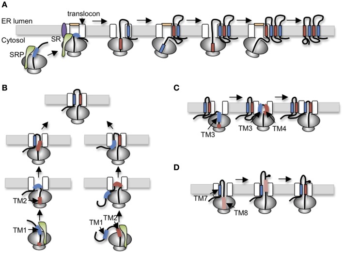
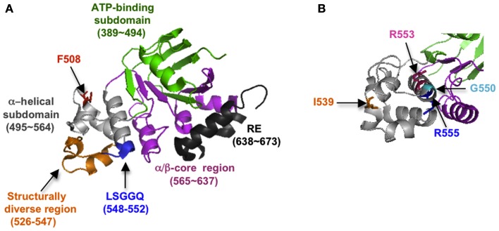
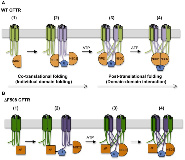
In the past decade much has been learned about how Cystic Fibrosis Transmembrane Conductance Regulator (CFTR) folds and misfolds as the etiologic cause of cystic fibrosis (CF). CFTR folding is complex and hierarchical, takes place in multiple cellular compartments and physical environments, and involves several large networks of folding machineries. Insertion of transmembrane (TM) segments into the endoplasmic reticulum (ER) membrane and tertiary folding of cytosolic domains begin cotranslationally as the nascent polypeptide emerges from the ribosome, whereas posttranslational folding establishes critical domain-domain contacts needed to form a physiologically stable structure. Within the membrane, N- and C-terminal TM helices are sorted into bundles that project from the cytosol to form docking sites for nucleotide binding domains, NBD1 and NBD2, which in turn form a sandwich dimer for ATP binding. While tertiary folding is required for domain assembly, proper domain assembly also reciprocally affects folding of individual domains analogous to a jig-saw puzzle wherein the structure of each interlocking piece influences its neighbors. Superimposed on this process is an elaborate proteostatic network of cellular chaperones and folding machineries that facilitate the timing and coordination of specific folding steps in and across the ER membrane. While the details of this process require further refinement, we finally have a useful framework to understand key folding defect(s) caused by ΔF508 that provides a molecular target(s) for the next generation of CFTR small molecule correctors aimed at the specific defect present in the majority of CF patients.

Keywords: ABC transporter; CFTR; cotranslational folding; cystic fibrosis; membrane protein biogenesis; nucleotide binding domain; protein translocation.


Figures

Figure 1
Figure 1
Cystic fibrosis transmembrane conductance regulator structural organization. (A) Schematic diagram of CFTR showing transmembrane topology and domain organization. (B) A predicted human CFTR structure based on homology model from Sav1866.
Figure 2
Figure 2
Variations of polytopic protein topogenesis. (A) Simplest cotranslational topogenesis model in which ER targeting begins as signal recognition particle (SRP) recognizes an emerging signal sequence (TM), binds its receptor (SR) at the ER membrane, and transfers the RNC to the Sec61 translocon. TM topology is achieved through alternating signal and stop transfer activities that sequentially open and close the translocon pore. Careful orchestration of ribosome translocon junction ensures delivery of soluble domains into either cytosol or ER lumen and integration of TMs into the bilayer. (B) For CFTR, topology of TM1 and TM2 is established by two alternate pathways in which translocation is initiated by either TM1 (left) or TM2 (right). Most CFTR nascent chains utilized a posttranslational mechanism in which TM2 insertion drags TM1 into the translocon. (C) The short loop between TM3 and TM4 (five residues) suggests that TM3 and TM4 simultaneously insert into the translocon as a helical hairpin. A similar mechanism is also proposed for TM5-6, TM9-10, and TM11-12. (D) Stop transfer activity of TM8 is weakened by Asp924 which results in transient exposure of TM8 in the ER lumen before acquiring its final membrane spanning topology.
Figure 3
Figure 3
(A) Crystal structure of NBD1 showing subdomain organization and location of key structural elements (2BBO). (B) Slightly different view of (A), showing location of suppressor mutations (1XMI).
Figure 4
Figure 4
Step-wise CFTR folding pathway. (A) CFTR folding begins cotranslationally as individual domains are synthesized, and proceeds as domains assemble into a mature tertiary structure. (B) The ΔF508 mutation destabilizes NBD1 structure, interferes with the TMD1, TMD2, and NBD2 folding, and disturbs interactions between NBD1 and ICL4, compromising domain–domain assembly.
Figure 5
Figure 5
Chaperones assist CFTR folding and target misfolded CFTR for degradation. (A) As CFTR is synthesized, numerous chaperones and co-chaperones (some depicted here) decorate the nascent polypeptide on both lumenal and cytosolic sides of the ER membrane. Hsp70 and co-chaperones interact with NBD1, followed by calnexin association with TMD2. Hsp70-Hop interactions recruits Hsp90 complexes which likely aid domain assembly in conjunction with calnexin. (B) Failure to achieve productive folding at any step in the folding pathway is detected by persistent binding of Hsp70, which serves to recruit E3 ligases (i.e., RMA1 and CHIP) that ubiquitinate CFTR and target it to the 26S proteasome.

References

    1. Alder N. N., Shen Y., Brodsky J. L., Hendershot L. M., Johnson A. E. (2005). The molecular mechanisms underlying BiP-mediated gating of the Sec61 translocon of the endoplasmic reticulum. J. Cell Biol. 168, 389–39910.1083/jcb.200409174 - DOI - PMC - PubMed
    1. Aleksandrov A. A., Kota P., Aleksandrov L. A., He L., Jensen T., Cui L., et al. (2010). Regulatory insertion removal restores maturation, stability and function of deltaF508 CFTR. J. Mol. Biol. 401, 194–21010.1016/j.jmb.2010.06.019 - DOI - PMC - PubMed
    1. Aleksandrov A. A., Kota P., Cui L., Jensen T., Alekseev A. E., Reyes S., et al. (2012). Allosteric modulation balances thermodynamic stability and restores function of deltaF508 CFTR. J. Mol. Biol. 419, 41–6010.1016/j.jmb.2012.03.001 - DOI - PMC - PubMed
    1. Aller S. G., Yu J., Ward A., Weng Y., Chittaboina S., Zhuo R., et al. (2009). Structure of P-glycoprotein reveals a molecular basis for poly-specific drug binding. Science 323, 1718–172210.1126/science.1168750 - DOI - PMC - PubMed
    1. Amaral M. D. (2004). CFTR and chaperones: processing and degradation. J. Mol. Neurosci. 23, 41–4810.1385/JMN:23:1-2:041 - DOI - PubMed