

Mechanisms underlying the tissue-specific and regulated activity of the Gnrhr promoter in mammals
- PMID: 23248618
- PMCID: PMC3521148
- DOI: 10.3389/fendo.2012.00162
Mechanisms underlying the tissue-specific and regulated activity of the Gnrhr promoter in mammals
Abstract
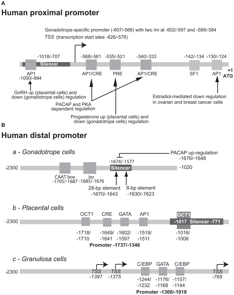
The GnRH receptor (GnRHR) plays a central role in the development and maintenance of reproductive function in mammals. Following stimulation by GnRH originating from the hypothalamus, GnRHR triggers multiple signaling events that ultimately stimulate the synthesis and the periodic release of the gonadotropins, luteinizing-stimulating hormone (LH) and follicle-stimulating hormones (FSH) which, in turn, regulate gonadal functions including steroidogenesis and gametogenesis. The concentration of GnRHR at the cell surface is essential for the amplitude and the specificity of gonadotrope responsiveness. The number of GnRHR is submitted to strong regulatory control during pituitary development, estrous cycle, pregnancy, lactation, or after gonadectomy. These modulations take place, at least in part, at the transcriptional level. To analyze this facet of the reproductive function, the 5' regulatory sequences of the gene encoding the GnRHR have been isolated and characterized through in vitro and in vivo approaches. This review summarizes results obtained with the mouse, rat, human, and ovine promoters either by transient transfection assays or by means of transgenic mice.
Keywords: GnRH receptor; gonadotrope cell lines; homeodomain proteins; promoter regions; steroidogenic factor 1; transcription; transgenic mice.
Figures
Similar articles
-
Activity of the porcine gonadotropin-releasing hormone receptor gene promoter is partially conferred by a distal gonadotrope specific element (GSE) within an upstream enhancing region, two proximal GSEs and a retinoid X receptor binding site.Reprod Biol Endocrinol. 2015 May 17;13:45. doi: 10.1186/s12958-015-0033-0. Reprod Biol Endocrinol. 2015. PMID: 25981521 Free PMC article.
-
GnRH Receptor Expression and Reproductive Function Depend on JUN in GnRH Receptor‒Expressing Cells.Endocrinology. 2018 Mar 1;159(3):1496-1510. doi: 10.1210/en.2017-00844. Endocrinology. 2018. PMID: 29409045 Free PMC article.
-
Identification and analysis of two novel sites of rat GnRH receptor gene promoter activity: the pineal gland and retina.Neuroendocrinology. 2013;97(2):115-31. doi: 10.1159/000337661. Epub 2012 Aug 25. Neuroendocrinology. 2013. PMID: 22414758
-
Molecular mechanisms of gonadotropin-releasing hormone receptor gene regulation.J Soc Gynecol Investig. 1999 Jul-Aug;6(4):169-78. doi: 10.1016/s1071-5576(99)00022-2. J Soc Gynecol Investig. 1999. PMID: 10486777 Review.
-
[An ambiguous role of steroidogenic factor 1 in the rat GnRH receptor gene expression. Lessons from transgenic mice].J Soc Biol. 2004;198(1):73-9. J Soc Biol. 2004. PMID: 15146959 Review. French.
Cited by
-
Gonadotropin-releasing hormone stimulates biliary proliferation by paracrine/autocrine mechanisms.Am J Pathol. 2015 Apr;185(4):1061-72. doi: 10.1016/j.ajpath.2014.12.004. Am J Pathol. 2015. PMID: 25794706 Free PMC article.
-
The relationship between basal and regulated Gnrhr expression in rodent pituitary gonadotrophs.Mol Cell Endocrinol. 2016 Dec 5;437:302-311. doi: 10.1016/j.mce.2016.08.040. Epub 2016 Aug 26. Mol Cell Endocrinol. 2016. PMID: 27569529 Free PMC article.
-
Evidence for divergent endocrine regulation of the murine and ovine GnRH receptor gene promoters.Front Endocrinol (Lausanne). 2025 Jul 29;16:1597028. doi: 10.3389/fendo.2025.1597028. eCollection 2025. Front Endocrinol (Lausanne). 2025. PMID: 40801031 Free PMC article.
-
Molecular basis for the evolved instability of a human G-protein coupled receptor.Cell Rep. 2021 Nov 23;37(8):110046. doi: 10.1016/j.celrep.2021.110046. Cell Rep. 2021. PMID: 34818554 Free PMC article.
-
The Effects of Zearalenone on the Localization and Expression of Reproductive Hormones in the Ovaries of Weaned Gilts.Toxins (Basel). 2021 Sep 7;13(9):626. doi: 10.3390/toxins13090626. Toxins (Basel). 2021. PMID: 34564630 Free PMC article.
References
-
- Achermann J. C., Ito M., Ito M., Hindmarsh P. C., Jameson J. L. (1999). A mutation in the gene encoding steroidogenic factor-1 causes XY sex reversal and adrenal failure in humans. Nat. Genet. 22 125–126 - PubMed
-
- Achermann J. C., Weiss J., Lee E. J., Jameson J. L. (2001). Inherited disorders of the gonadotropin hormones. Mol. Cell. Endocrinol. 179 89–96 - PubMed
-
- Alarid E. T., Windle J. J., Whyte D. B., Mellon P. L. (1996). Immortalization of pituitary cells at discrete stages of development by directed oncogenesis in transgenic mice. Development 122 3319–3329 - PubMed
-
- Albarracin C. T., Frosch M. P., Chin W. W. (1999). The gonadotropin-releasing hormone receptor gene promoter directs pituitary-specific oncogene expression in transgenic mice. Endocrinology 140 2415–2421 - PubMed
-
- Albarracin C. T., Kaiser U. B., Chin W. W. (1994). Isolation and characterization of the 5′-flanking region of the mouse gonadotropin-releasing hormone receptor gene. Endocrinology 135 2300–2306 - PubMed
LinkOut - more resources
Full Text Sources