. 2012:2:978.
doi: 10.1038/srep00978. Epub 2012 Dec 17.

Cdc42-dependent structural development of auditory supporting cells is required for wound healing at adulthood

Affiliations

Cdc42-dependent structural development of auditory supporting cells is required for wound healing at adulthood

Tommi Anttonen et al. Sci Rep. 2012.

Abstract

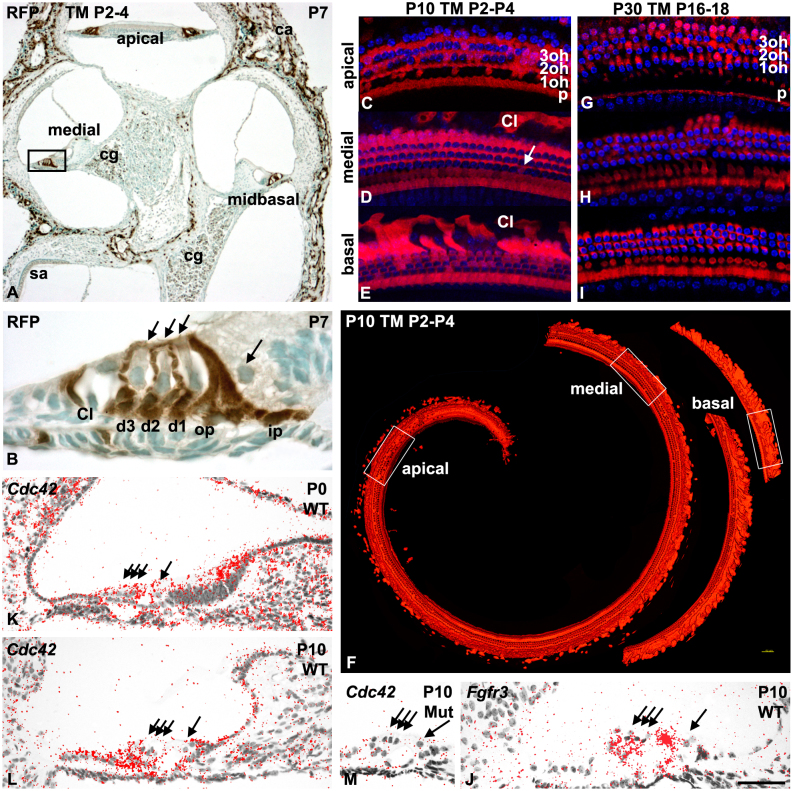
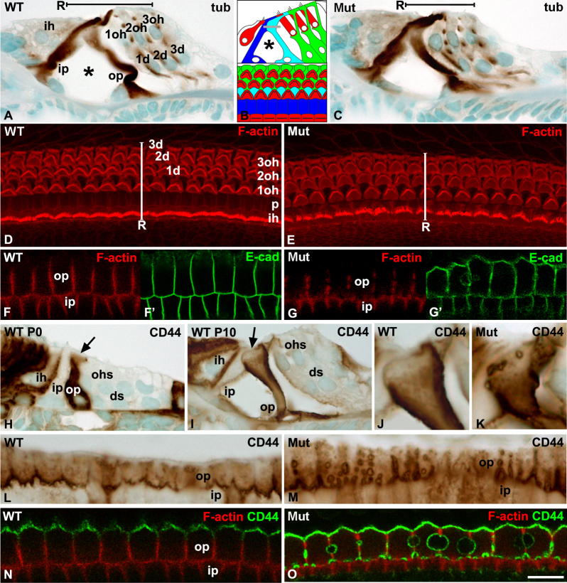
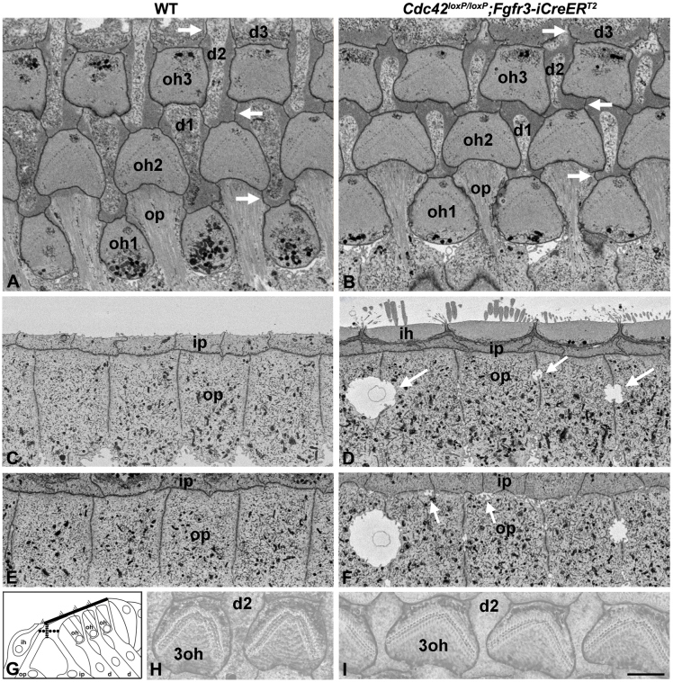
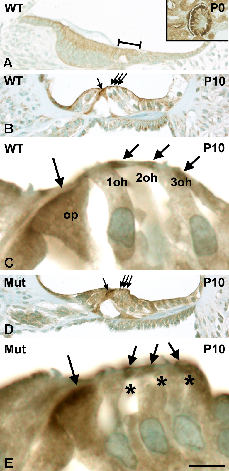
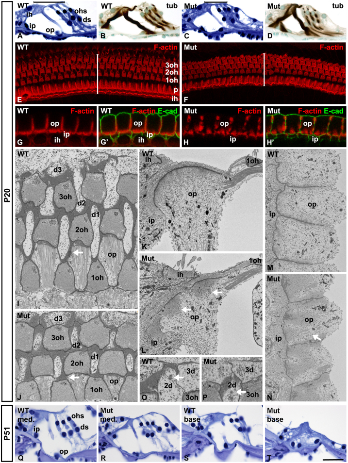
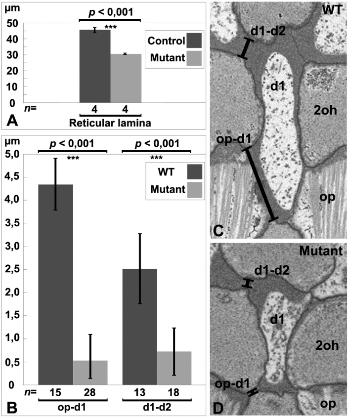
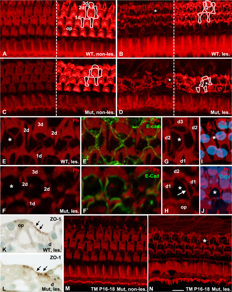
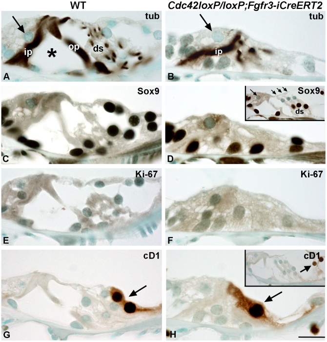
Cdc42 regulates the initial establishment of cytoskeletal and junctional structures, but only little is known about its role at later stages of cellular differentiation. We studied Cdc42's role in vivo in auditory supporting cells, epithelial cells with high structural complexity. Cdc42 inactivation was induced early postnatally using the Cdc42(loxP/loxP);Fgfr3-iCre-ER(T2) mice. Cdc42 depletion impaired elongation of adherens junctions and F-actin belts, leading to constriction of the sensory epithelial surface. Fragmented F-actin belts, junctions containing ectopic lumens and misexpression of a basolateral membrane protein in the apical domain were observed. These defects and changes in aPKCλ/ι expression suggested that apical polarization is impaired. Following a lesion at adulthood, supporting cells with Cdc42 loss-induced maturational defects collapsed and failed to remodel F-actin belts, a process that is critical to scar formation. Thus, Cdc42 is required for structural differentiation of auditory supporting cells and this proper maturation is necessary for wound healing in adults.


Figures

Figure 1
Figure 1. Recombination in the cochlea of Fgfr3-iCre-ERT2 mice. Cdc42 expression in the organ of Corti.
(A,B) Fgfr3-iCre-ERT2;Ai14(tdTomato) mice treated with tamoxifen between P2 and P4 show recombination in the organ of Corti, cochlear ganglion and surrounding bone, as revealed by RFP immunohistochemistry in paraffin sections at P7. The organ of Corti of the medial turn boxed in (A) is shown in higher magnification in (B). Hair cells are negative, as opposed to the adjacent supporting cells. (C–F) At P10, tdTomato fluorescence is found in supporting cells throughout the cochlea of Fgfr3-iCre-ERT2;Ai14(tdTomato) mice treated with tamoxifen between P2 and P4. Boxed areas in the 3D isosurface view of the cochlea (F) are shown in z-projections (C–E). DAPI marks nuclei. In addition to supporting cells, a part of hair cells, especially in the apical, but also in the most basal part of the cochlea are recombined. Note the single recombined outer hair cell (arrow) in the medial turn (D). (G–I) At P30, tdTomato fluorescence at different levels of the cochlea of Fgfr3-iCre-ERT2;Ai14(tdTomato) mice injected with tamoxifen between P16 and P18. Recombination is restricted to supporting cells. (J) Fgfr3 is expressed in supporting cells of the organ of Corti at P10. (K,L) Cdc42 is expressed throughout the cochlea at P0. Expression is more regionalized at P10. (M) Absence of Cdc42 expression in the organ of Corti of Cdc42loxP/loxP;Fgfr3-iCre-ERT2 mice at P10. Thin arrows mark outer hair cells and thick arrow an inner hair cell in (B,J-M). Abbreviations: cg, cochlear ganglion; ca, otic capsule; ip, inner pillar cell; op, outer pillar cell; d, Deiters' cell; p, pillar cell; Cl, Claudius cell; oh, outer hair cell; ih, inner hair cell; sa, saccule; WT, wildtype; Mut, mutant; TM, tamoxifen. Scale bar (in J): A,F, 160 μm; B, 20 μm; C–E, G–I, 40 μm; J–M, 50 μm.
Figure 2
Figure 2. Impaired differentiation of the apical domain of supporting cells of Cdc42loxP/loxP;Fgfr3-iCre-ERT2 mice.
Tamoxifen treatment between P2 and P4. Paraffin sections (A,C,H–M) and wholemount confocal specimens (D–G′,N,O) represent the medial part of cochlea from wildtype control and mutant mice at P10. (A,B) β-tubulin staining and schematic representation of the cytoarchitecture of normal organ of Corti in transverse plane and in a surface view (light blue, outer pillar cells; dark blue, inner pillar cells; green, Deiters' cells; red, hair cells; asterisk, tunnel of Corti). Heads of outer pillar cells are covered by heads of inner pillar cells. The grey line in (B) marks the level of confocal z-projections used in (F–G′,N,O). (C) β-tubulin staining is unaltered in mutant mice. (D,E) Phalloidin labeling shows constriction of the reticular lamina of mutants. (F–G′) Adherens junctions and F-actin belts of outer pillar cells of mutants are abnormal, as revealed by double-labeling for F-actin and E-cadherin. (H,I) Progression of differentiation of outer pillar cells during the first postnatal week, as shown by CD44 staining. Arrows mark adherens junctions. (J,K) At P10, outer pillar cells of mutant mice display CD44-positive ectopic lumens at apical junctions. (L,M) A paraffin section from a mutant cochlea cut in the plane parallel to the epithelial surface shows an abundance of CD44-positive ectopic lumens at outer pillar cells junctions. (N,O) In mutants at P10, F-actin belts are fragmented at the site of the lumens, as revealed by double-labeling for F-actin and CD44. Abbreviations: tub, β-tubulin; R, reticular lamina; ip, inner pillar cell; op, outer pillar cell; d, Deiters' cell; oh, outer hair cell; ih, inner hair cell, p, pillar cell; WT, wildtype; Mut, mutant; E-cad, E-cadherin. Scale bar (in O): A,C,H,I, 20 μm; D,E, 15 μm; F–G′,J–O, 8 μm.
Figure 3
Figure 3. Subcellular abnormalities in the apical domain of supporting cells of Cdc42loxP/loxP;Fgfr3-iCre-ERT2 mice.
Tamoxifen treatment between P2 and P4. SBF-SEM analysis of wildtype control (A,C,E,H) and mutant (B,D,F,I) mice at P10. (A,B) Supporting cells of mutant specimens show unelongated adherens junctions and F-actin belts (arrows). (C–D) Ectopic lumens (arrows) are seen between outer pillar cells of mutant specimens. (G) Schematic representation of the organ of Corti shows the planes of images (A,B; solid), (C,D; dashed) and (E,F; dots). (H,I) 3D reconstruction of stereociliary bundles (arrows) of outer hair cells shows unaltered morphology in mutant mice. Abbreviations: op, outer pillar cell; ip, inner pillar cell; d, Deiters' cell; oh, outer hair cell; ih, inner hair cell; WT, wildtype. Scale bar (in I): A–F, 3.5 μm; H,I, 2.5 μm.
Figure 4
Figure 4. Expression of the aPKCλ/ι polarity protein in the organ of Corti.
Tamoxifen treatment between P2 and P4. Paraffin sections from the medial part of the cochlea. (A) aPKCλ/ι expression is absent from the organ of Corti at birth. The sensory epithelial surface is marked by a line. (B,C) In wildtype mice at P10, apices of supporting cells show aPKCλ/ι expression. Note the sharp expression in the adherens junction of pillar cells, shown in a high-magnification view (C). (D,E) In Cdc42loxP/loxP;Fgfr3-iCre-ERT2 mutant mice, aPKCλ/ι expression is delocalized from the junctions and has a diffuse pattern. The three parallel arrows mark the heads of Deiters' cells and the single arrow the adherens junction of pillar cells. Asterisks mark the phalangeal processes of Deiters' cells located between outer hair cells. Abbreviations: oh, outer hair cell; op, outer pillar cell; WT, wildtype; Mut, mutant. Scale bar (in E): A,B,D, 160 μm; C,E, 10 μm.
Figure 5
Figure 5. Adult phenotype of the organ of Corti of Cdc42loxP/loxP;Fgfr3-iCre-ERT2 mice.
Tamoxifen was administered between P2 and P4. Paraffin sections (A–D, Q,R), confocal views (E–H′) and SBF-SEM images (I–P) represent the medial part of cochlea at P20. Paraffin sections (S,T) from the basal part at P51. (A–D) Hematoxylin- and β-tubulin stainings show largely comparable histology in wildtype control and mutant mice. (E,F) Phalloidin staining reveals constricted organization of the reticular lamina of mutant mice. (G-H′) Double-staining for phalloidin and E-cadherin shows discontinuous F-actin belts of pillar cells of mutants. (I,J) Junctions and F-actin belts between supporting cells (arrows) are unelongated in mutant mice, as shown in surface plane. (K–N). Outer pillar cells of mutant mice display fragmented F-actin belts (arrows), as revealed in transverse (K,L) and surface planes (M,N). Note that the anchoring of microtubule bundles (arrow) follows F-actin fragmentation in mutants (N). (O,P) The centrosome (arrows) has a comparable localization in the heads of Deiters' cells of wildtype and mutant mice. (Q,R) Hematoxylin staining shows that, in the cochleas of mutant mice at P51, cell numbers and the cytoarchitecture are preserved in the medial turn, but there is cellular degeneration in the basal turn. Abbreviations: tub, β-tubulin; ih, inner hair cell; oh, outer hair cell; ip, inner pillar cell; op, outer pillar cell; p, pillar cell; d, Deiters' cell; E-cad, E-cadherin; WT, wildtype; Mut, mutant; med, medial turn. Scale bar (in T): A–D, Q–T, 60 μm; E,F, 25 μm; G–H′ 10 μm; I–N, 4 μm; O,P, 3 μm.
Figure 6
Figure 6. Quantification of the width of the reticular lamina and the length of supporting cell junctions in wildtype control and Cdc42loxP/loxP;Fgfr3-iCre-ERT2 mice at P20.
Both the reticular lamina (A) and the junctions between outer pillar cells and the 1st row of Deiters' cells, and between the 1st and 2nd rows of Deiters' cells (B) show highly significant shortening (p < 0.001) in mutants. The former quantification was based on phalloidin-labeled wholemounts, the latter on SBF-SEM specimens. Mean ± standard deviation (s.d.) and the number of junctions (n) measured are shown. (C,D) Examples of the lines drawn to perform quantification of junctional lengths, shown both in wildtype and mutant specimens (see Methods). Abbreviations: op, outer pillar cell; d, Deiters' cell; oh, outer hair cell; WT, wildtype.
Figure 7
Figure 7. Cdc42 depletion leads to collapse of the organ of Corti upon ototoxic insult.
Tamoxifen treatment between P2 and P4. Transverse sections from the medial part of the cochlea between days 2 to 28 post-lesion. (A,B) RFP-stained pillar and Deiters' cells in the organ of Corti from Fgfr3-iCre-ERT2;Ai14(tdTomato) mice, as revealed at days 3 (A) and 28 (B) post-lesion. The arrow marks the inner hair cell. (C–J) Hematoxylin-stained sections at days 2, 7, 14 and 28 post-lesion show gradual collapse of the organ of Corti of Cdc42loxP/loxP;Fgfr3-iCre-ERT2 mice, in contrast to wildtype control animals. In mutants, note the disappearance of the tunnel of Corti (asterisk). Outer hair cells are absent at all post-lesion time points in both genotypes. Abbreviations: ip, inner pillar cell; op, outer pillar cell; ds, Deiters' cells; He, Hensen cell; d.pl, days post-lesion; WT, wildtype. Scale bar (in J): A–J, 20 μm.
Figure 8
Figure 8. Effects of Cdc42 depletion on wound healing in the organ of Corti.
Wildtype control and Cdc42loxP/loxP;Fgfr3-iCre-ERT2 mutant mice were treated with tamoxifen between P2 and P4 (A–L) or between P16 and P18 (M,N), and the medial cochlear turn was analyzed. Ototoxically lesioned animals were analyzed at day 2 post-lesion. Asterisks mark examples of the sites of lost outer hair cells. Phalloidin-labeled (red) images are partially represented as isosurface views in (A–D). Outlines of Deiters' cells at the site of lost outer hair cells are shown in (A–D). (A) The normal organization of hair cells and supporting cells in the organ of Corti of wildtype non-lesioned mice. (B) Following lesion, lost outer hair cells are replaced by supporting cell scars in wildtype mice. (C) In mutant non-lesioned mice, the width of the reticular lamina is decreased due to shortening of supporting cell junctions. (D) In mutant lesioned mice, the F-actin network appears shrunken and no signs of scars at the sites of lost hair cells can be seen. (E–F′) Double-labeling for F-actin and E-cadherin shows the expansion of the plasma membranes and cortical F-actin of neigbouring Deiters' cells to the site of a lost outer hair cell, in the form of a bridge-like structure. This remodeling process is not seen in the mutant specimen. (G) A higher magnification view shows a well-formed F-actin bridge in a control specimen. (H) Mutant specimens occasionally show weak attempts (arrow) towards formation of this structure. (I,J) Supporting cell scars are formed at the sites of sporadic outer hair cell loss, marked by the absence of nuclear DAPI staining (blue), in non-lesioned control, but not in mutant animals. (K,L) Sections show ZO-1 staining (arrows) at the reticular lamina of both control and mutant animals after the lesion. The staining has an irregular pattern in mutants. (M) Tamoxifen treatment at a mature stage does not affect the cytoarchitecture of the organ of Corti of mutant mice (compare to Fig. 8A,C). (N) Scar formation is unaffected in mutant mice treated with tamoxifen at a mature stage (compare to Fig. 8B,D). Abbreviations: d, Deiters' cell; op, outer pillar cell; ph, phalloidin; E-cad, E-cadherin; WT les., wildtype lesioned; Mut les., mutant lesioned; TM, tamoxifen. Scale bar (in N): A–D,M,N, 8 μm; E–H, 5 μm; I,J, 6 μm; K,L, 30 μm.
Figure 9
Figure 9. The phenotype of auditory supporting cells of Cdc42loxP/loxP;Fgfr3-iCre-ERT2 mice challenged with ototoxins.
Tamoxifen treatment between P2 and P4. Paraffin sections from the medial part of the cochlea at day 28 post-lesion. Ototoxic drugs induce outer hair cell loss while inner hair cells (large arrows) are maintained. (A,B) In mutant mice, β-tubulin staining shows the collapse of outer pillar cells and Deiters' cells and the absence of the tunnel of Corti (asterisk). (C,D) Both in wildtype and mutant mice, Sox9 is expressed in supporting cells of the organ of Corti and in non-sensory cells of flanking regions. The inset shows Sox9 expression in supporting cells of a wildtype, non-lesioned cochlea. Large arrow marks the inner hair cell, thin arrows outer hair cells. (E,F) Ki-67 is absent from the organ of Corti of wildtype and mutant mice. (G,H) Cyclin D1 is expressed in Claudius cells (arrows) of wildtype, non-lesioned mice (inset) as well as in ototoxically challenged animals of both genotypes. Abbreviations: ip, inner pillar cell; ip, outer pillar cell; Ds, Deiters' cells; WT, wildtype; tub, β-tubulin; cD1, cyclin D1. Scale bar (in H): A–H, 10 μm.

References

    1. Forge A. & Wright T. The molecular architecture of the inner ear. Br. Med. Bull 63, 5–24 (2002). - PubMed
    1. Raphael Y. & Altschuler R. A. Structure and innervation of the cochlea. Brain Res Bull 60, 397–422 (2003). - PubMed
    1. Colvin J. S., Bohne B. A., Harding G. W., McEwen D. G. & Ornitz D. M. Skeletal overgrowth and deafness in mice lacking fibroblast growth factor receptor 3. Nat. Genet 12, 390–397 (1996). - PubMed
    1. Gulley R. L. & Reese T. S. Intercellular junctions in the reticular lamina of the organ of Corti. J. Neurocytol 5, 479–507 (1976). - PubMed
    1. Leonova E. V. & Raphael Y. Organization of cell junctions and cytoskeleton in the reticular lamina in normal and ototoxically damaged organ of Corti. Hear. Res. 113, 14–28 (1997). - PubMed

Publication types

Substances

LinkOut - more resources