Review
. 2013 Mar;46(2):102-14.
doi: 10.3109/08916934.2012.757597. Epub 2013 Jan 18.

Somatic mutagenesis in autoimmunity

Affiliations
Review

Somatic mutagenesis in autoimmunity

Thiago Detanico et al. Autoimmunity. 2013 Mar.

Abstract

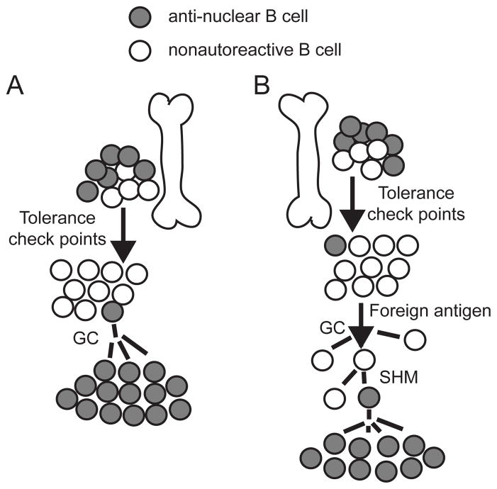
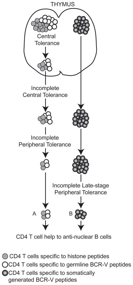
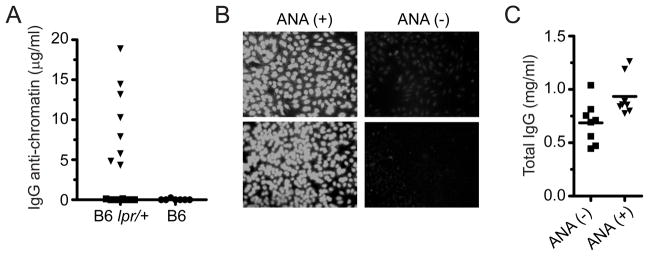
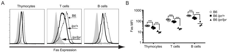
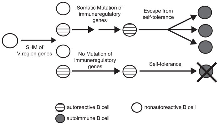
Our laboratory investigates systemic autoimmune disease in the context of mouse models of systemic lupus erythematosus (SLE). SLE is associated with high titers of serum autoantibodies of the IgG class that are predominantly directed against nuclear antigens, with pathological manifestations that are considered by many to be characteristic of an immune-complex mediated disease. In this review, we focus on the known and potential roles of somatic mutagenesis in SLE. We will argue that anti-nuclear antibodies (ANA) arise predominantly from nonautoreactive B cells that are transformed into autoreactive cells by the process of somatic hypermutation (SHM), which is normally associated with affinity maturation during the germinal center reaction. We will also discuss the role of SHM in creating antigenic peptides in the V region of the B cell receptor (BCR) and its potential to open an avenue of unregulated T cell help to autoreactive B cells. Finally, we will end this review with new experimental evidence suggesting that spontaneous somatic mutagenesis of genes that regulate B cell survival and activation is a rate-limiting causative factor in the development of ANA.


Conflict of interest statement

The authors declare no conflict of interest.

Figures

Figure 1
Figure 1. Origin of anti-nuclear B cells in SLE
(A) Germline-founder hypothesis: an anti-nuclear B cell, generated upon VDJ recombination in the bone marrow, evades tolerance checkpoints and enters the peripheral circulation. Upon recognition of a self-antigen, this autoreactive cells enters a germinal center (GC) and initiates an autoimmune response. (B) Mutation-founder hypothesis: a B cell with a normal, nonautoreactive receptor is recruited into a GC by an immunogen. Somatic hypermutation (SHM) generates an anti-nuclear B cell, which is the antecedent of an autoimmune lineage.
Figure 2
Figure 2. Origin of T cell help for anti-nuclear B cells
Source of T cell help to antinuclear B cells in SLE. (A) Defect in central and/or peripheral T cell tolerance in a lupus-prone background allows autoreactive CD4 T cells reactive with peptides from histones and/or germline-encoded BCR-V regions to escape self-tolerance and help anti-nuclear B cells. (B) Self-tolerance in CD4 T cells is intact with respect to peptides from histones and germline-encoded BCR-V regions; however CD4 T cells that recognize somatically generated BCR-V region-derived peptides provide a source of help to autoreactive B cells. Autoimmune prone-genetic backgrounds might have a defective regulation of this type of T-B interaction, resulting in the production of anti-nuclear antibodies by an autoimmune B cell.
Figure 3
Figure 3. Stochastic ANA and hypergammaglobulinemia in B6lpr/+ mice
(A) Total IgG anti-chromatin in B6lpr/+ (n=16) and wildtype B6 mice (n=7) at 6 months of age. (B) Representative IgG HEp-2 stain of B6lpr/+ anti-chromatin positive [ANA(+)] or negative [ANA(-)] observed in A. (C) Total IgGκ in sera of B6lpr/+ mice. Mice were segregated on the basis of the presence (▼) or absence (■) of IgG anti-chromatin antibodies. Anti-chromatin assays: 96 well trays were coated with 10 μg/ml of calf-chromatin or bovine serum albumin (BSA). Bound IgG Abs were detected with goat anti-mouse IgG heavy-chain specific antibody (SouthernBiotech). Specific IgG anti-chromatin Abs were calculated by subtracting counts to BSA-coated wells. Concentrations were calculated using a standard curve generated with the 3H9/Vκ4 mAb. Total IgG Abs were assayed in a similar manner, except that trays were coated with goat anti-mouse IgG (heavy chain-specific) at 1 μg/ml and bound antibodies were detected with biotin-goat anti-mouse κ.
Figure 4
Figure 4. Allelic exclusion of Fas in lymphocytes
(A) Left panel, ex vivo expression of Fas on thymocytes. Middle panel, Fas expression on CD4 T cells after 48 hours of in vitro stimulation with PMA/ionomycin. Right panel, expression of Fas on B cells after in vitro stimulation with anti-μ and anti-CD40. Solid histograms, thick line and dotted line indicate Fas expression by cells from B6lpr/lpr, B6lpr/+ and wildtype B6 mice respectively. (B) Mean fluorescence intensity (MFI) for stains involving multiple animals (*** p<0.01).
Figure 5
Figure 5. Equivalent NP antibody responses by wildtype B6 and B6lpr/+ mice
Mice were immunized with 100 μg of NP-chicken gamma globulin (CGG) in alum i.p. at day 0. Sera from indicated time points were tested for IgG anti-(NP-BSA). Relative affinity was calculated using the ratio of binding to NP4-BSA versus NP30-BSA. Values represented by each point were obtained from binding curves generated with serial 2-fold dilutions of sera. Secondary injection was performed i.p. on day 22 with 100 μg of NP-CGG in PBS and is indicated in graph by a red arrow. Day 7b refers to sera obtained 7 days after the secondary injection. Total NP Abs were calculated using the B1–8 mAb as a standard. (A) Impaired affinity maturation in B6lpr/lpr compared to B6 mouse, immunized with NP-CGG. (B–C) Normal kinetics, quantities and affinity maturation in serum from B6lpr/+ immunized with NP-CGG.
Figure 6
Figure 6. Stochastic ANA in Cd22 and Pten-conditional heterozygous deficient mice
(A–B) IgG anti-chromatin antibodies and total IgG in sera of Cd22+/−, Pten flox/+ Cd19-Cre and B6 mice. Mice were segregated into anti-chromatin positive (▼) and anti-chromatin negative (■) groups as defined by a solid phase immunoassay. Assays were performed as described in Fig. 3. (C) Sera of ANA positive mice were incubated in calf chromatin-coated wells in the presence of competing soluble chromatin at the indicated concentrations. Bound IgG anti-chromatin Abs were detected using a goat anti-mouse IgG (SouthernBiotech). The high-affinity, anti-nuclear mAb 3H9/Vκ4 was included for comparison.
Figure 7
Figure 7. Stochastic autoimmunity due to somatic mutations in immunoregulatory genes
A nonautoreactive B cell gives rise to anti-nuclear B cells in the GC. Most such B cells are censored by self-tolerance mechanisms unless they acquire additional critical somatic mutations that alter or ablate the function of immunoregulatory genes involved in self-tolerance. Somatic mutations in immuneregulatory genes could arise in an AID-dependent and/or independent manner.

References

    1. Kotzin BL. Systemic lupus erythematosus. Cell. 1996;85:303–306. - PubMed
    1. Theofilopoulos AN, Dixon FJ. Murine models of systemic lupus erythematosus. Advances in immunology. 1985;37:269–390. - PubMed
    1. Flesher DL, Sun X, Behrens TW, Graham RR, Criswell LA. Recent advances in the genetics of systemic lupus erythematosus. Expert Rev Clin Immunol. 2010;6:461–479. - PMC - PubMed
    1. Goodnow CC. Multistep pathogenesis of autoimmune disease. Cell. 2007;130:25–35. - PubMed
    1. Wardemann H, Yurasov S, Schaefer A, Young JW, Meffre E, Nussenzweig MC. Predominant autoantibody production by early human B cell precursors. Science. 2003;301:1374–1377. - PubMed

Publication types

Substances