Review
. 2013 Sep 20;19(9):1026-35.
doi: 10.1089/ars.2012.5065. Epub 2013 Feb 4.

Cytochromes b561: ascorbate-mediated trans-membrane electron transport

Affiliations
Review

Cytochromes b561: ascorbate-mediated trans-membrane electron transport

Han Asard et al. Antioxid Redox Signal. .

Abstract

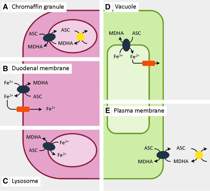
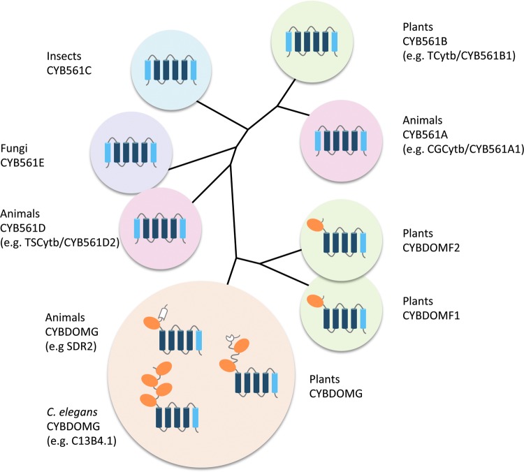
Significance: Cytochromes b561 (CYB561s) constitute a family of trans-membrane (TM), di-heme proteins, occurring in a variety of organs and cell types, in plants and animals, and using ascorbate (ASC) as an electron donor. CYB561s function as monodehydroascorbate reductase, regenerating ASC, and as Fe³⁺-reductases, providing reduced iron for TM transport. A CYB561-core domain is also associated with dopamine β-monooxygenase redox domains (DOMON) in ubiquitous CYBDOM proteins. In plants, CYBDOMs form large protein families. Physiological functions supported by CYB561s and CYBDOMs include stress defense, cell wall modifications, iron metabolism, tumor suppression, and various neurological processes, including memory retention. CYB561s, therefore, significantly broaden our view on the physiological roles of ASC.

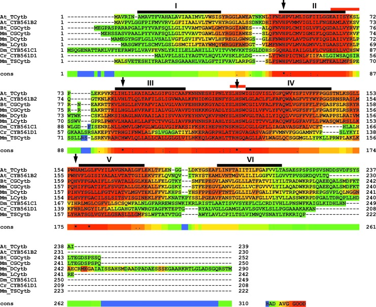
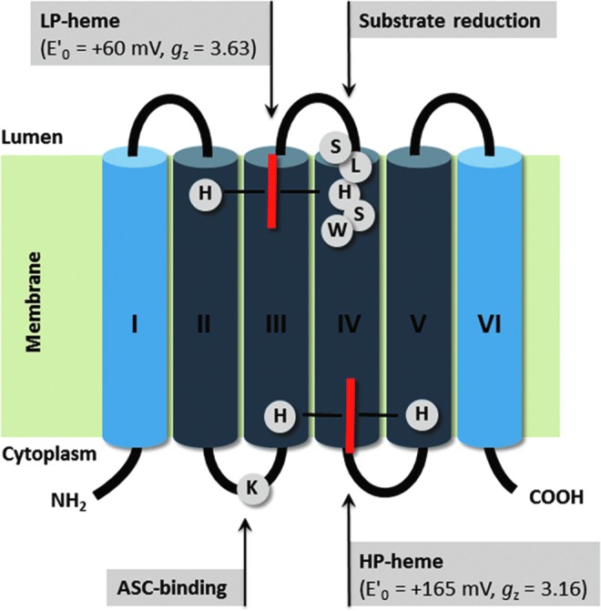
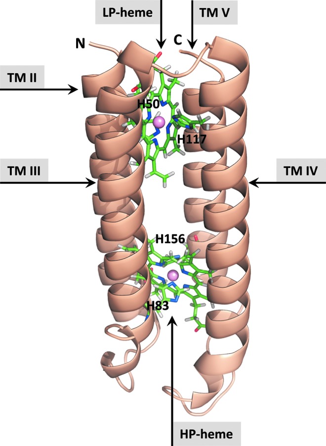
Recent advances: The ubiquitous nature of CYB561s is only recently being recognized. Significant advances have been made through the study of recombinant CYB561s, revealing structural and functional properties of a unique "two-heme four-helix" protein configuration. In addition, the DOMON domains of CYBDOMs are suggested to contain another heme b.

Critical issues: New CYB561 proteins are still being identified, and there is a need to provide an insight and overview on the various roles of these proteins and their structural properties.

Future directions: Mutant studies will reveal in greater detail the mechanisms by which CYB561s and CYBDOMs participate in cell metabolism in plants and animals. Moreover, the availability of efficient heterologous expression systems should allow protein crystallization, more detailed (atomic-level) structural information, and insights into the intra-molecular mechanism of electron transport.


Figures

FIG. 1.
FIG. 1.
Subcellular localization and orientation of CYB561 family members in plant and animal cells. CYB561s (blue ovals) catalyze trans-membrane electron transport (TMET) through membranes, separating cytoplasm and acid compartments (secretory vesicles, vacuole, and extracellular matrix). Localization, orientation, and electron donor/acceptor couples are based on experimental evidence (on native and recombinant proteins), and ‘most plausible’ interpretations (see text). Ascorbate (ASC) appears a principal electron donor for all CYB561s, whereas both monodehydroascorbate (MDHA) and Fe3+ may act as electron acceptors. Animal CYB561s: (A) Chromaffin granules: CGCytb/CYB561A1 in chromaffin granule vesicle membranes, involved in catecholamine synthesis operating in concert with ASC-dependent oxidases (yellow circles); (B) duodenal DCytb/CYB561A2 in the duodenal cell membrane, reducing intestinal Fe3+-chelates before uptake through Fe2+-transporters (orange rectangles); (C) lysosomal membrane LCytb/CYB561A3. Plant CYB561: (D) plant tonoplast TCytb/CYB561B1 may reduce vacuolar Fe3+ for transport to the cytoplasm; (E) (putative) plant plasma membrane CYB561B acting in concert with extracellular oxidases (e.g., ASC oxidase). To see this illustration in color, the reader is referred to the web version of this article at www.liebertpub.com/ars
FIG. 2.
FIG. 2.
Multiple alignment of CYB561s, showing TM domains, conserved residues, and substrate-binding domains. ClustalW2.1 alignment of selected CYB561 sequences, representing isoforms from most evolutionary clusters (59) (see Fig. 5), showing TM domains (I–VI, black bars), conserved His residues (arrows), and putative substrate interacting sites (red bars). Sequences are as follows: (i) Arabidopsis thaliana tonoplast CYB561 (At_TCytb/CYB561B1; Q8L856); (ii) At_CYB561B2 (Q9SWS1); (iii) Bostaurus chromaffin granule CYB561 (Bt_CGCytb/CYB561A1; P10897); (iv) Mus musculus chromaffin granule CYB561 (Mm_CGCytb/CYB561A1; Q60720); (v) M. musculus duodenal CYB561 (Mm_DCytb/CYB561A2; Q925G2); (vi) M. musculus lysosomal CYB561 (Mm_LCytb/CYB561A3; Q6P1H1); (vii) Drosophila melanogaster CYTB561 (Dm_CYB561C1; Q9W4U9); (viii) Chlamydomonas reinhardtii CYB561 (Cr_CYB561E1; XP_001702111.1); and (ix) M. musculus tumor suppressor CYB561(Mm_TSCytb/CYB561D2; Q9WUE3). Color coding of sequence similarity is performed using the T-Coffee alignment evaluation (17). To see this illustration in color, the reader is referred to the web version of this article at www.liebertpub.com/ars
FIG. 3.
FIG. 3.
Schematic 2D structure of the Arabidopsis TCytb/CYB561B1 TM organization, including hemes, heme coordination, and putative substrate binding sites. CYB561s are composed of six TM domains (I-VI), with two heme-b molecules (red bars), coordinated by four conserved His on TM helices II-V. The high potential (HP)-heme is located on the cytoplasmic protein side, accepting electrons from ASC (or other electron donors). The heme coordination and orientation is confirmed through electron paramagnetic resonance studies on site-directed-mutant proteins. Also shown is a Lys residue, possibly involved in substrate (ASC) binding, and a well-conserved putative substrate-binding site (SLHSW). Dark blue: CYB561 four helix core domain. To see this illustration in color, the reader is referred to the web version of this article at www.liebertpub.com/ars
FIG. 4.
FIG. 4.
Calculated 3D atomic structure model of the Arabidopsis TCytb/CYB561B1-core domain. Based on extensive CYB561s sequence analysis, structural constraints were identified to predict 3D structural models, using molecular modeling tools. Counterclockwise helix topologies are shown, including the HP- and low-potential (LP) hemes (ball-and-stick). Balls represent the Fe-atoms [modified after (6)]. To see this illustration in color, the reader is referred to the web version of this article at www.liebertpub.com/ars
FIG. 5.
FIG. 5.
Evolutionary relationship between CYB561s and CYB561-core-containing CYBDOM proteins. CYB561 and CYBDOM proteins, cluster in various subgroups. The CYB561-core domain (four TM helices containing four conserved His and two hemes in bis-His coordination) is dark blue (see also Fig. 3). In the CYBDOM protein family, the CYB561 domain constituted by a CYB561-core plus the C-terminal α-helix of CYB561s is combined with one or more dopamine β-monooxygenase N-terminal domain (DOMON) domains (brown ovals). Cluster identifications are based on (59), except that clusters D and E were exchanged, for nomenclature reasons (see Supplementary Table S1). Each CYB561 cluster contains sequences from only animals or plants or insects or fungi, with the notable exception of Chlamydomonas reinhardtii CYB561 (XP_001702111.1, see also Supplementary Table S1) that clusters with the ‘fungal’ cluster CYB561E. CYBDOMF is divided into F1 and F2 clusters with diverging DOMON sequences. CYBDOMG includes sequences with one DOMON plus a reelin domain (white), sequences with four DOMONs, and plant sequences with two DOMONs and a DM13 domain (white). To see this illustration in color, the reader is referred to the web version of this article at www.liebertpub.com/ars
FIG. 6.
FIG. 6.
DOMON domain of P. chrysosporium cellobiose dehydrogenase (CDH), showing the uncommon b-type heme coordination by a Met-His pair. The DOMON domains of CYBDOM proteins are predicted to have the same 3D-fold based on two antiparallel β-sheets forming a β-sandwich structure as the DOMON domain of CDH (26). Although the sequence similarity between CYBDOM and CDH-DOMONs is limited, the pair of heme-coordinating residues Met65 and His163 (P. chrysosporium numbering) is strictly conserved. The structure was drawn from pdb file 1d7b (21). Heme and side chains of heme-coordinating residues are represented in sticks with the following color codes C (green), N (blue), O (red), and S (yellow). To see this illustration in color, the reader is referred to the web version of this article at www.liebertpub.com/ars

References

    1. Aravind L. Domon: an ancient extracellular domain in dopamine beta-monooxygenase and other proteins. Trends Biochem Sci. 2001;26:524–526. - PubMed
    1. Asard H. Horemans N. Caubergs RJ. Transmembrane electron transport in ascorbate-loaded plasma membrane vesicles from higher plants involves a b-type cytochrome. FEBS Lett. 1992;306:143–146. - PubMed
    1. Asard H. Terol-Alcayde J. Preger V. Del Favero J. Verelst W. Sparla F. Pérez-Alonso M. Trost P. Arabidopsis thaliana sequence analysis confirms the presence of cyt b-561 in plants: evidence for a novel protein family. Plant Physiol Biochem. 2000;38:905–912.
    1. Asard H. Venken M. Caubergs R. Reijnders W. Oltmann FL. De Greef JA. b-Type cytochromes in higher plant plasma membranes. Plant Physiol. 1989;90:1077–1083. - PMC - PubMed
    1. Askerlund P. Larsson C. Widell S. Cytochromes of plant plasma membranes. Characterization by absorbance difference spectroscopy and redox titration. Physiol Plant. 1989;76:123–134.

Publication types

LinkOut - more resources