Review
. 2012 Oct-Dec;8(4):103-13.
doi: 10.4161/org.23307. Epub 2012 Oct 1.

Craniosynostosis: molecular pathways and future pharmacologic therapy

Affiliations
Review

Craniosynostosis: molecular pathways and future pharmacologic therapy

Kshemendra Senarath-Yapa et al. Organogenesis. 2012 Oct-Dec.

Abstract

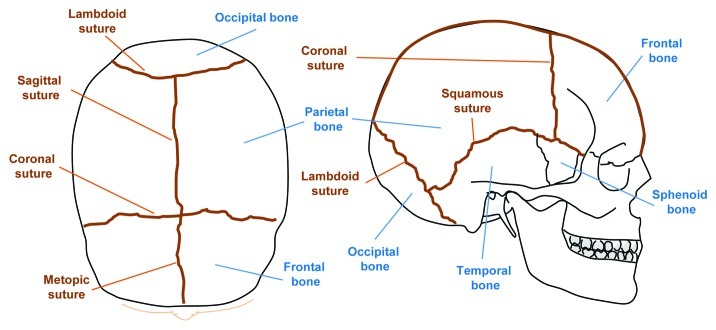
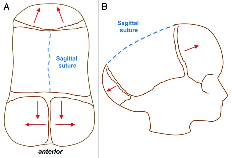
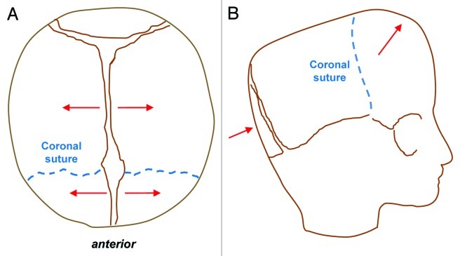
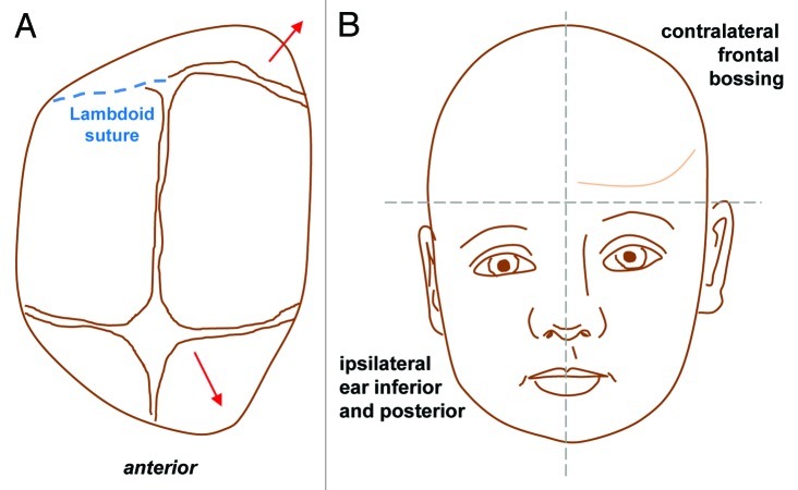
Craniosynostosis describes the premature fusion of one or more cranial sutures and can lead to dramatic manifestations in terms of appearance and functional impairment. Contemporary approaches for this condition are primarily surgical and are associated with considerable morbidity and mortality. The additional post-operative problems of suture refusion and bony relapse may also necessitate repeated surgeries with their own attendant risks. Therefore, a need exists to not only optimize current strategies but also to develop novel biological therapies which could obviate the need for surgery and potentially treat or even prevent premature suture fusion. Clinical studies of patients with syndromic craniosynostosis have provided some useful insights into the important signaling pathways and molecular events guiding suture fate. Furthermore, the highly conserved nature of craniofacial development between humans and other species have permitted more focused and step-wise elucidation of the molecular underpinnings of craniosynostosis. This review will describe the clinical manifestations of craniosynostosis, reflect on our understanding of syndromic and non-syndromic craniosynostoses and outline the different approaches that have been adopted in our laboratory and elsewhere to better understand the pathogenesis of premature suture fusion. Finally, we will assess to what extent our improved understanding of the pathogenesis of craniosynostosis, achieved through laboratory-based and clinical studies, have made the possibility of a non-surgical pharmacological approach both realistic and tangible.

Keywords: FGF; calvarium; craniosynostosis; signaling; suture.


Figures

None
Figure 1. Schematic representation of the major bones and sutures of the adult human cranium. Top view (left) and lateral view (right) of the calvarium, showing bones (blue) and sutures (brown). By the second year of life the metopic suture is normally closed.
None
Figure 2. Sagittal synostosis (Scaphocepaly). Top view (A) and lateral view (B) schematics showing the direction of aberrant compensatory bone growth (red lines) following premature sagittal suture fusion leading to an elongated, scaphocephalic skull, derived from the Greek word scaphos for boat and cephalos for head.
None
Figure 3. Bilateral coronal craniosynostosis. Top view (B) and lateral view (B) schematics showing the direction of compensatory calvarial growth (red lines) secondary to premature fusion of both coronal sutures (blue dashed lines) leading to shortening in the anteroposterior direction, increased height of the vault and therefore a turribrachycephalic deformity.
None
Figure 4. Metopic Synostosis. Top view (A) and lateral view (B) schematics demonstrating trigonocephaly, or a triangular keel-shaped head. Compensatory calvarial growth (red lines) can lead to widening and increased height in the parietal region due to compensatory growth in a posterior direction following premature fusion of the metopic suture (blue dashed line).
None
Figure 5. Lambdoid synostosis. Top view (A) and antero-posterior view (B) showing the direction of compensatory bone growth (red lines) following premature fusion of the lambdoid suture (blue dashed line). Lambdoid synostosis leads to ipsilateral flattening of the occipital region with a compensatory mastoid bulge which is a key distinguishing feature from positional (sometimes called positional or deformational) plagiocephaly from which it can be clinically difficult to differentiate. Contralateral growth in the parietal region is also noted and the cranial base becomes tilted.

References

    1. Otto. A. Lehrbuch der Pathologischen Anatomie. Berlin, Germany: Rucher., 1830.
    1. Lenton KA, Nacamuli RP, Wan DC, Helms JA, Longaker MT. Cranial suture biology. Curr Top Dev Biol. 2005;66:287–328. doi: 10.1016/S0070-2153(05)66009-7. - DOI - PubMed
    1. Slater BJ, Lenton KA, Kwan MD, Gupta DM, Wan DC, Longaker MT. Cranial sutures: a brief review. Plast Reconstr Surg. 2008;121:170e–8e. doi: 10.1097/01.prs.0000304441.99483.97. - DOI - PubMed
    1. Virchow R. Berh Phyd Med Gesellsch Wuerzburg. 1851;2:231–71.
    1. Moss ML. The pathogenesis of premature cranial synostosis in man. Acta Anat (Basel) 1959;37:351–70. doi: 10.1159/000141479. - DOI - PubMed

LinkOut - more resources