Comparative Study
. 2013 Jan 17;54(1):455-66.
doi: 10.1167/iovs.12-11152.

QLT091001, a 9-cis-retinal analog, is well-tolerated by retinas of mice with impaired visual cycles

Affiliations
Comparative Study

QLT091001, a 9-cis-retinal analog, is well-tolerated by retinas of mice with impaired visual cycles

Tadao Maeda et al. Invest Ophthalmol Vis Sci. .

Abstract

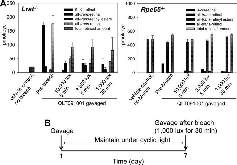
Purpose: Investigate whether retinas of mice with impaired retinal cycles exposed to light or kept in the dark tolerate prolonged high-dose administration of QLT091001, which contains as an active ingredient, the 9-cis-retinal precursor, 9-cis-retinyl acetate.

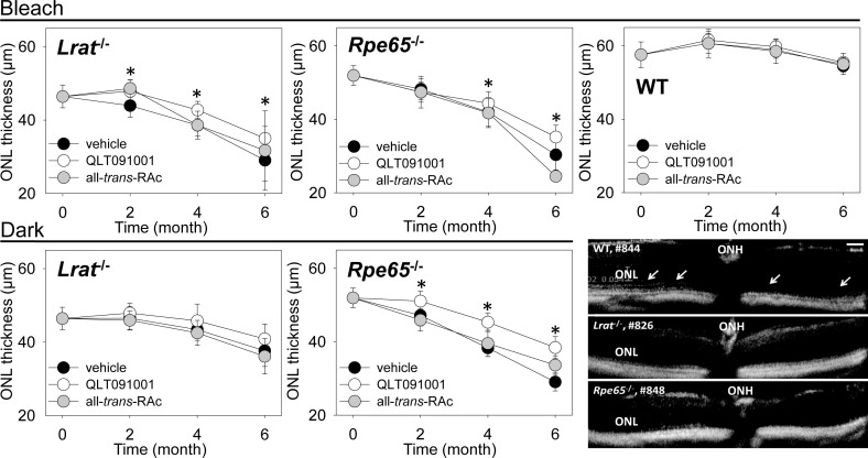
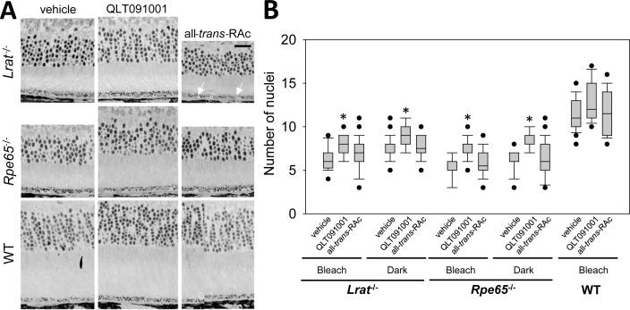
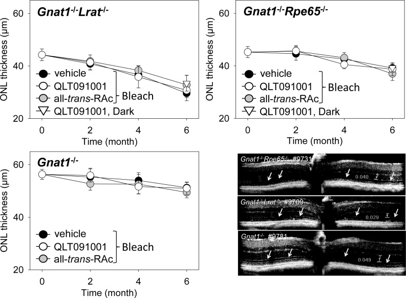
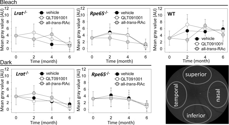
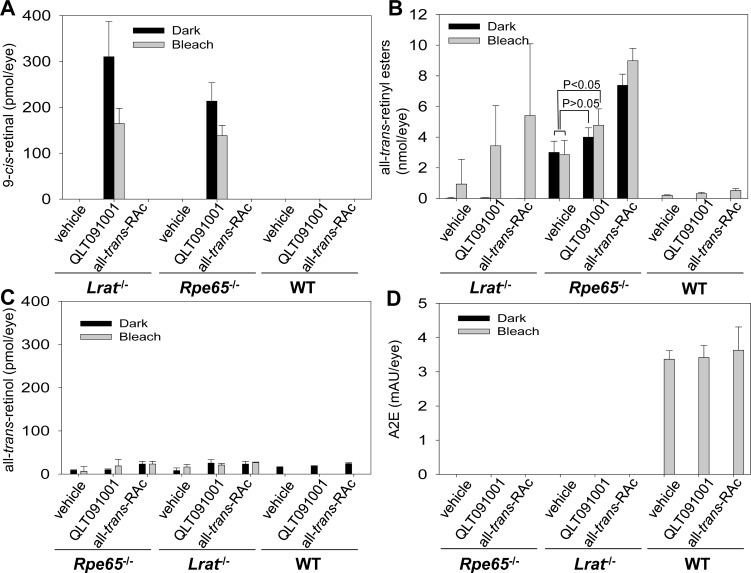
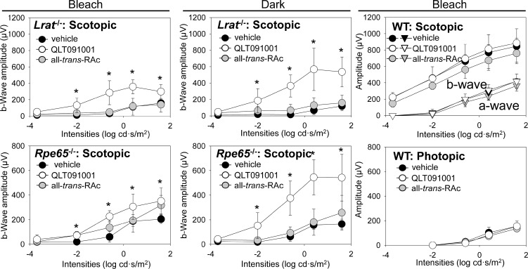
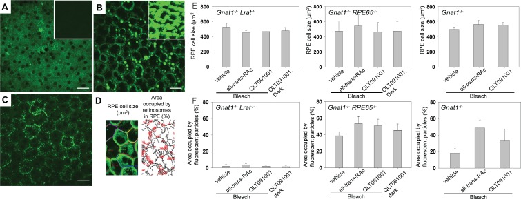
Methods: Four- to six-week-old Lrat(-/-) and Rpe65(-/-) mice (n = 126) as well as crossbred Gnat1(-/-) mice lacking rod phototransduction (n = 110) were gavaged weekly for 6 months with 50 mg/kg QLT091001, either after being kept in the dark or after light bleaching for 30 min/wk followed by maintenance in a 12-hour light ≤ 10 lux)/12-hour dark cycle. Retinal health was monitored by spectral-domain optical coherent tomography (SD-OCT) and scanning laser ophthalmoscopy (SLO) every other month and histological, biochemical, and visual functional analyses were performed at the end of the experiment. Two-photon microscopy (TPM) was used to observe retinoid-containing retinosome structures in the RPE.

Results: Retinal thickness and morphology examined by SD-OCT were well maintained in all strains treated with QLT091001. No significant increases of fundus autofluorescence were detected by SLO imaging of any strain. Accumulation of all-trans-retinyl esters varied with genetic background, types of administered compounds and lighting conditions but retinal health was not compromised. TPM imaging clearly revealed maintenance of retinosomes in the RPE of all mouse strains tested.

Conclusions: Retinas of Lrat(-/-), Rpe65(-/-), and crossbred Gnat1(-/-) mice tolerated prolonged high-dose QLT091001 treatment well.


Conflict of interest statement

Disclosure: T. Maeda, QLT, Inc. (F, C), Polgenix, Inc. (C), P; Z. Dong, QLT, Inc. (F); H. Jin, QLT, Inc. (F); O. Sawada, QLT, Inc. (F); S. Gao, QLT, Inc. (F); D. Utkhede, QLT, Inc. (F, E); W. Monk, QLT, Inc. (F, E); G. Palczewska, QLT, Inc. (F); K. Palczewski, QLT, Inc. (C), Polgenix, Inc. (F, C), P

Figures

Figure 1
Figure 1
A bleaching protocol tests the effect of QLT091001 on retinas of Lrat−/− and Rpe65−/− mice. (A) Optimization of bleaching conditions to eliminate residual 9-cis-retinal from retinas of Lrat−/− or Rpe65−/− mice after a single gavage with 50 mg/kg of QLT091001. Four-week-old Lrat−/− or Rpe65−/− mice were used for this experiment. Three days after retinoid/vehicle administration, mice were exposed to three different lighting conditions to photoisomerize residual iso-rhodopsin. After exposure to 1000 lux light for 30 minute that bleached more than 70% of the retina, 9-cis-retinal was released from photoreceptor cells in both mouse strains. (B) Schematic protocol for weekly light exposure with retinoid/vehicle gavage. Based on the results shown in (A), the experimental weekly light exposure protocol included a 30 minute bleach with 1000 lux light followed by retinoid/vehicle gavage in addition to maintenance of mice in a 12-hour light (≤10 lux)/12-hour dark environment for 6 months. Dark control groups of mice were gavaged weekly while maintained in the dark. Error bars indicate SDs, n ≥ 3.
Figure 2
Figure 2
ONL thickness assessed by SD-OCT in Lrat−/−, Rpe65−/−, and WT mice after treatment with either vehicle, QLT091001, or all-trans-RAc. ONL thicknesses were measured at regions 500 μm away from the optic nerve head (ONH) in the superior, inferior, temporal, and nasal retina in SD-OCT images obtained at 0° and 90° every second month. ONL thicknesses from these 4 different regions of the retina were averaged and plotted. Protective effects of QLT091001 became statistically significant at 2 months in Lrat−/− mice maintained under light cycle and bleach and Rpe65−/− mice maintained in dark, and at 4 months in Rpe65−/− mice maintained in the dark. These positive effects were maintained after 6 months treatment. Effects were more prominent in Rpe65−/− mice maintained in dark, whereas only mild protective effects were observed in Lrat−/− mice maintained in the dark. There were no significant differences noted in WT mice. Representative SD-OCT images from WT, Lrat−/− and Rpe65−/− after 6 months treatment are shown in the right bottom corner. The ELM was clearly evident in SD-OCT images of WT retina (white arrows), but not those from Lrat−/− and Rpe65−/− mice. Error bars indicate SDs. Magnification bar indicates 50 μm, n ≥3 per group. *P < 0.05.
Figure 3
Figure 3
ONL thickness assessed by histology in Lrat−/−, Rpe65−/−, and WT mice after treatment with either vehicle, QLT091001, or all-trans-RAc. (A) Nuclei present in photoreceptor populations of representative retinal plastic cross sections from Lrat−/−, Rpe65−/−, and WT mice treated with either vehicle, QLT091001 or all-trans-retinyl esters (all-trans-RAc) are shown. Oil-droplet like structures were observed in the RPE of some Lrat−/− mice maintained under a regular light cycle plus bleach protocol (white arrows). (B) Numbers of photoreceptor nuclei counted at 4 different regions in the retina similar to those assessed for ONL thickness by SD-OCT in each mouse strain treated with retinoids/vehicle were averaged and plotted. Numbers of nuclei were significantly preserved in the eyes of mice treated with QLT091001 (P < 0.05) whereas numbers were comparable in vehicle- or all-trans-RAc-treated eyes regardless of mouse strain. Boxes denote interquartile range, lines within boxes denote medians, whiskers denote 10th and 90th percentiles, and symbols denote outliers (A). Magnification bar indicates 20 μm (B), n ≥3 per group at each time point. *P < 0.05.
Figure 4
Figure 4
ONL thickness assessed by SD-OCT in retinas of Gnat1−/−Lrat−/−, Gnat1−/−, and Gnat1−/−Rpe65−/− mice after 6 months of treatment with vehicle, QLT091001, or all-trans-RAc. Mice were 4-weeks-old at the beginning of treatment. Averaged thicknesses of the ONL measured from SD-OCT images obtained at 0 and 90 degrees from four different regions of the retina were not affected by the compounds administered to Gnat1−/−Lrat−/− or Gnat1−/−Rpe65−/− mice. Representative SD-OCT images after 6 months of treatment are shown (right-bottom panel). ELMs were discerned in SD-OCT images from all strains (white arrows). Error bars indicate SDs, n ≥3 per group at each time point. *P < 0.05.
Figure 5
Figure 5
AF measurements by SLO during 6 months of vehicle, QLT091001 or all-trans-RAc treatment of Lrat−/−, Rpe65−/−, or WT mice. AF levels in SLO fundus images were quantified every other month during weekly retinoid/vehicle administration to Lrat−/−, Rpe65−/− and WT mice that were either maintained under a regular light cycle plus bleach protocol or kept in the dark. Mean gray values obtained from four different regions of the fundus (right-bottom panel) were averaged and plotted. No significant differences were noted between vehicle-, QLT091001- or all-trans-RAc-treated Lrat−/−, RPE65−/−, and WT mice maintained in either a normal light/dark cycle with bleach or in a dark environment. Error bars indicate SDs, n = 3 to 5 per group at each time point.
Figure 6
Figure 6
Eye retinoid content in Lrat−/−, Rpe65−/−, or WT mice treated with either vehicle, QLT091001 or all-trans-RAc for 6-months. RPE65−/−, Lrat−/−, and WT mice were 4 to 6 weeks old at the beginning of treatment. Eye retinoids were extracted and quantified after separation by normal phase HPLC. 9-cis-Retinal was detected in eyes of both Lrat−/− and RPE65−/− mice treated with QLT091001 under both normal lighting plus bleach conditions and maintenance in the dark ([A], left and center). Several Lrat−/− light-exposed mice showed an unexpected all-trans-retinyl ester accumulation enhanced by treatment with either QLT091001 or all-trans-RAc ([B], left). High levels of all-trans-retinyl esters in Rpe65−/− mice were not affected by lighting conditions ([B], center). The ester levels in this strain maintained in the dark and treated with QLT091001 were comparable to those of the vehicle control (P > 0.05) whereas ester levels of mice maintained in the dark treated with all-trans-RAc and mice maintained under light cycle plus bleach treated with QLT091001 and all-trans-RAc were significantly increased compared to ester levels of those treated with vehicle ([B], center, P < 0.05). Levels of all-trans-retinyl esters were mildly increased in eyes of WT mice treated by QLT091001 and all-trans-RAc (B). The levels of all-trans-retinol varied but were higher in the eyes of all mice treated with either QLT091001 or all-trans-RAc (C). Age-dependent accumulation of the retinoid byproduct, A2E was observed only in WT mice maintained under normal light with bleach and this was not altered by exposure to either QLT091001 or all-trans-RA (D). Error bars indicate SDs, n ≥3 in each group.
Figure 7
Figure 7
ERG recordings of visual function in Lrat−/−, Rpe65−/−, and WT mice after 6 months of treatment with either vehicle, QLT091001 or all-trans-RAc. Animals were 4 weeks old at the beginning of treatment. ERG recordings were obtained from Lrat−/− and Rpe65−/− mice under scotopic conditions and from WT mice under both scotopic and photopic conditions. Plots of b-wave amplitudes from Lrat−/− and Rpe65−/− mice and scotopic a- and b-wave and photopic b-wave amplitudes from WT mice are shown. ERG amplitudes of QLT091001-treated groups were significantly higher than those of all-trans-RAc- or vehicle-treated Lrat−/− and Rpe65−/− mice. ERG responses of WT mice were comparable between the different regimens. Error bars indicate SDs, n >3 in each group. *P < 0.05.
Figure 8
Figure 8
Characterization of the RPE from Lrat−/−, Rpe65−/− or WT mice by TPM after 6 months of treatment with vehicle, all-trans-RAc or QLT091001. Animals were 4 weeks-old at the beginning of treatment. Representative TPM images are shown for albino Gnat1−/−Lrat−/− (A), albino Gnat1−/−Rpe65−/− (B), and albino Gnat1−/− (C) mice. Imaging conditions were optimized to visualize the subcellular structure of RPE cells. Insets in (A) and (B) show images obtained under the same conditions as those used in (C). Areas of individual RPE cells and areas occupied by fluorescent deposits in the RPE were estimated as shown in (D). There was no significant difference in the size of RPE cells between strains and treatments in these mice (E). As shown in (F), the fraction of an area occupied by fluorescent deposits was consistently greatest in mice treated with all-trans-RAc. Error bars in (E) and (F) show SDs, n ≥3 in each group. *P < 0.05.

References

    1. Cideciyan AV. Leber congenital amaurosis due to RPE65 mutations and its treatment with gene therapy. Prog Retin Eye Res. 2010; 29: 398–427. - PMC - PubMed
    1. Gu SM, Thompson DA, Srikumari CR, et al. Mutations in RPE65 cause autosomal recessive childhood-onset severe retinal dystrophy. Nat Genet. 1997; 17: 194–197. - PubMed
    1. Palczewski K. Blind dogs that can see: pharmacological treatment of Leber congenital amaurosis caused by a defective visual cycle. Arch Ophthalmol. 2010; 128: 1483–1485. - PMC - PubMed
    1. Palczewski K. G protein-coupled receptor rhodopsin. Annu Rev Biochem. 2006; 75: 743–767. - PMC - PubMed
    1. Palczewski K. Retinoids for treatment of retinal diseases. Trends Pharmacol Sci. 2010; 31: 284–295. - PMC - PubMed

Publication types

MeSH terms