. 2012 Dec 18:8:88.
doi: 10.1186/1744-8069-8-88.

Differential lumbar spinal cord responses among wild type, CD4 knockout, and CD40 knockout mice in spinal nerve L5 transection-induced neuropathic pain

Affiliations

Differential lumbar spinal cord responses among wild type, CD4 knockout, and CD40 knockout mice in spinal nerve L5 transection-induced neuropathic pain

Ling Cao et al. Mol Pain. .

Abstract

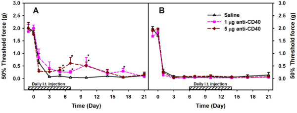
Background: Our previous studies have indicated that both lumbar spinal cord-infiltrating CD4+ T cells and microglial CD40 contribute to the maintenance of mechanical hypersensitivity in a murine model of neuropathic pain spinal nerve L5 transection (L5Tx). To further delineate the CD4 and CD40-mediated mechanisms involved in the development of L5Tx-induced neuropathic pain behaviors, we examined the lumbar spinal cord mononuclear cells of wild type (WT) BALB/c, BALB/c-CD4 knockout (KO), and BALB/c-CD40 KO mice via flow cytometry.

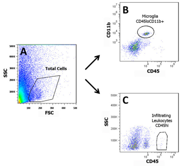
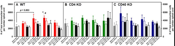
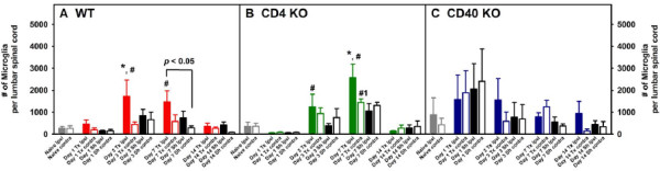
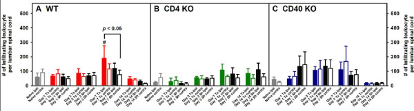
Results: In WT mice, L5Tx induced significant but transient (at day 3 and/or day 7) increases of the total numbers of mononuclear cells, microglial cells (CD45loCD11b+), and infiltrating leukocytes (CD45hi) in the ipsilateral side of the spinal cord. In CD4 KO mice, significant elevation of microglia was detected only on day 7 post-L5Tx, while no significant increase in infiltrating leukocytes post-L5Tx was observed. CD40 KO mice did not exhibit any of the changes observed in WT mice. Furthermore, neutralizing CD40 antibody treatment indicated an early involvement of CD40 signaling in the development of L5Tx-induced mechanical hypersensitivity.

Conclusions: Altogether, data indicate that both CD4 and CD40 play a role in L5Tx-induced leukocyte infiltration into the lumbar spinal cord but have differential contributions to spinal cord microglial activation following peripheral nerve injury.


Figures

Figure 1
Figure 1
Flow cytometric analysis of lumbar spinal cord mononuclear cells. Total mononuclear cells collected from each sample (pooled ipsilateral or contralateral side of lumbar spinal cords from 3–4 mice) were labeled with mAbs against CD11b and CD45. All samples were examined with an Accuri C6 flow cytometer and analyzed with FlowJo. For each sample, total cell population was first identified (A), and then microglia (CD45loCD11b+) (B) and infiltrating leukocytes (CD45hi) (C) were identified within the gated total cell population. The percentage of each of these populations within the total cells was recorded. The total number of microglia and infiltrating leukocytes per spinal cord were calculated based on the recorded percentages, the total mononuclear cells collected, and the number of mice used in each sample. Representative plots shown here are from a “WT day 3 L5Tx ipsilateral” spinal cord mononuclear cell sample.
Figure 2
Figure 2
Numbers of total mononuclear cells in lumbar spinal cord following L5Tx in WT, CD4KO, and CD40KO mice. WT, CD4 KO, and CD40 KO mice were subjected to sham or L5Tx surgery. Lumbar spinal cord mononuclear cells from separate groups of mice of each genotype were collected at indicated times post-surgery. The temporal changes of the numbers of total lumbar spinal cord mononuclear cells in WT (A, n = 5–8), CD4 KO (B, n = 4) and CD40 KO (C, n = 4) mice are shown here (mean ± SEM). One-way ANOVA was performed to examine the basal level genotypic differences among naïve mice and no significant differences were found. Two-way ANOVA for data sets in each graph were performed. * indicates significant differences between the indicated group and all other groups at the same time point. # indicates significant differences between the indicated group and the corresponding Day 0 group. An additional significant result from statistical comparison is also shown within the graph. “Tx” = L5Tx, “Sh” = sham operation, “ipsi” = ipsilateral side, and “contra” = contralateral side. For naïve mice, “ipsi” = left and “contra” = right.
Figure 3
Figure 3
Numbers of microglia (CD45loCD11b+) in lumbar spinal cord following L5Tx in WT, CD4KO, and CD40KO mice. Total mononuclear cells collected as described in Figure 2 were analyzed for their microglial content. The temporal changes of the numbers of lumbar spinal cord microglia in WT (A, n = 5–8), CD4 KO (B, n = 4) and CD40 KO (C, n = 4) mice are shown here (mean ± SEM). One-way ANOVA was performed to examine the basal level genotypic differences among naïve mice and no significant differences were found. Two-way ANOVA for data sets in each graph were performed. * indicates significant differences between the indicated group and all other groups at the same time point. # indicates significant differences between the indicated group and all other groups within the same treatment group (including the corresponding Day 0 group). #1 indicates significant differences between the indicated group and days 0, 1 and 14 groups within the same treatment group. An additional significant result from statistical comparison is also shown within the graph. “Tx” = L5Tx, “Sh” = sham operation, “ipsi” = ipsilateral side, and “contra” = contralateral side. For naïve mice, “ipsi” = left and “contra” = right
Figure 4
Figure 4
Numbers of infiltrating leukocytes (CD45hi) in lumbar spinal cord following L5Tx in WT, CD4KO, and CD40KO mice. Total mononuclear cells collected as described in Figure 2 were analyzed for their content of infiltrating leukocytes. The temporal changes of the numbers of lumbar spinal cord infiltrating leukocytes in WT (A, n = 5–8), CD4 KO (B, n = 4) and CD40 KO (C, n = 4) mice are shown here (mean ± SEM). One-way ANOVA was performed to examine the basal level genotypic differences among naïve mice and no significant differences were found. Two-way ANOVA for data sets in each graph were performed. # indicates significant differences between the indicated group and all other groups within the same treatment group (including the corresponding Day 0 group). An additional significant result from statistical comparison is also shown within the graph. “Tx” = L5Tx, “Sh” = sham operation, “ipsi” = ipsilateral side, and “contra” = contralateral side. For naïve mice, “ipsi” = left and “contra” = right.
Figure 5
Figure 5
Mechanical hypersensitivity of WT mice treated with anti-CD40. WT mice were subjected to L5Tx. Neutralizing CD40 antibody was i.t. administered to WT mice daily either from day −1 to day 7 (A) or from day 6 to day 14 (B). Mechanical sensitivity of each mouse was tested before L5Tx and repeatedly after L5Tx using a series of von Frey filaments via the up-down method. All data are presented as mean ± SEM (n = 6). Two-way RM ANOVA was performed for data sets within each graph. * indicates the significant differences between the indicated group and the saline treated group within the same time point. In addition, within each treatment group, p < 0.05 between any time point post-L5Tx and both day 0 and day −1 time points (not indicated in the graph).

Similar articles

Cited by

References

    1. Jensen TS. et al.A new definition of neuropathic pain. Pain. 2011;152(10):2204–2205. doi: 10.1016/j.pain.2011.06.017. - DOI - PubMed
    1. DeLeo JA, Tanga FY, Tawfik VL. Neuroimmune activation and neuroinflammation in chronic pain and opioid tolerance/hyperalgesia. Neuroscientist. 2004;10(1):40–52. doi: 10.1177/1073858403259950. - DOI - PubMed
    1. Milligan ED, Watkins LR. Pathological and protective roles of glia in chronic pain. Nat Rev Neurosci. 2009;10(1):23–36. doi: 10.1038/nrn2533. - DOI - PMC - PubMed
    1. Ren K, Dubner R. Interactions between the immune and nervous systems in pain. Nat Med. 2010;16(11):1267–1276. doi: 10.1038/nm.2234. - DOI - PMC - PubMed
    1. Lindenlaub T, Sommer C. Cytokines in sural nerve biopsies from inflammatory and non-inflammatory neuropathies. Acta Neuropathol. 2003;105(6):593–602. - PubMed

Publication types