. 2013 Feb;19(2):167-76.
doi: 10.1261/rna.036269.112. Epub 2012 Dec 17.

Modulation of quaternary structure and enhancement of ligand binding by the K-turn of tandem glycine riboswitches

Affiliations

Modulation of quaternary structure and enhancement of ligand binding by the K-turn of tandem glycine riboswitches

Nathan J Baird et al. RNA. 2013 Feb.

Abstract

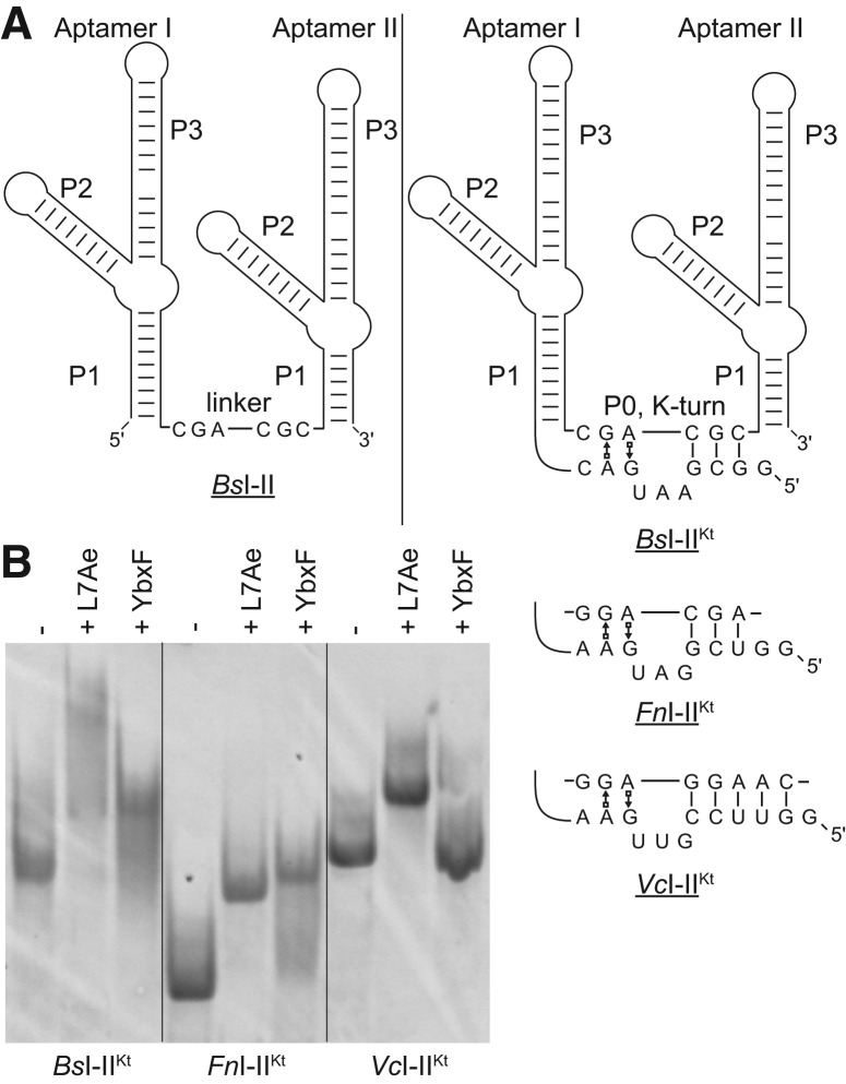
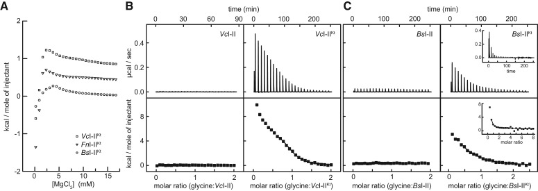
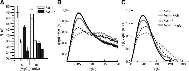
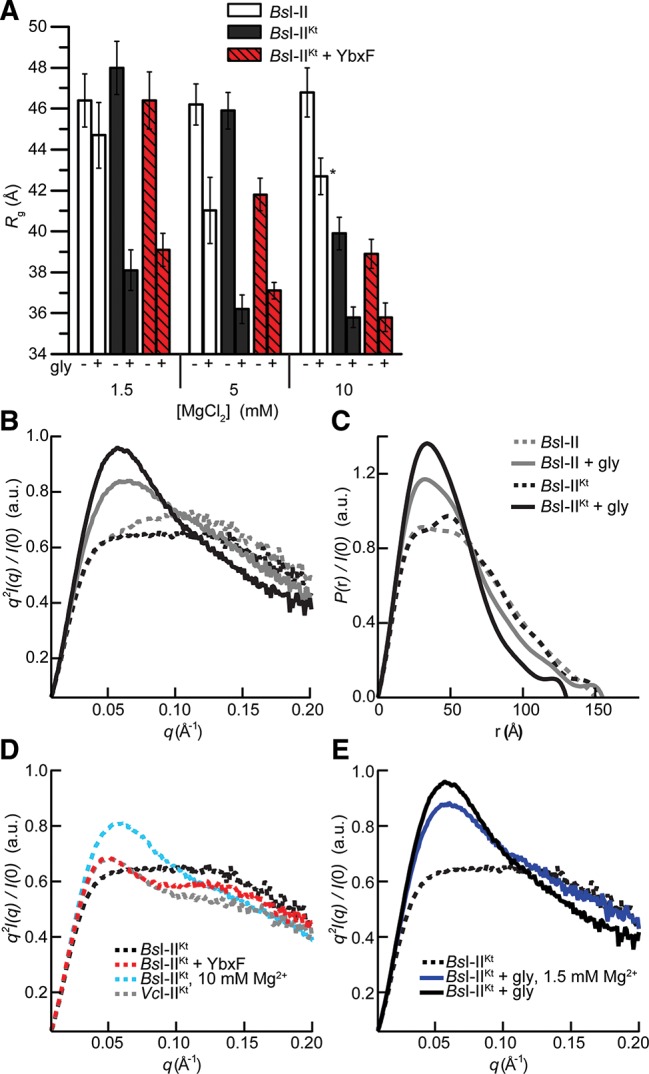
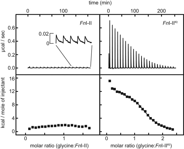
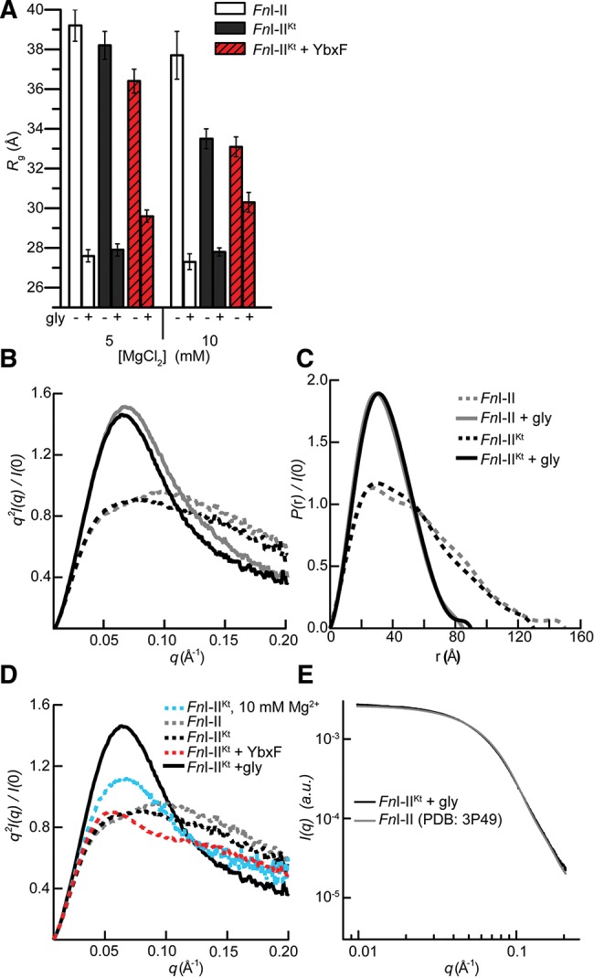
Most known glycine riboswitches have two homologous aptamer domains arranged in tandem and separated by a short linker. The two aptamers associate through reciprocal "quaternary" interactions that have been proposed to result in cooperative glycine binding. Recently, the interaptamer linker was found to form helix P0 with a previously unrecognized segment 5' to the first aptamer domain. P0 was shown to increase glycine affinity, abolish cooperativity, and conform to the K-turn motif consensus. We examine the global thermodynamic and structural role of P0 using isothermal titration calorimetry (ITC) and small-angle X-ray scattering (SAXS), respectively. To evaluate the generality of P0 function, we prepared glycine riboswitch constructs lacking and including P0 from Bacillus subtilis, Fusobacterium nucleatum, and Vibrio cholerae. We find that P0 indeed folds into a K-turn, supports partial pre-folding of all three glycine-free RNAs, and is required for ITC observation of glycine binding under physiologic Mg(2+) concentrations. Except for the unusually small riboswitch from F. nucleatum, the K-turn is needed for maximally compacting the glycine-bound states of the RNAs. Formation of a ribonucleoprotein complex between the B. subtilis or the F. nucleatum RNA constructs and the bacterial K-turn binding protein YbxF promotes additional folding of the free riboswitch, and enhances glycine binding. Consistent with the previously reported loss of cooperativity, P0-containing B. subtilis and V. cholerae tandem aptamers bound no more than one glycine molecule per riboswitch. Our results indicate that the P0 K-turn helps organize the quaternary structure of tandem glycine riboswitches, thereby facilitating ligand binding under physiologic conditions.


Figures

FIGURE 1.
FIGURE 1.
The P0 helix of glycine riboswitches is a K-turn. (A) Tandem glycine riboswitch aptamers joined by a single-stranded linker, denoted I–II (left), and by the recently discovered (Kladwang et al. 2012; Sherman et al. 2012) K-turn, denoted I–IIKt (right). The sequence of the K-turn is given for each of the three I–IIKt examined. (B) EMSA analysis of three I–IIKt constructs using the L7Ae and YbxF K-turn binding proteins.
FIGURE 2.
FIGURE 2.
ITC analyses of RNA folding and glycine binding. (A) Mg2+ titration of the three I–IIKt RNAs used in this study. For clarity, data for FnI–IIKt and VcI–IIKt are offset by 0.4 kcal/mol and 0.8 kcal/mol, respectively, in this panel only. (B) ITC titrations of glycine into VcI–II (left) and VcI–IIKt (right) in 5 mM Mg2+. (C) ITC titrations of glycine into BsI–II (left) and BsI–IIKt (right) in 5 mM Mg2+, including a titration in which the final molar ratio (glycine:RNA) in the solution is >8 (inset).
FIGURE 3.
FIGURE 3.
SAXS analyses of VcI–II and VcI–IIKt. (A) Comparison of Rg in the absence and presence of saturating glycine in 5 mM and 10 mM Mg2+. (B) Kratky and (C) P(r) plots for experiments performed in 5 mM Mg2+ with VcI–II and VcI–IIKt in the absence and presence of glycine.
FIGURE 4.
FIGURE 4.
SAXS analyses of B. subtilis tandem glycine aptamers. (A) Rg in the absence and presence of saturating glycine in 1.5 mM, 5 mM, and 10 mM Mg2+ for BsI–II, BsI–IIKt, and BsI–IIKt + YbxF. Sample marked with asterisk exhibited partial oligomerization (Materials and Methods). (B) Kratky and (C) P(r) plots for experiments performed in 5 mM Mg2+ with BsI–II and BsI–IIKt in the absence and presence of glycine. (D) Kratky plots (in 5 mM Mg2+, except where noted) demonstrating the effect of Mg2+ and YbxF on the structure of BsI–IIKt in the absence of glycine. VcI–IIKt plot is reproduced from Figure 3D for additional comparison. (E) Kratky plots demonstrating the effect of glycine in 1.5 mM and 5 mM Mg2+.
FIGURE 5.
FIGURE 5.
ITC analyses of FnI–II and FnI–IIKt. (Left) Titration of glycine into FnI–II. A small heat signal is observed upon injection of glycine (inset) and is very slow to re-equilibrate (>10 min). (Right) Glycine binding by FnI–IIKt results in a binding isotherm.
FIGURE 6.
FIGURE 6.
SAXS analyses of FnI–II, FnI–IIKt, and FnI-IIKt + YbxF. (A) Rg in the absence and presence of saturating glycine in 5 mM and 10 mM Mg2+. (B) Kratky and (C) P(r) plots from data collected at 5 mM Mg2+ demonstrate glycine-binding-induced compaction. (D) Kratky plots comparing the effect of Mg2+, YbxF, and glycine on folding of FnI–IIKt (in 5 mM Mg2+, except where noted). (E) Comparison of the experimental X-ray scattering profile for FnI–IIKt with the scattering profile calculated (Materials and Methods) from the crystal structure (Butler et al. 2011) of FnI–II.

Similar articles

Cited by

References

    1. Alatossava T, Jutte H, Kuhn A, Kellenberger E 1985. Manipulation of intracellular magnesium content in polymyxin B nonapeptide-sensitized Escherichia coli by ionophore A23187. J Bacteriol 162: 413–419 - PMC - PubMed
    1. Altschul SF, Madden TL, Schaffer AA, Zhang J, Zhang Z, Miller W, Lipman DJ 1997. Gapped BLAST and PSI-BLAST: A new generation of protein database search programs. Nucleic Acids Res 25: 3389–3402 - PMC - PubMed
    1. Ames TD, Breaker RR 2011. Bacterial aptamers that selectively bind glutamine. RNA Biol 8: 82–89 - PMC - PubMed
    1. Baird NJ, Ferré-D’Amaré AR 2010. Idiosyncratically tuned switching behavior of riboswitch aptamer domains revealed by comparative small-angle X-ray scattering analysis. RNA 16: 598–609 - PMC - PubMed
    1. Baird NJ, Westhof E, Qin H, Pan T, Sosnick TR 2005. Structure of a folding intermediate reveals the interplay between core and peripheral elements in RNA folding. J Mol Biol 352: 712–722 - PubMed

Publication types

MeSH terms

LinkOut - more resources