Comparative Study
. 2013 Feb;19(2):280-93.
doi: 10.1261/rna.034850.112. Epub 2012 Dec 18.

Potent microRNA suppression by RNA Pol II-transcribed 'Tough Decoy' inhibitors

Affiliations
Comparative Study

Potent microRNA suppression by RNA Pol II-transcribed 'Tough Decoy' inhibitors

Rasmus O Bak et al. RNA. 2013 Feb.

Abstract

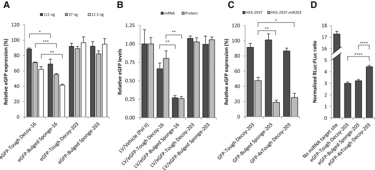
MicroRNAs (miRNAs) are key regulators of gene expression and modulators of diverse biological pathways. Analyses of miRNA function as well as therapeutic managing of miRNAs rely on cellular administration of miRNA inhibitors which may be achieved by the use of viral vehicles. This study explores the miRNA-suppressive capacity of inhibitors expressed intracellularly from lentivirus-derived gene vectors. Superior activity of two decoy-type inhibitors, a "Bulged Sponge" with eight miRNA recognition sites and a hairpin-shaped "Tough Decoy" containing two miRNA recognition sites, is demonstrated in a side-by-side comparison of seven types of miRNA inhibitors transcribed as short RNAs from an RNA Pol III promoter. We find that lentiviral vectors expressing Tough Decoy inhibitors are less vulnerable than Bulged Sponge-encoding vectors to targeting by the cognate miRNA and less prone, therefore, to reductions in transfer efficiency. Importantly, it is demonstrated that Tough Decoy inhibitors retain their miRNA suppression capacity in the context of longer RNA transcripts expressed from an RNA Pol II promoter. Such RNA Pol II-transcribed Tough Decoy inhibitors are new tools in managing of miRNAs and may have potential for temporal and spatial regulation of miRNA activity as well as for therapeutic targeting of miRNAs that are aberrantly expressed in human disease.


Figures

FIGURE 1.
FIGURE 1.
Overview of miRNA inhibition strategies. (A) Schematic representations of the seven miRNA inhibitors compared in the current study. (i) AntagomiR with a single perfect miRNA target site (Scherr et al. 2007); (ii) a single perfect miRNA target site stabilized with terminal hairpin structures (Vermeulen et al. 2007); (iii) a Tough Decoy with two bulged miRNA target sites (Haraguchi et al. 2009); (iv) an shRNA targeting the mature miRNA (only the guide strand of the processed shRNA is depicted) (Wang et al. 2011); (v) a miRNA sponge with eight consecutive miRNA perfect target sites (Ebert et al. 2007; Gentner et al. 2009); (vi) a sponge with eight consecutive bulged miRNA target sites (Ebert et al. 2007; Gentner et al. 2009); (vii) a miRNA mask that binds and protects the miRNA target sites on the mRNA (Choi et al. 2007). (B) Schematic representation of the lentiviral plasmid used for expressing miRNA inhibitors from the H1 RNA Pol III promoter (shown in white). (CMV) Cytomegalovirus promoter, (Ψ) viral RNA packaging signal, (RRE) rev response element, (cPPT) central polypurine tract, (PGK) phosphoglycerate kinase promoter, (Puror) puromycin resistance gene (puromycin N-acetyl-transferase [PAC]), (WPRE) woodchuck hepatitis virus posttranscriptional regulatory element. (C) miRNA levels in HEK-293T, HEK-293, and HeLa cells. qPCR threshold cycles (Ct) were determined for miR-16 and miR-203 in HEK-293T, HEK-293, and HeLa cells. Additionally, Ct values for the U48 small nuclear RNA (RNU48) were determined as a reference small RNA. Data are depicted as mean + SEM.
FIGURE 2.
FIGURE 2.
Tough Decoy inhibitors perform best among seven miRNA inhibition strategies when delivered by plasmid transfection or lentiviral transduction. A dual-luciferase assay was used to screen the potency of seven vector-encoded miRNA inhibitors targeting miR-16 (A,C) or miR-203 (B,D). miRNA inhibitor expression cassettes were delivered either by plasmid transfection (A,B) or by lentiviral transduction (C,D). A miR203-encoding plasmid was included in all assays evaluating miR-203 inhibitors. Data are depicted as mean + SEM. (*) P < 0.05, (**) P < 0.01, (***) P < 0.001, (****) P < 0.0001.
FIGURE 3.
FIGURE 3.
Transfer of Bulged Sponge-encoding lentiviral vectors are markedly affected by endogenous miRNAs targeting the vector. (A) Reduced transductional titer of Sponge-encoding vectors as determined by the number of puromycin-resistant colony-forming units per mL (CFU/mL). (B) Reduced integration capacity of Bulged Sponge-encoding lentiviral vectors as determined by the number of lentiviral integrations quantified by qPCR. Data are depicted as mean + SEM. (*) P < 0.05, (**) P < 0.01, (***) P < 0.001, (****) P < 0.0001.
FIGURE 4.
FIGURE 4.
Tough Decoy inhibitors retain their functionality when fused to an RNA Pol II eGFP transcript. (A) Schematic overview of the lentiviral vector plasmid used for expression of miRNA inhibitors in fusion with eGFP. (B) Schematic representations of the expressed fusion transcripts containing the Bulged Sponge and Tough Decoy inhibitors, respectively. (CG) Dual-luciferase assays comparing Tough Decoy and Bulged Sponge inhibitors transcribed from RNA Pol II and Pol III promoters targeting miR-16 (C), miR-203 (D), miR-143 (E), miR-145 (F), and miR-195 (G). Data are depicted as mean + SEM. (*) P < 0.05, (**) P < 0.01, (***) P < 0.001, (****) P < 0.0001.
FIGURE 5.
FIGURE 5.
Lentiviral vectors encoding RNA Pol II-transcribed Tough Decoys are functional and not prone to reductions in transductional titers. (A,B) Dual-luciferase assays evaluating the potency of lentivirally delivered RNA Pol II-transcribed Tough Decoys and Bulged Sponge inhibitors targeting miR-16 (A) or miR-203 (B). (C) Transductional titers of lentiviral vectors encoding RNA Pol II-transcribed miR16- and miR203-directed Tough Decoy and Bulged Sponge inhibitors as determined by the number of lentiviral integrations quantified by qPCR. (D,E) Endogenous miRNA levels were quantified by qPCR following lentiviral delivery of RNA Pol II-driven Tough Decoy and Bulged Sponge inhibitors. For miR-16 inhibition, MCF-7 cells were transduced (D), and for miR-203 inhibition, HaCaT cells were transduced (E). (F) TNFα mRNA levels were quantified by qPCR in HaCaT cells transduced with lentiviral vectors encoding RNA Pol II-driven miR-203 inhibitors. Data are depicted as mean + SEM. (*) P < 0.05, (**) P < 0.01, (***) P < 0.001, (****) P < 0.0001.
FIGURE 6.
FIGURE 6.
Analysis of miRNA targeting of the inhibitor-encoding transcript. (A) Flow cytometric analysis of eGFP expression in HEK-293 cells 2 d after transfection with different amounts of plasmids encoding RNA Pol II-transcribed eGFP fused with Tough Decoy or Bulged Sponge inhibitors targeting either miR-16 or miR-203. eGFP intensity (relative eGFP expression) is presented relative to the eGFP intensity measured in cells transfected with the same amount of the standard unfused eGFP-encoding plasmid. (B) Analysis of eGFP-inhibitor transcript levels and eGFP protein levels following lentiviral delivery of eGFP-inhibitors. Transcript and protein levels were evaluated by qPCR and flow cytometry, respectively. (C) Flow cytometric analysis of eGFP expression in naive HEK-293T cells and HEK-293T cells overexpressing miR203 (HEK-293T-miR203) 2 d after transfection with 12.3 ng of the plasmid encoding RNA Pol II-transcribed eGFP fused with Tough Decoy, Bulged Sponge, or 4 × Tough Decoy (eight miRNA target sites in four consecutive Tough Decoys) inhibitors targeting miR-203. eGFP intensity (relative eGFP expression) is presented relative to the eGFP intensity measured in cells transfected with the same amount of the standard unfused eGFP-encoding plasmid. (D) Dual-luciferase assay comparing Tough Decoy, Bulged Sponge, and 4 × Tough Decoy inhibitors targeting miR-203 and transcribed from RNA Pol II promoters. Data are depicted as mean + SEM. (*) P < 0.05, (**) P < 0.01, (***) P < 0.001, (****) P < 0.0001.

References

    1. Ameres SL, Martinez J, Schroeder R 2007. Molecular basis for target RNA recognition and cleavage by human RISC. Cell 130: 101–112 - PubMed
    1. An W, Telesnitsky A 2001. Frequency of direct repeat deletion in a human immunodeficiency virus type 1 vector during reverse transcription in human cells. Virology 286: 475–482 - PubMed
    1. Bak RO, Stenderup K, Rosada C, Petersen LB, Moldt B, Dagnaes-Hansen F, Jakobsen M, Kamp S, Jensen TG, Dam TN, et al. 2011. Targeting of human interleukin-12B by small hairpin RNAs in xenografted psoriatic skin. BMC Dermatol 11: 5. - PMC - PubMed
    1. Care A, Catalucci D, Felicetti F, Bonci D, Addario A, Gallo P, Bang ML, Segnalini P, Gu Y, Dalton ND, et al. 2007. MicroRNA-133 controls cardiac hypertrophy. Nat Med 13: 613–618 - PubMed
    1. Chang K, Elledge SJ, Hannon GJ 2006. Lessons from Nature: MicroRNA-based shRNA libraries. Nat Methods 3: 707–714 - PubMed

Publication types