Clinical Trial
. 2013 Mar;33(3):322-9.
doi: 10.1038/jcbfm.2012.196. Epub 2012 Dec 19.

In vivo detection of cerebral cortical microinfarcts with high-resolution 7T MRI

Affiliations
Clinical Trial

In vivo detection of cerebral cortical microinfarcts with high-resolution 7T MRI

Susanne J van Veluw et al. J Cereb Blood Flow Metab. 2013 Mar.

Abstract

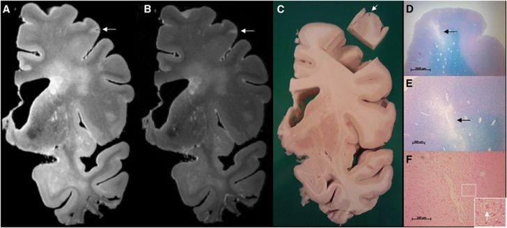
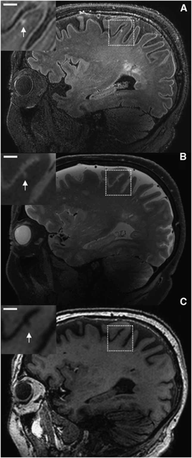
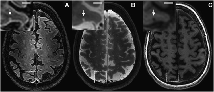
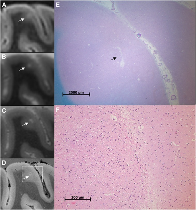
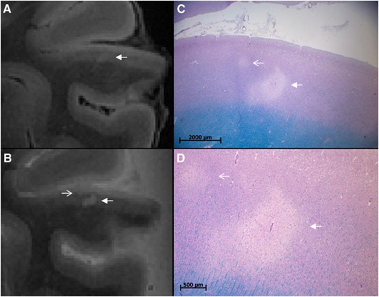
Cerebrovascular disease has an important role in cognitive decline and dementia. In this context, cerebral microinfarcts are attracting increasing attention, but these lesions could thus far not be detected in vivo. The aim of this study was to try to identify possible cortical microinfarcts on high-resolution 7T in vivo magnetic resonance imaging (MRI) and to perform a histopathologic validation study on similar appearing lesions on 7T ex vivo MRI of postmortem brain tissue. The study population consisted of 22 elderly subjects, who underwent 7T MRI. The fluid attenuated inversion recovery, T(2), and T(1) weighted scans of these subjects were examined for possible cortical microinfarcts. In the ex vivo MRI study, 15 formalin-fixed coronal brain slices of 6 subjects with Alzheimer and vascular pathology were examined and subjected to histopathologic verification. On the in vivo scans, 15 cortical lesions could be identified that were likely to be microinfarcts in 6 subjects. In the postmortem tissue, 6 similar appearing lesions were identified of which 5 were verified as cortical microinfarcts on histopathology. This study provides strong evidence that cortical microinfarcts can be detected in vivo, which will be of great value in further studies into the role of vascular disease in cognitive decline and dementia.


Figures

Figure 1
Figure 1
Sampling procedure: correlation of ex vivo magnetic resonance imaging (MRI) and histology. The sampling procedure was developed in three brain slices from anonymous donors. In one of these brain slices a cortical lesion of interest, presumed to be ischemic, was identified on ex vivo MRI. This lesion appeared as a hyperintense cortical lesion with a hypointense center on a fluid attenuated inversion recovery (FLAIR) (A; 400 × 400 × 400 μm3), and as a hyperintense cortical lesion on a T2 weighted (B; 400 × 400 × 400 μm3) magnetic resonance (MR) image. After dissection of the tissue for sampling a cavity in the cortex was visible (C; tissue block is turned over). On histopathologic examination, this cortical lesion was identified as a cerebral microinfarct (CMI) with cavitation and gliosis in the surrounding tissue (D, and in greater detail E) (Luxol fast blue & Periodic acid-Schiff (L&P) stain). On higher magnification, gliosis is visible (F; arrow indicates a reactive astrocyte) (hematoxylin/eosin (HE) stain). Tissue was submerged in Fomblin.
Figure 2
Figure 2
Possible cerebral microinfarct (CMI) at 7.0 T in vivo magnetic resonance imaging (MRI). A possible cortical CMI in a 76-year-old nondemented male is represented as a hyperintense cortical lesion on a fluid attenuated inversion recovery (FLAIR) (A; 0.8 × 0.8 × 0.8 mm3) and a T2 weighted (B; 0.7 × 0.7 × 0.7 mm3), and as a hypointense lesion on a T1 weighted (C; 1.0 × 1.0 × 1.0 mm3) magnetic resonance (MR) image on a sagittal view of the brain. Scale bar indicates 4 mm.
Figure 3
Figure 3
Possible cerebral microinfarct (CMI) at 7.0 T in vivo magnetic resonance imaging (MRI). A possible cortical CMI in a 66-year-old nondemented female is represented as a hyperintense cortical lesion on a fluid attenuated inversion recovery (FLAIR) (A; 0.8 × 0.8 × 0.8 mm3) and a T2 weighted (B; 0.7 × 0.7 × 0.7 mm3), and as a hypointense lesion on a T1 weighted (C; 1.0 × 1.0 × 1.0 mm3) magnetic resonance (MR) image on a transversal view of the brain. Scale bar indicates 4 mm.
Figure 4
Figure 4
A cerebral microinfarct (CMI) identified with ex vivo magnetic resonance imaging (MRI). A CMI in a formalin-fixed coronal brain slice of a 64-year-old patient with a neuropathologic diagnosis of Alzheimer's disease (AD). The CMI is presented as a hyperintense cortical lesion on a clinical resolution fluid attenuated inversion recovery (FLAIR) (A; 0.8 × 0.8 × 0.8 mm3) and a clinical resolution T2 weighted image (B; 0.7 × 0.7 × 0.7 mm3), and as a hyperintense cortical lesion in greater detail on an ultra-high resolution T2 weighted (C; 400 × 400 × 400 μm3) magnetic resonance (MR) image. On an ultra-high resolution T2* weighted (D; 180 × 180 × 180 μm3) MR image, the CMI can be observed in even greater detail and in close resemblance to the microscopic images in (E) and (F). On histopathologic examination, this cortical lesion was established as a CMI with moderate gliosis in the surrounding tissue (E; Luxol fast blue & Periodic acid-Schiff (L&P) stain, and in greater detail F; hematoxylin/eosin (HE) stain).
Figure 5
Figure 5
An additional cerebral microinfarct (CMI) identified with histology. A CMI in a formalin-fixed coronal brain slice of a 79-year-old patient with Alzheimer's disease (AD) pathology. The CMI is presented as a hyperintense cortical lesion on an ultra-high resolution fluid attenuated inversion recovery (FLAIR) (A; arrow; 400 × 400 × 400 μm3) and an ultra-high resolution T2 weighted (B; arrow; 400 × 400 × 400 μm3) magnetic resonance (MR) image. On histopathologic examination, this cortical lesion was established as a CMI (C, Luxol fast blue & Periodic acid-Schiff (L&P) stain, and in greater detail D, L&P stain). Note the smaller CMI in (C) and (D) (open arrow) which was initially solely identified on histopathologic examination, but at reinspection proved to be visible on the ultra-high resolution T2 weighted MR image as well (B; open arrow).

Comment in

References

    1. Pendlebury ST, Rothwell PM. Prevalence, incidence, and factors associated with pre-stroke and post-stroke dementia: a systematic review and meta-analysis. Lancet Neurol. 2009;8:1006–1018. - PubMed
    1. Kalaria RN, Kenny RA, Ballard CG, Perry R, Ince P, Polvikoski T. Towards defining the neuropathological substrates of vascular dementia. J Neurol Sci. 2004;226:75–80. - PubMed
    1. Schneider JA, Arvanitakis Z, Bang W, Bennett DA. Mixed brain pathologies account for most dementia cases in community-dwelling older persons. Neurology. 2007;69:2197–2204. - PubMed
    1. Pantoni L. Cerebral small vessel disease: from pathogenesis and clinical characteristics to therapeutic challenges. Lancet Neurol. 2010;9:689–701. - PubMed
    1. Smith EE, Schneider JA, Wardlaw JM, Greenberg SM. Cerebral microinfarcts: the invisible lesions. Lancet Neurol. 2012;11:272–282. - PMC - PubMed

Publication types