. 2013 Sep;6(5):900-10.
doi: 10.1038/mi.2012.128. Epub 2012 Dec 19.

Th17 cells confer long-term adaptive immunity to oral mucosal Candida albicans infections

Affiliations

Th17 cells confer long-term adaptive immunity to oral mucosal Candida albicans infections

N Hernández-Santos et al. Mucosal Immunol. 2013 Sep.

Abstract

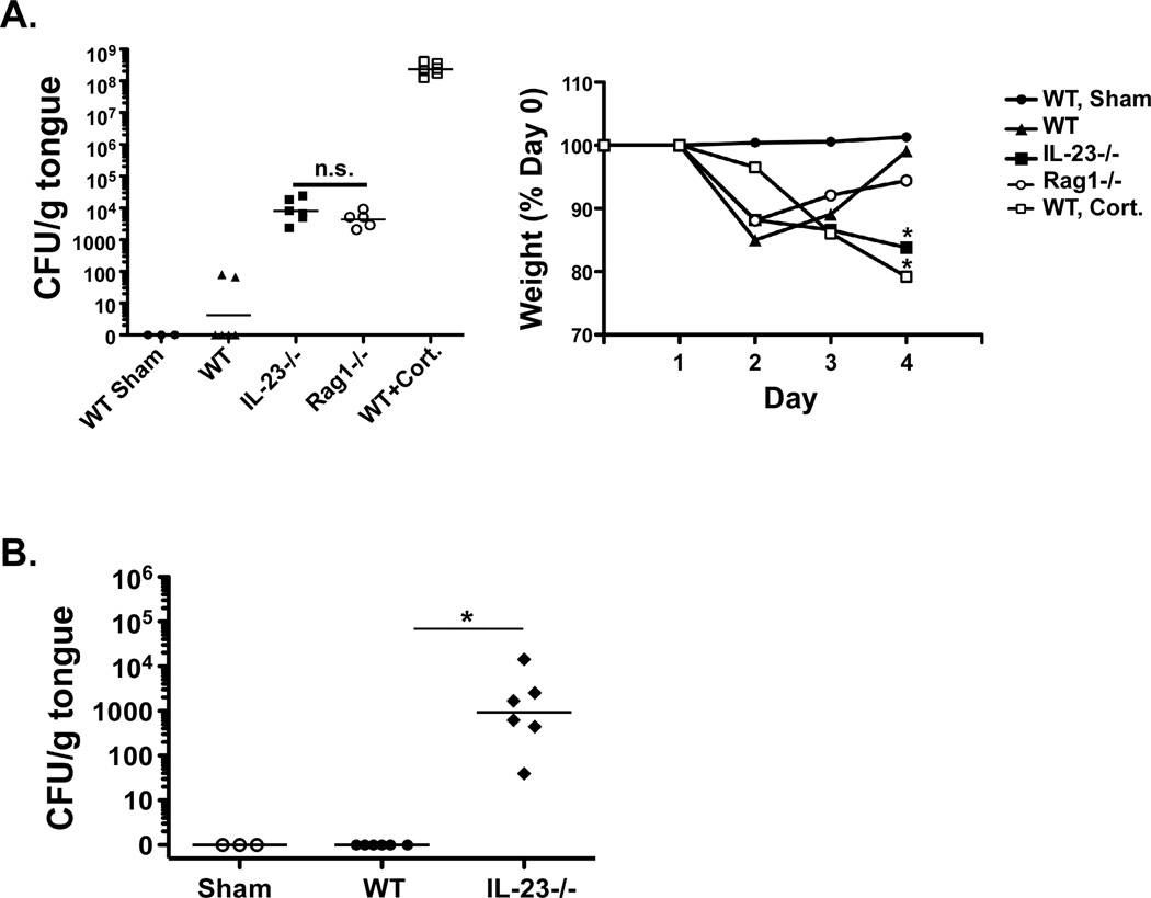
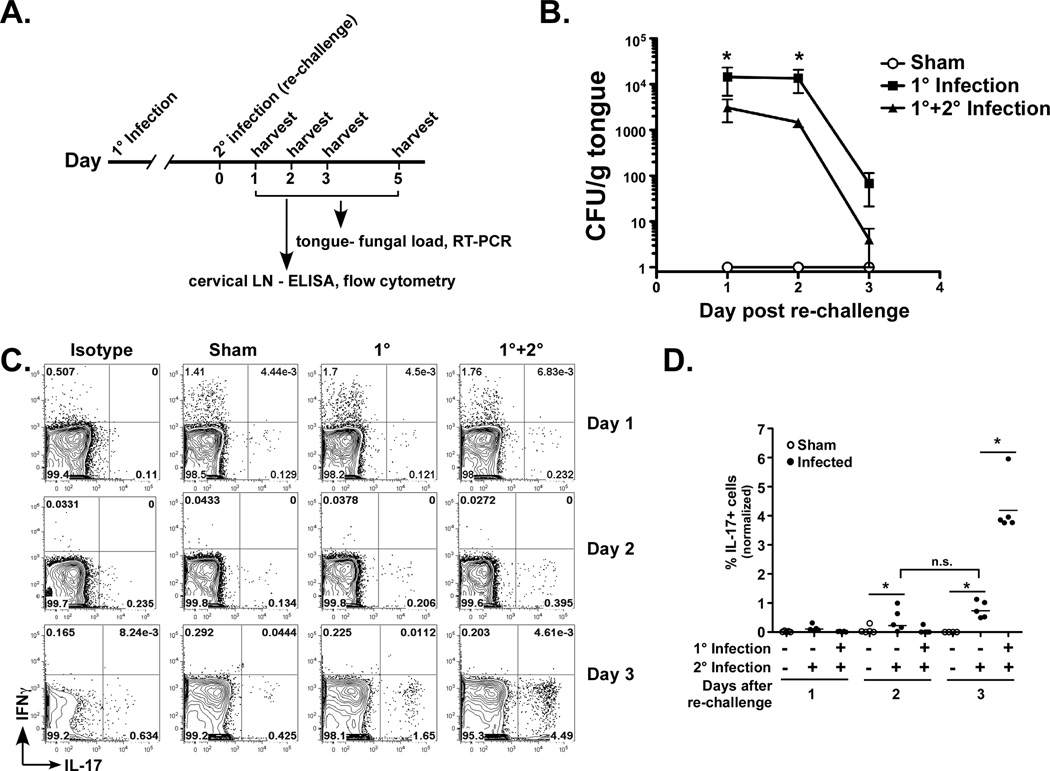
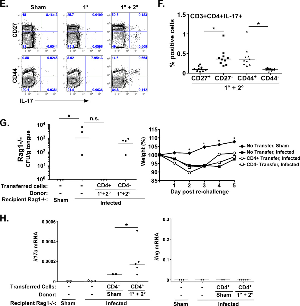
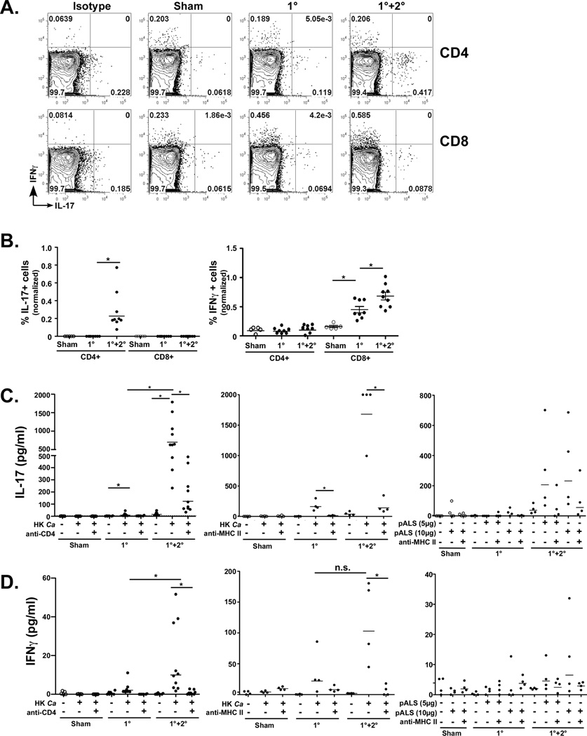
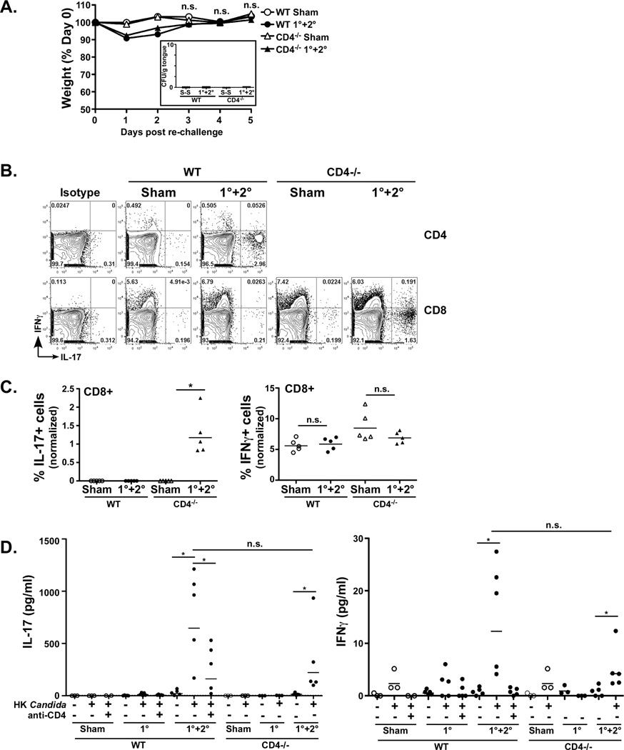
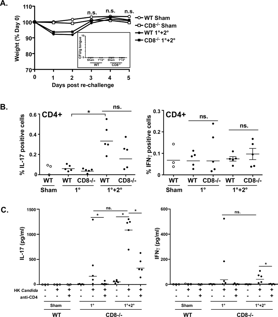
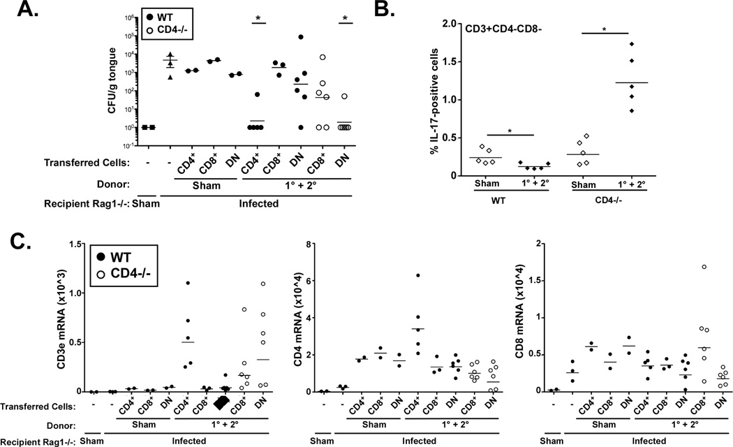
Oropharyngeal candidiasis (OPC) is an opportunistic infection caused by Candida albicans. Despite its prevalence, little is known about C. albicans-specific immunity in the oral mucosa. Vaccines against Candida generate both T helper type 1 (Th1) and Th17 responses, and considerable evidence implicates interleukin (IL)-17 in immunity to OPC. However, IL-17 is also produced by innate immune cells that are remarkably similar to Th17 cells, expressing the same markers and localizing to similar mucosal sites. To date, the relative contribution(s) of Th1, Th17, and innate IL-17-producing cells in OPC have not been clearly defined. Here, we sought to determine the nature and function of adaptive T-cell responses to OPC, using a new recall infection model. Mice subjected to infection and re-challenge with Candida mounted a robust and stable antigen-specific IL-17 response in CD4+ but not CD8+ T cells. There was little evidence for Th1 or Th1/Th17 responses. The Th17 response promoted accelerated fungal clearance, and Th17 cells could confer protection in Rag1-/- mice upon adoptive transfer. Surprisingly, CD4 deficiency did not cause OPC but was instead associated with compensatory IL-17 production by Tc17 and CD3+CD4-CD8- cells. Therefore, classic CD4+Th17 cells protect from OPC but can be compensated by other IL-17-producing cells in CD4-deficient hosts.


Conflict of interest statement

Conflicts of interest: SLG has received a research grant from Amgen, honoraria and travel reimbursements from Novartis, and consults for Lycera Corp.

Figures

Figure 1
Figure 1. Rag1−/− mice are susceptible to OPC
WT, IL-23−/− or Rag1−/− mice were subjected to sublingual infection with C. albicans, and fungal load in tongue was assessed by plating onto YPD and colony enumeration in triplicate. Sham mice were inoculated with PBS only. Cortisone treated mice were given 225 mg/kg cortisone acetate at days −1, +1 and 3 relative to infection. CFU/g tongue tissue is indicated with geometric mean (bar). Weight loss (average per cohort) during infection is shown as a percentage of starting weight at day 0. Data represent 2 independent experiments. *p<0.05 compared to WT mice infected with Candida. B. IL-23−/− mice remain persistently infected with C. albicans. The indicated mice were subjected to 1° infection. 21 days later, fungal load in tongue was assessed by plating on YPD and colony enumeration in triplicate. *p<0.05 compared to WT.
Figure 2
Figure 2. Adaptive Candida albicans-responsive Th17 cells accelerate fungal clearance
A. New re-challenge OPC infection model. Diagram of infection timeline. B. Re-challenge is associated with accelerated Candida clearance. WT mice were subjected to a 1° infection with Candida or PBS (Sham). After 6 weeks, mice were re-challenged with Candida (1° + 2° infection) or PBS. Mice were sacrificed at days 1, 2 and 3 post re-challenge, and fungal load in tongue was assessed by plating and colony enumeration in triplicate. Data are pooled from 2 replicate experiments. C. CD4+ Th17 but not Th1 cells are generated upon Candida re-challenge. cLN cells from the experiment described in B were isolated 1–3 days after re-challenge and stained for CD4, IL-17 and IFNγ. Lymphocytes (determined by forward and side scatter profiles) were gated on CD4+, and IL-17+ and IFNγ+ cell frequencies are depicted. Representative FACS plots are shown. D. Summary of data from panel C. Note that data are normalized to ICS staining from isotype controls. E–F. IL-17+ cells exhibit an activated phenotype. cLN cells were isolated 3 days after 1° or re-challenge and gated on CD3 and CD4. Surface expression of CD44 and CD27 was examined relative to intracellular IL-17. Data from mice subjected to recall are summarized in panel F. * p<0.05 G. CD4+ T cells are sufficient to confer immunity to OPC in Rag1−/− mice. WT mice were subjected to Sham or re-challenge (1°+2°). On day 3 after re-challenge, CD4+ and CD4- cells were harvested from cLN by magnetic sorting. 1 × 10⁷ CD4+ or 2×10⁷ CD4- cells were transferred into Rag1−/− recipients. After 24 h, Rag1−/− mice were infected with Candida, and weight loss monitored for 5 d (right). Fungal load in tongue was assessed at day 5 (left). * p<0.05 by t-test with Mann-Whitney correction or ANOVA; n.s., not significant. H. IL-17 but not IFNγ is expressed locally upon transfer with re-challenged CD4+ cells. Rag1−/− mice were adoptively transferred with (or without) CD4+ cells from WT mice given a Sham infection or a recall (1° + 2°) infection. Expression of il17a and ifng mRNA in tongue were assessed in by qPCR. Note that these samples are from a different experiment than shown in panel G. * p<0.05 by t-test with Mann-Whitney correction.
Figure 3
Figure 3. IL-17 is produced by CD4+ but not CD8+ T cells in cervical LN upon re-challenge with Candida
A–B. CD4 but not CD8 IL-17+ T cells are induced following re-challenge. WT mice were subjected to the re-challenge model in Fig 2A. On day 3 post re-challenge, cLN cells were gated on CD3+ lymphocytes and CD4+ or CD8+ populations, and IL-17+ and IFNγ+ cell frequencies were assessed by ICS. Representative plots are shown. B. Summary of data from panel A, indicating percentages of IL-17+ (left) or IFNγ+ (right) cells from CD4+ and CD8+ compartments. Data are normalized to isotype control staining and are representative of 2 experiments. C–D. IL-17 and IFNγ levels in cLN cells following Candida re-challenge. Cells from cLN were isolated 3 d post re-challenge from the indicated mice and were then cultured in vitro for 4–5 d ± heat-killed C. albicans (HK Ca) (left and middle panels) or the ALS1/3 peptide (right) in the presence or absence of anti-CD4 neutralizing Abs (left) or anti-MHC Class II Abs (middle and right) (32). IL-17A or IFNγ was assessed by ELISA. * p<0.05 by Mann-Whitney t-test.
Figure 4
Figure 4. CD4-deficient mice are resistant to re-challenge with Candida, associated with compensatory IL-17 from CD8+ T cells
A. CD4−/− mice are resistant to OPC. WT or CD4−/− mice were subjected to the OPC re-challenge model. Daily weight measurements post re-challenge are indicated as a percentage of starting weight. N.s., not significant by ANOVA. Inset: Fungal burden on day 5 after re-challenge is indicated. B–C. CD4−/− but not WT mice induce IL-17+CD8+ T cells upon re-challenge. WT or CD4−/− mice were subjected to the re-challenge model. On day 5 post re-challenge, cLN cells were gated on CD3+ lymphocytes and CD4+ or CD8+ populations, and IL-17+ and IFNγ+ cell frequencies are depicted in CD4+ (WT only) or CD8+ cells. Representative plots are shown. B. Summary of flow cytometry data from panel A, indicating percentages of IL-17+ (left) or IFNγ+ (right) cells from CD4+ and CD8+ compartments. Data are normalized to isotype controls and are representative of 2 independent experiments. D. CD4−/− and WT mice produce IL-17 upon re-challenge. Cells from cLN were isolated 3 d post re-challenge and cultured in vitro for 5 d ± heat-killed (HK) Candida albicans ± anti-CD4 neutralizing Abs. Concentrations of IL-17A or IFNγ were assessed by ELISA.
Figure 5
Figure 5. CD8−/− mice respond to Candida similarly to WT
A. CD8−/− mice are resistant to OPC. WT or CD8−/− mice were subjected to the OPC re-challenge model. Daily weight measurements are indicated as a percent of starting weight. N.s., not significant by ANOVA. Inset: Fungal burdens on day 5 post re-challenge are indicated. B. CD8−/− mice enhance Th17 cells but not Th1 cells upon re-challenge. On day 3 post re-challenge, cLN cells from WT or CD8−/− mice were gated on lymphocytes, CD3+ cells and CD4+ cells. The frequency of IL-17+ and IFNγ+ cells among the CD4+ population is shown. Data are normalized to isotype controls. *p<0.05 by t-test with Mann-Whitney correction. C. CD8−/− mice secrete IL-17 upon re-challenge with Candida in a CD4-dependent manner. Cells from cLN of indicated mice were isolated 3 d post re-challenge and cultured in vitro for 5 d ± HK C. albicans ± anti-CD4 neutralizing Abs. IL-17A or IFNγ were assessed by ELISA. Note that this is from the same experiment presented in Fig 4D. *p<0.05 by Mann-Whitney t-test.
Figure 6
Figure 6. CD8+ and DN cells protect from OPC in the context of CD4-deficiency
A. WT (filled circles) or CD4−/− mice (open circles) were subjected to sham infection or re-challenge (1°+2° infections). CD4+, CD8+ and DN cells were purified by magnetic sorting and transferred into Rag1−/− recipients, which were then subjected to OPC or Sham infections, as indicated. Fungal load after 4 days was assessed by plating. B. CD3+CD4-CD8- (DN) cells express IL-17 upon re-challenge in CD4−/− but not WT mice. Percent of DN cells expressing IL-17 by ICS is indicated. * p<0.05 by Mann Whitney t-test. C. Expression of T cell markers in tongue correlates with disease protection. Rag1−/− recipients were adoptively transferred with the indicated cell populations (CD4+, CD8+ or DN) from WT sham or infected mice. Tongue mRNA was evaluated for cd3e, cd4 or cd8 by qPCR (multiplied by 104). Each data point indicates a single mouse analyzed in triplicate.

Similar articles

Cited by

References

    1. Dongari-Bagtoglou A, Fidel P. The host cytokine responses and protective immunity in oropharyngeal candidiasis. J Dent Res. 2005;84:966–977. - PubMed
    1. Klein RS, Harris CA, Small CB, Moll B, Lesser M, Friedland GH. Oral candidiasis in high-risk patients as the initial manifestation of the acquired immunodeficiency syndrome. N Engl J Med. 1984 Aug 9;311(6):354–358. - PubMed
    1. Dong C. Diversification of T-helper-cell lineages: finding the family root of IL-17-producing cells. Nat Rev Immunol. 2006 Apr;6(4):329–333. - PubMed
    1. Steinman L. A brief history of T(H)17, the first major revision in the T(H)1/T(H)2 hypothesis of T cell-mediated tissue damage. Nature Med. 2007 Feb;13(2):139–145. - PubMed
    1. Farah C, Hu Y, Riminton S, Ashman R. Distinct roles for interleukin-12p40 and tumour necrosis factor in resistance to oral candidiasis defined by gene targeting. Oral Microbiol Immunol. 2006;21:252–255. - PubMed

Publication types

MeSH terms