

Elastin, a novel extracellular matrix protein adhering to mycobacterial antigen 85 complex
- PMID: 23250738
- PMCID: PMC3567642
- DOI: 10.1074/jbc.M112.415679
Elastin, a novel extracellular matrix protein adhering to mycobacterial antigen 85 complex
Abstract
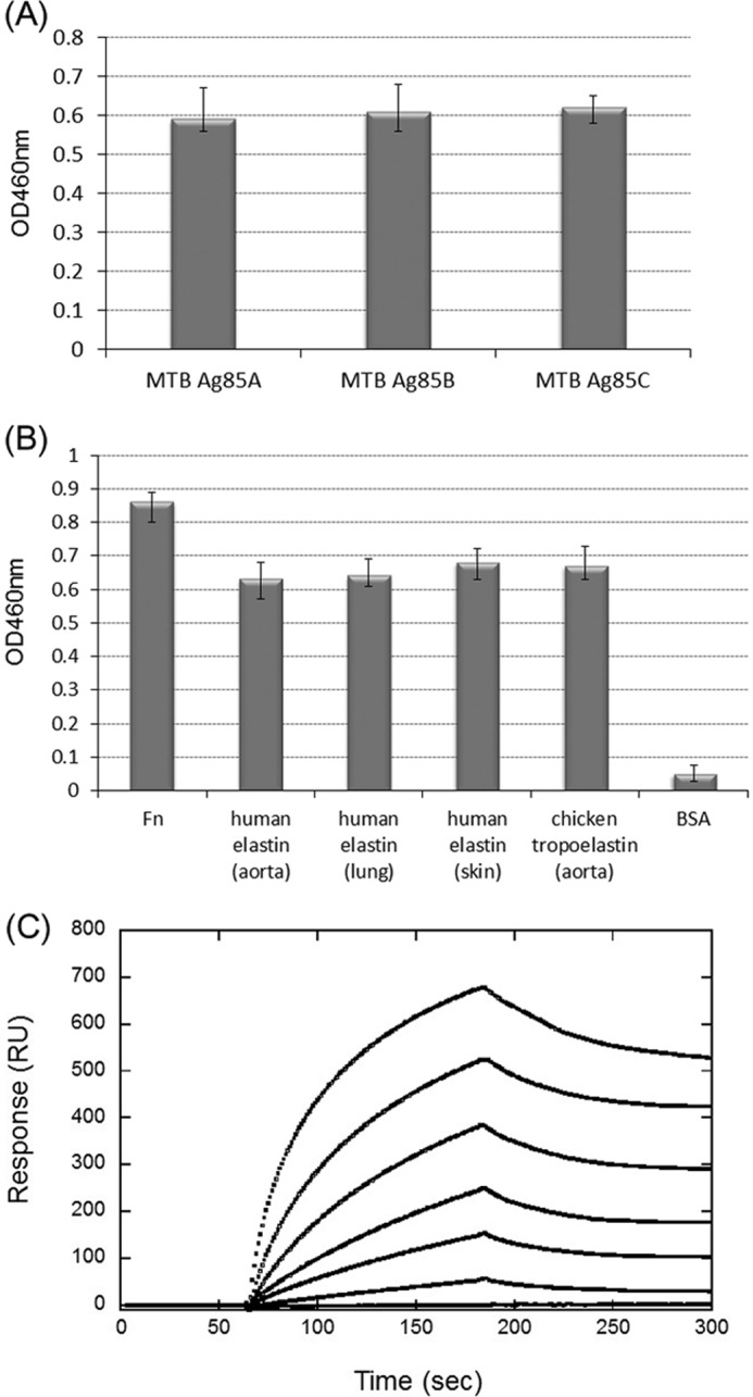
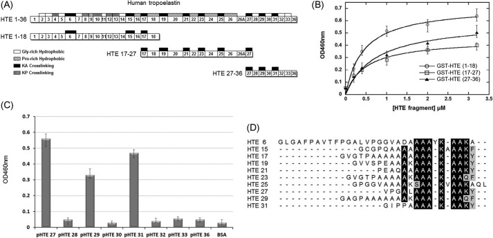
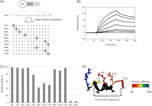
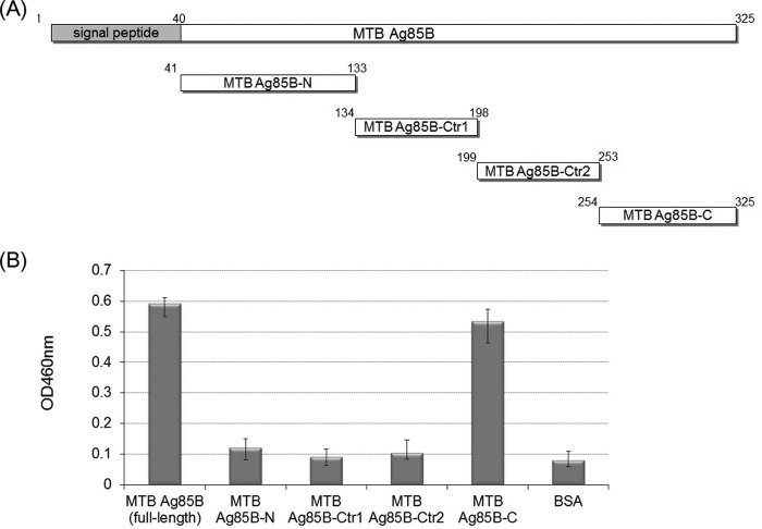
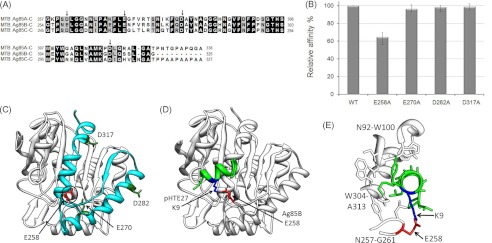
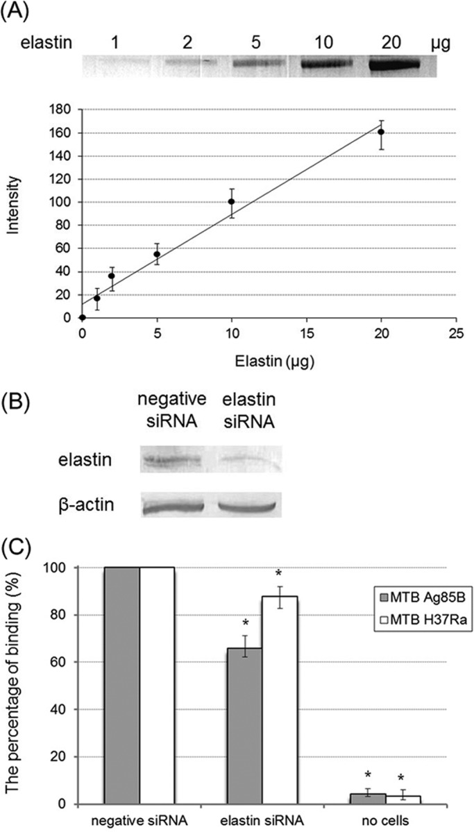
The antigen 85 complex (Ag85) consists of three predominantly secreted proteins (Ag85A, Ag85B, and Ag85C), which play a key role in the mycobacterial pathogenesis and also possess enzymatic mycolyltransferase activity involved in cell wall synthesis. Ag85 is not only considered to be a virulence factor because its expression is essential for intracellular survival within macrophages, but also because it contributes to adherence, invasion, and dissemination of mycobacteria in host cells. In this study, we report that the extracellular matrix components, elastin and its precursor (tropoelastin) derived from human aorta, lung, and skin, serve as binding partners of Ag85 from Mycobacterium tuberculosis. The binding affinity of M. tuberculosis Ag85 to human tropoelastin was characterized (K(D) = 0.13 ± 0.006 μm), and a novel Ag85-binding motif, AAAKAA(K/Q)(Y/F), on multiple tropoelastin modules was identified. In addition, the negatively charged Glu-258 of Ag85 was demonstrated to participate in an electrostatic interaction with human tropoelastin. Moreover, binding of Ag85 on elastin siRNA-transfected Caco-2 cells was significantly reduced (34.3%), implying that elastin acts as an important ligand contributing to mycobacterial invasion.
Figures
Similar articles
-
Novel mycobacteria antigen 85 complex binding motif on fibronectin.J Biol Chem. 2012 Jan 13;287(3):1892-902. doi: 10.1074/jbc.M111.298687. Epub 2011 Nov 29. J Biol Chem. 2012. PMID: 22128161 Free PMC article.
-
Cyclipostins and cyclophostin analogs inhibit the antigen 85C from Mycobacterium tuberculosis both in vitro and in vivo.J Biol Chem. 2018 Feb 23;293(8):2755-2769. doi: 10.1074/jbc.RA117.000760. Epub 2018 Jan 4. J Biol Chem. 2018. PMID: 29301937 Free PMC article.
-
The M. tuberculosis antigen 85 complex and mycolyltransferase activity.Lett Appl Microbiol. 2002;34(4):233-7. doi: 10.1046/j.1472-765x.2002.01091.x. Lett Appl Microbiol. 2002. PMID: 11940150
-
Novel insights into Mycobacterium antigen Ag85 biology and implications in countermeasures for M. tuberculosis.Crit Rev Eukaryot Gene Expr. 2012;22(3):179-87. doi: 10.1615/critreveukargeneexpr.v22.i3.10. Crit Rev Eukaryot Gene Expr. 2012. PMID: 23140159 Review.
-
Antigen 85 complex as a powerful Mycobacterium tuberculosis immunogene: Biology, immune-pathogenicity, applications in diagnosis, and vaccine design.Microb Pathog. 2017 Nov;112:20-29. doi: 10.1016/j.micpath.2017.08.040. Epub 2017 Sep 20. Microb Pathog. 2017. PMID: 28942172 Review.
Cited by
-
Leptospira Immunoglobulin-Like Protein B Interacts with the 20th Exon of Human Tropoelastin Contributing to Leptospiral Adhesion to Human Lung Cells.Front Cell Infect Microbiol. 2017 May 9;7:163. doi: 10.3389/fcimb.2017.00163. eCollection 2017. Front Cell Infect Microbiol. 2017. PMID: 28536676 Free PMC article.
-
Exploring the Potential Role of Moonlighting Function of the Surface-Associated Proteins From Mycobacterium bovis BCG Moreau and Pasteur by Comparative Proteomic.Front Immunol. 2019 Apr 26;10:716. doi: 10.3389/fimmu.2019.00716. eCollection 2019. Front Immunol. 2019. PMID: 31080447 Free PMC article.
-
Role of elastic fiber degradation in disease pathogenesis.Biochim Biophys Acta Mol Basis Dis. 2023 Jun;1869(5):166706. doi: 10.1016/j.bbadis.2023.166706. Epub 2023 Mar 29. Biochim Biophys Acta Mol Basis Dis. 2023. PMID: 37001705 Free PMC article. Review.
-
A possible complementary tool for diagnosing tuberculosis: a feasibility test of immunohistochemical markers.Int J Clin Exp Pathol. 2015 Nov 1;8(11):13900-10. eCollection 2015. Int J Clin Exp Pathol. 2015. PMID: 26823702 Free PMC article.
-
Evaluation of peptide designing strategy against subunit reassociation in mucin 1: A steered molecular dynamics approach.PLoS One. 2017 Aug 17;12(8):e0183041. doi: 10.1371/journal.pone.0183041. eCollection 2017. PLoS One. 2017. PMID: 28817726 Free PMC article.
References
-
- Raviglione M. C., Snider D. E., Jr., Kochi A. (1995) Global epidemiology of tuberculosis. Morbidity and mortality of a worldwide epidemic. JAMA 273, 220–226 - PubMed
-
- Vannberg F. O., Chapman S. J., Hill A. V. (2011) Human genetic susceptibility to intracellular pathogens. Immunol. Rev. 240, 105–116 - PubMed
-
- World Health Organization (WHO) (2012) Global Tuberculosis Report World Health Organization, Geneva, Switzerland
-
- Tiruviluamala P., Reichman L. B. (2002) Tuberculosis. Annu. Rev. Public Health 23, 403–426 - PubMed
-
- Baldwin S. L., D'Souza C. D., Orme I. M., Liu M. A., Huygen K., Denis O., Tang A., Zhu L., Montgomery D., Ulmer J. B. (1999) Immunogenicity and protective efficacy of DNA vaccines encoding secreted and non-secreted forms of Mycobacterium tuberculosis Ag85A. Tuber. Lung Dis. 79, 251–259 - PubMed
Publication types
MeSH terms
Substances
LinkOut - more resources
Full Text Sources
Other Literature Sources
