. 2013 Feb 1;288(5):2923-32.
doi: 10.1074/jbc.M112.419978. Epub 2012 Dec 18.

Palmitate and lipopolysaccharide trigger synergistic ceramide production in primary macrophages

Affiliations

Palmitate and lipopolysaccharide trigger synergistic ceramide production in primary macrophages

Joel D Schilling et al. J Biol Chem. .

Abstract

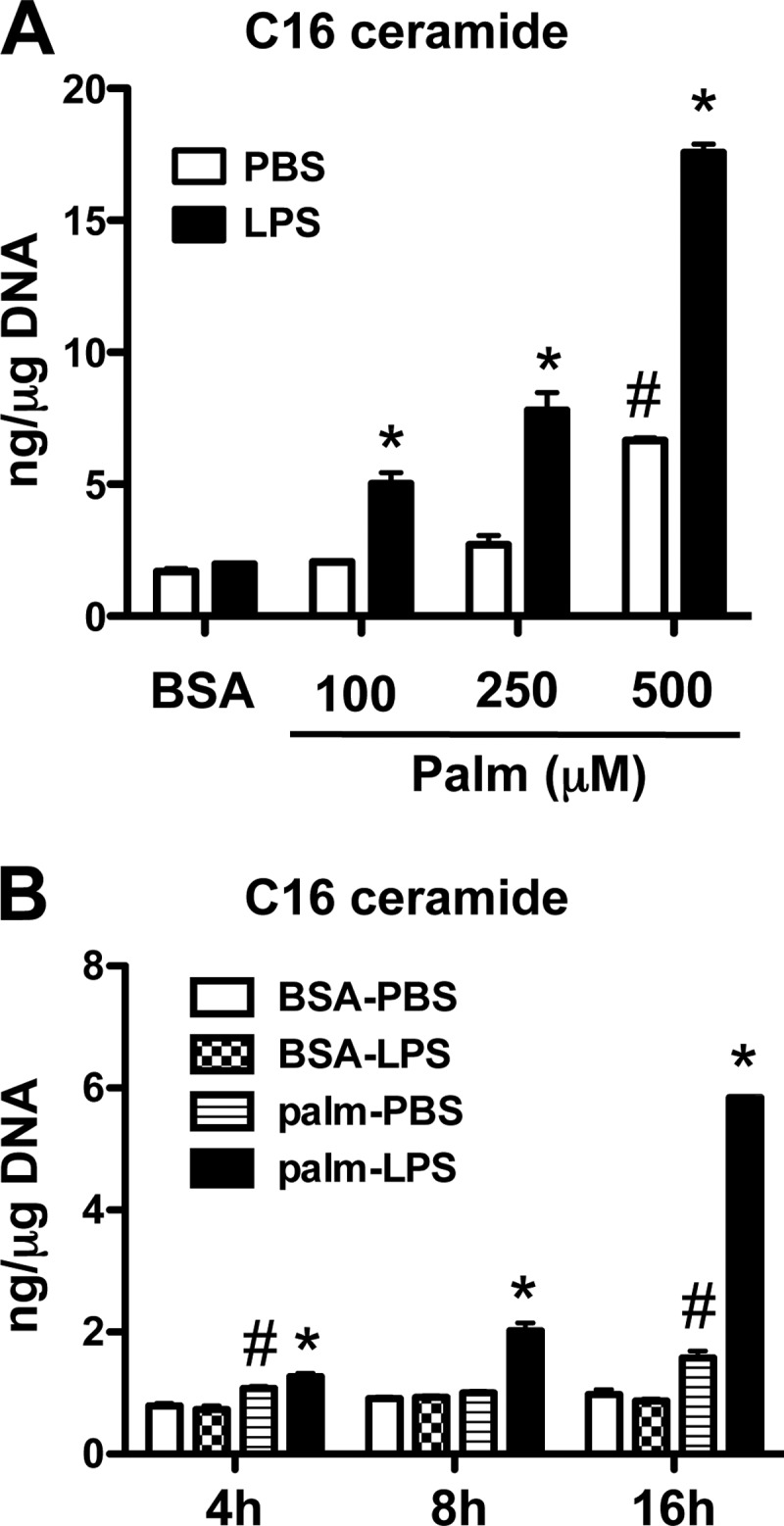
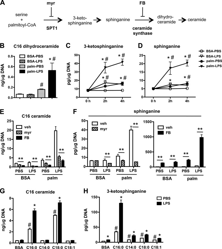
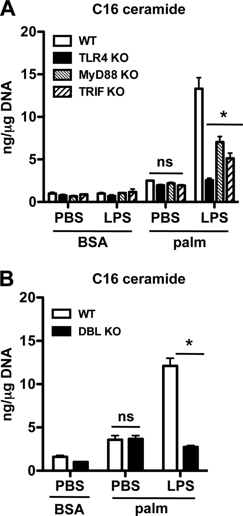
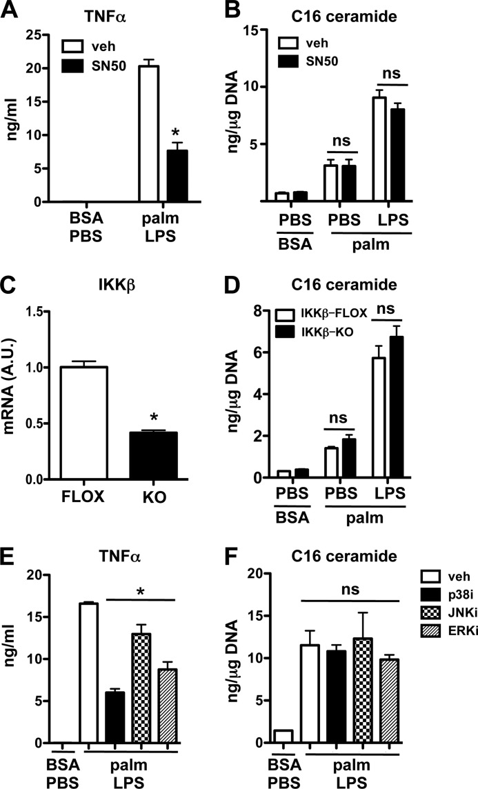
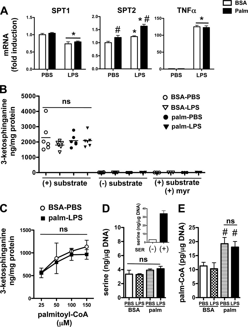
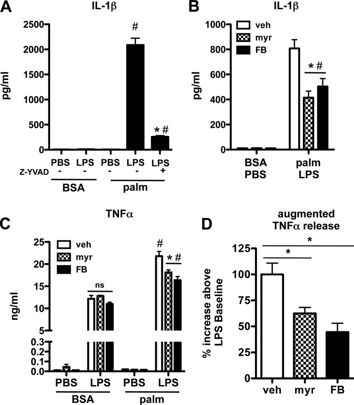
Macrophages play a key role in host defense and in tissue repair after injury. Emerging evidence suggests that macrophage dysfunction in states of lipid excess can contribute to the development of insulin resistance and may underlie inflammatory complications of diabetes. Ceramides are sphingolipids that modulate a variety of cellular responses including cell death, autophagy, insulin signaling, and inflammation. In this study we investigated the intersection between TLR4-mediated inflammatory signaling and saturated fatty acids with regard to ceramide generation. Primary macrophages treated with lipopolysaccharide (LPS) did not produce C16 ceramide, whereas palmitate exposure led to a modest increase in this sphingolipid. Strikingly, the combination of LPS and palmitate led to a synergistic increase in C16 ceramide. This response occurred via cross-talk at the level of de novo ceramide synthesis in the ER. The synergistic response required TLR4 signaling via MyD88 and TIR-domain-containing adaptor-inducing interferon beta (TRIF), whereas palmitate-induced ceramide production occurred independent of these inflammatory molecules. This ceramide response augmented IL-1β and TNFα release, a process that may contribute to the enhanced inflammatory response in metabolic diseases characterized by dyslipidemia.


Figures

FIGURE 1.
FIGURE 1.
LPS synergizes with palmitate to increase ceramide in macrophages. A, peritoneal macrophages (pMACs) were incubated for 16 h with the indicated concentrations of palmitate (Palm) (or with BSA as control) alone or in combination with 50 ng/ml of LPS (or with PBS as control). C16 ceramide concentrations were determined by LC-MS/MS and normalized per μg DNA. B, pMACs were stimulated with 500 μm palm ± LPS for the indicated time points, and C16 ceramide was quantified by LC-MS/MS. Bar graphs report the means ± S.E. (S.E.) for a minimum of three experiments, each performed in triplicate. *, p < 0.05 for LPS versus PBS; #, p < 0.05 for palm versus BSA. n.s., not significant.
FIGURE 2.
FIGURE 2.
Palmitate and LPS synergize to induce de novo ceramide biosynthesis. A, schematic shows the de novo ceramide synthesis pathway and the site of action of chemical inhibitors myriocin (myr) and fumonisin B1 (FB). B, C16:0 dihydroceramide levels were determined by LC-MS/MS in pMACs stimulated with 500 μm palm ± LPS for 16 h and normalized per μg of DNA. C and D, the de novo ceramide synthesis intermediates 3-ketosphinganine (C) and sphinganine (D) were quantified using LC-MS/MS at 2 and 4 h after treatment of pMACs with 500 μm palm ± LPS. E and F, pMACs were pretreated with vehicle (veh; white bars), myr (1 μm, hatched bars), or FB (5 μm, black bars) for 30 min before co-treatment with the indicated stimuli. C16 ceramide (E) and sphinganine (F) were determined at 16 h by LC-MS/MS. G and H, macrophages were treated with the indicated fatty acids ± LPS, and C16:0 ceramide (G) or 3-ketosphinganine (H) levels were determined at 16 h. Bar graphs report the mean ± S.E. for a minimum of 3 experiments, each performed in triplicate. *, p < 0.05 for LPS versus PBS; #, p < 0.05 for palm or other FA versus BSA; **, p < 0.05 for inhibitor versus vehicle.
FIGURE 3.
FIGURE 3.
Synergistic ceramide production requires signaling via TLR4, MyD88, and TRIF. A, macrophages from WT, TLR4 KO, MyD88 KO, or TRIF KO mice were treated as indicated, and C16 ceramide was quantified at 16 h. B, pMACs from WT or MyD88/TRIF double KO mice (DBL KO) were treated with palm ± LPS, and C16 ceramide levels were quantified at 16 h. Bar graphs report the mean ± S.E. for a minimum of three experiments, each performed in triplicate. *, p < 0.05 for KO versus WT; ns, not significant.
FIGURE 4.
FIGURE 4.
The induction of ceramide in response to palmitate and LPS occurs through a NF-κB and MAP kinase-independent mechanism. A and B, macrophages were pretreated with the NF-κB translocation inhibitor SN50 (25 μm) and then with palmitate ± LPS and SN50 for 16 h. TNFα secretion (A) and C16 ceramide levels (B) were quantified by ELISA and LC-MS/MS, respectively. C and D, mRNA expression of IKKβ relative to 36B4 was quantified by quantitative real-time-PCR (C), and ceramide levels were quantified after 16 h of treatment as indicated (D) in macrophages from IKKβ flox mice (FLOX) and from IKKβ flox mice crossed with LysM-Cre mice (KO). E and F, MAP kinase inhibitors for p38 (p38i SB203580, 20 μm), JNK (JNKi SP600125, 20 μm), and ERK (ERKi PD98059,10 μm) were preincubated with pMACs before stimulation with palm and LPS. TNFα secretion (E) and C16 ceramide levels (F) were determined 16 h after treatment. Bar graphs report the mean ± S.E. for a minimum of three experiments, each performed in triplicate. *p < 0.05 for KO/TG versus WT and for inhibitor versus vehicle. ns, not significant.
FIGURE 5.
FIGURE 5.
SPT expression and activity in cell extracts is unchanged after LPS and palmitate treatment of macrophages. A, pMACs were treated PBS or LPS (50 ng/ml) in the presence of BSA (white bars) or 500 μm palm (black bars), and mRNA was harvested 2 h after stimulation. Gene expression levels for the 2 subunits of SPT (SPT1, SPT2) and TNFα were determined by quantitative real-time-PCR and normalized to 36B4 expression. B, cell lysates were prepared from pMACs treated for 2 h with the indicated stimuli and assayed in vitro for SPT activity by quantification of 3-ketosphinganine production using LC-MS/MS. Control reactions were performed without the addition of substrate or in the presence of myr. ns, not significant. C, palmitoyl CoA dose-response curves for SPT activity are shown for lysates prepared from cells treated with BSA-PBS or palm-LPS. D and E, pMACs were treated with the indicated stimuli for 2 h after which intracellular serine (D) or palmitoyl-CoA (E) concentrations were determined by LC-MS/MS. As a control for serine detection, intracellular serine concentrations were determined in pMACs after incubation with media containing (+) or not containing (−) 10 mm serine (SER) for 2 h (D, inset). All experiments were performed in triplicate a minimum of three times. *, p < 0.05 for LPS versus PBS; #, p < 0.05 for palm versus BSA.
FIGURE 6.
FIGURE 6.
Augmented de novo ceramide synthesis in macrophages exposed to palmitate and LPS leads to excessive IL-1β and TNFα secretion. A, pMACS were treated with BSA or palm (250 μm) ± LPS with or without 20 μm Z-YVAD or vehicle control, and IL-1β levels in the supernatant were determined at 24 h. B, macrophages were stimulated with LPS and palm in the presence of vehicle (white bars), myr (hatched bars), or FB (black bars), and IL-1β secretion was quantified by ELISA 24 h after treatment. C, TNFα secretion by pMACs was determined by ELISA 24 h after the indicated treatments in the presence of myr or FB. ns, not significant. D, augmented TNFα secretion, defined as the difference in TNFα release from palm-LPS versus LPS treated macrophages, was quantified in macrophages treated with veh, myr, or FB (vehicle-treated samples = 100%). All experiments were performed three times in triplicate. *, p < 0.05 for inhibitor versus veh; #, p < 0.05 for palm versus BSA. ns, not significant.

References

    1. Schwartz E. A., Zhang W. Y., Karnik S. K., Borwege S., Anand V. R., Laine P. S., Su Y., Reaven P. D. (2010) Nutrient modification of the innate immune response. A novel mechanism by which saturated fatty acids greatly amplify monocyte inflammation. Arterioscler. Thromb. Vasc. Biol. 30, 802–808 - PubMed
    1. Cascio G., Schiera G., Di Liegro I. (2012) Dietary fatty acids in metabolic syndrome, diabetes, and cardiovascular diseases. Curr. Diabetes Rev. 8, 2–17 - PubMed
    1. Shi H., Kokoeva M. V., Inouye K., Tzameli I., Yin H., Flier J. S. (2006) TLR4 links innate immunity and fatty acid-induced insulin resistance. J. Clin. Invest. 116, 3015–3025 - PMC - PubMed
    1. Assimacopoulos-Jeannet F. (2004) Fat storage in pancreas and in insulin-sensitive tissues in pathogenesis of type 2 diabetes. Int. J. Obes. Relat. Metab. Disord. 28, S53–S57 - PubMed
    1. Joshi N., Caputo G. M., Weitekamp M. R., Karchmer A. W. (1999) Infections in patients with diabetes mellitus. N. Engl. J. Med. 341, 1906–1912 - PubMed

Publication types

MeSH terms

LinkOut - more resources