. 2012 Dec;135(Pt 12):3584-98.
doi: 10.1093/brain/aws292.

Oxidative stress modulates mitochondrial failure and cyclophilin D function in X-linked adrenoleukodystrophy

Affiliations

Oxidative stress modulates mitochondrial failure and cyclophilin D function in X-linked adrenoleukodystrophy

Jone López-Erauskin et al. Brain. 2012 Dec.

Abstract

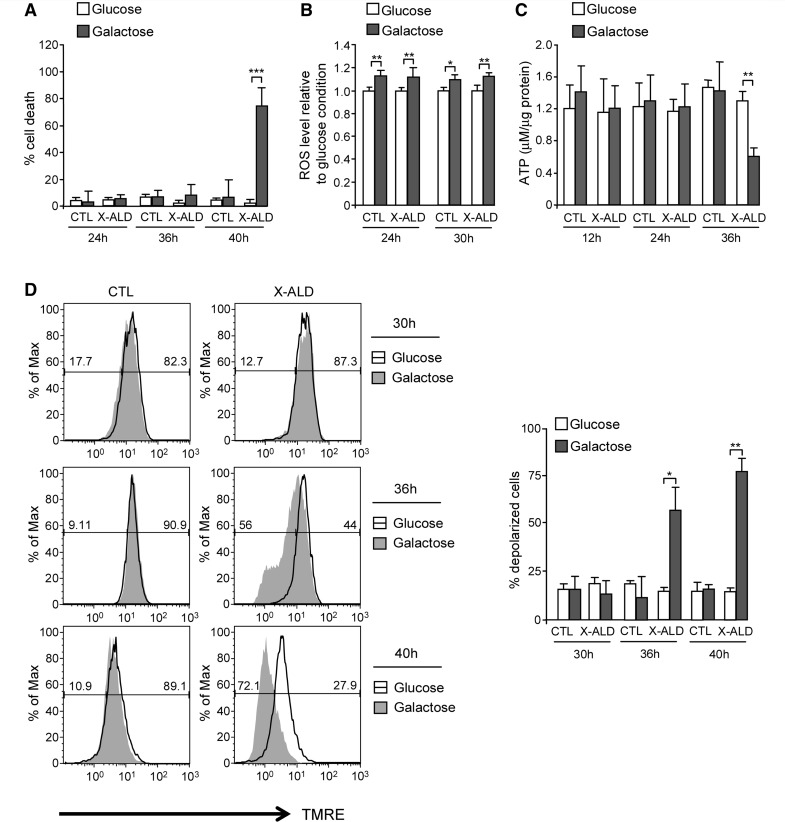
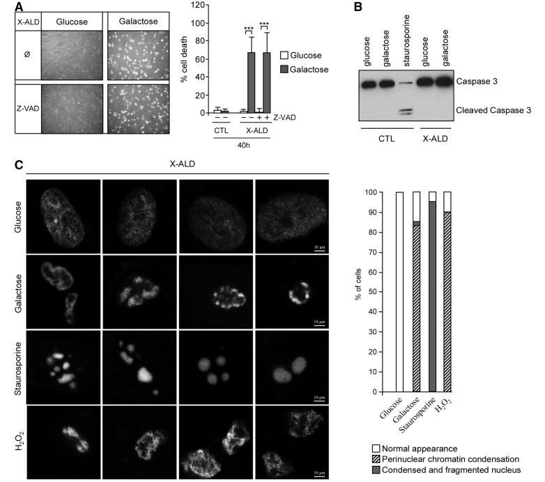
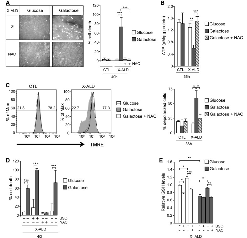
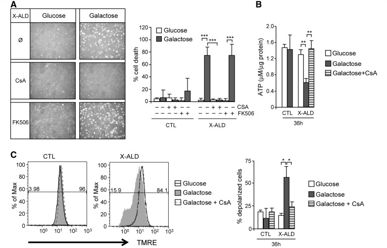
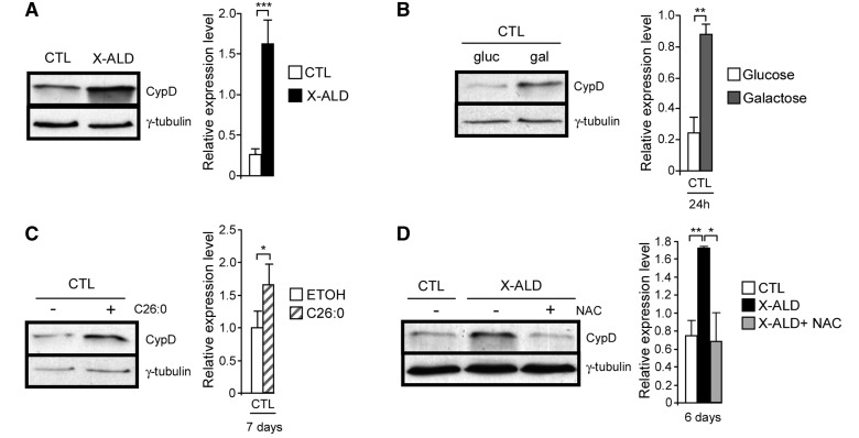
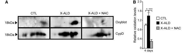
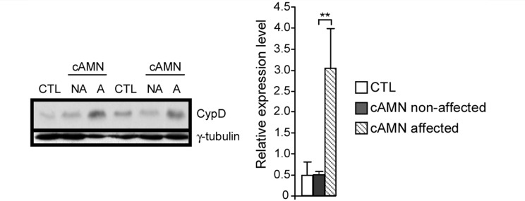
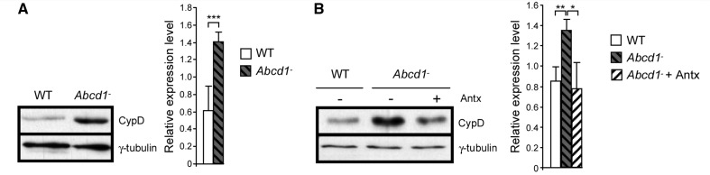
A common process associated with oxidative stress and severe mitochondrial impairment is the opening of the mitochondrial permeability transition pore, as described in many neurodegenerative diseases. Thus, inhibition of mitochondrial permeability transition pore opening represents a potential target for inhibiting mitochondrial-driven cell death. Among the mitochondrial permeability transition pore components, cyclophilin D is the most studied and has been found increased under pathological conditions. Here, we have used in vitro and in vivo models of X-linked adrenoleukodystrophy to investigate the relationship between the mitochondrial permeability transition pore opening and redox homeostasis. X-linked adrenoleukodystrophy is a neurodegenerative condition caused by loss of function of the peroxisomal ABCD1 transporter, in which oxidative stress plays a pivotal role. In this study, we provide evidence of impaired mitochondrial metabolism in a peroxisomal disease, as fibroblasts in patients with X-linked adrenoleukodystrophy cannot survive when forced to rely on mitochondrial energy production, i.e. on incubation in galactose. Oxidative stress induced under galactose conditions leads to mitochondrial damage in the form of mitochondrial inner membrane potential dissipation, ATP drop and necrotic cell death, together with increased levels of oxidative modifications in cyclophilin D protein. Moreover, we show increased expression levels of cyclophilin D in the affected zones of brains in patients with adrenomyeloneuropathy, in spinal cord of a mouse model of X-linked adrenoleukodystrophy (Abcd1-null mice) and in fibroblasts from patients with X-linked adrenoleukodystrophy. Notably, treatment with antioxidants rescues mitochondrial damage markers in fibroblasts from patients with X-linked adrenoleukodystrophy, including cyclophilin D oxidative modifications, and reverses cyclophilin D induction in vitro and in vivo. These findings provide mechanistic insight into the beneficial effects of antioxidants in neurodegenerative and non-neurodegenerative cyclophilin D-dependent disorders.


Figures

Figure 1
Figure 1
Mitochondrial dysfunction in X-linked adrenoleukodystrophy fibroblasts. Cell death (A), intracellular radical oxygen species levels (B), ATP content (C) and inner mitochondrial membrane potential (D) were quantified in control and X-linked adrenoleukodystrophy fibroblasts cultured in presence of 1 g/l glucose or glucose-free medium containing 1 g/l galactose (*P < 0.05; **P < 0.01; ***P < 0.001; n = 4/genotype and condition). In D, the numeric values above the line correspond to the percentage of polarized cells on the right-hand and depolarized cells on the left-hand of each panel. CTL = control; ROS = radical oxygen species; X-ALD = X-linked adrenoleukodystrophy.
Figure 2
Figure 2
Galactose induces necrotic cell death in X-linked adrenoleukodystrophy fibroblasts. Control and X-linked adrenoleukodystrophy fibroblasts cultured in glucose or galactose were treated with general caspase inhibitor Z-VAD, and cell death was measured (A). Cleaved caspase 3 was analysed in different conditions by western blot (B). Staurosporine (1 microM during 14 h) was used as a positive control of caspase 3 activator (***P < 0.001; n = 4/genotype and condition). The pattern of chromatin condensation induced by galactose was analysed using Hoescht 33342 staining (C). H2O2 (10 mM for 3 h) and staurosporine (2 µM for 6 h) were used as positive control for necrotic and apoptotic cell death, respectively. X-linked adrenoleukodystrophy fibroblasts of six different patients were analysed, and 25 cells per condition were scored. Representative pictures and quantification are shown. CTL = control; X-ALD = X-linked adrenoleukodystrophy.
Figure 3
Figure 3
X-linked adrenoleukodystrophy fibroblasts show necrotic features during galactose-induced cell death. X-linked adrenoleukodystrophy fibroblasts were cultured in glucose (A) (shown at larger magnification in C); or in glucose-free medium containing galactose (B) (shown at larger magnification in D). After 40 h, cells were collected and analysed by electron microscopy. V = vacuole; arrows indicate lipid inclusions; arrowheads indicate morphologically normal appearing mitochondria; asterisks indicate swollen mitochondria (n = 4/condition).
Figure 4
Figure 4
X-linked adrenoleukodystrophy fibroblast cell death and metabolic failure in galactose is oxidative stress dependent. Control and X-linked adrenoleukodystrophy fibroblasts cultured in glucose or galactose were treated with 1 mM of N-acetylcysteine. Cell death (A), ATP content (B) and inner mitochondrial membrane potential (C) were measured. In C, the numeric values above the line correspond to the percentage of polarized cells on the right-hand and depolarized cells on the left-hand of each panel. Fibroblasts cell death was quantified under the same conditions with L-buthionine-sulfoximine (500 µM) treatment (D). Reduced glutathione amount was quantified fluorimetrically using monochlorobimane in X-linked adrenoleukodystrophy fibroblasts cultured in glucose or in galactose under N-acetylcysteine and/or L-buthionine-sulfoximine treatments (E) (*P < 0.05; **P < 0.01; ***P < 0.001; n = 4/condition). BSO = L-buthionine-sulfoximine; CTL = control; GSH = reduced glutathione; NAC = N-acetylcysteine; X-ALD = X-linked adrenoleukodystrophy.
Figure 5
Figure 5
Galactose-induced mitochondrial dysfunction and cell death in X-linked adrenoleukodystrophy fibroblasts is caused by mitochondrial permeability transition pore opening. Control and X-linked adrenoleukodystrophy fibroblasts cultured in glucose or galactose were treated with 5 µM of cyclosporin A or 5 µM of FK506, and cell death was measured (A). In the same conditions with cyclosporin A treatment, ATP content (B) and inner mitochondrial membrane potential (C) were quantified (*P < 0.05; **P < 0.01; ***P < 0.001; n = 4/genotype and condition). In C, the numeric values above the line correspond to the percentage of polarized cells on the right-hand and depolarized cells on the left-hand of each panel. CsA = cyclosporin A; CTL = control; X-ALD = X-linked adrenoleukodystrophy.
Figure 6
Figure 6
Cyclophilin D expression is modulated by oxidative stress. Cyclophilin D levels were quantified in control and X-linked adrenoleukodystrophy fibroblasts at basal levels (A), in control fibroblasts after culture in galactose for 24 h (B) and with C26:0 (50 µM) for 7 days (C), and after treating X-linked adrenoleukodystrophy fibroblasts with N-acetylcysteine (0.5 mM) for 6 days (D) (*P < 0.05; **P < 0.01; ***P < 0.001; n = 5/genotype and condition). CTL = control; CypD = cyclophilin D; ETOH = ethanol; NAC = N-acetylcysteine; X-ALD = X-linked adrenoleukodystrophy.
Figure 7
Figure 7
Antioxidant treatment halts cyclophilin D oxidation in X-linked adrenoleukodystrophy fibroblasts. Oxyblot and western blot with specific antibody against cyclophilin D were performed to compare control and X-linked adrenoleukodystrophy fibroblasts treated or not with 1 mM of N-acetylcysteine. Three independent experiments were performed for each condition. Representative blots are shown (A). Relative protein oxidation levels were quantified and expressed as a percentage of control and referred to cyclophilin D expression (B) (*P < 0.05; ***P < 0.001; n = 3/genotype and condition). CTL = control; CypD = cyclophilin D; NAC = N-acetylcysteine; X-ALD = X-linked adrenoleukodystrophy.
Figure 8
Figure 8
Cyclophilin D is accumulated in affected white matter brain zones of cerebral adrenomyeloneuropathy patients. Cyclophilin D levels were analysed in control and in different areas of brains in patients with cerebral adrenomyeloneuropathy (**P < 0.01; n = 4/genotype and condition). A = affected; cAMN = cerebral adrenomyeloneuropathy; CTL = control; CypD = cyclophilin D; NA = non-affected.
Figure 9
Figure 9
Antioxidant treatment prevented cyclophilin D increase in Abcd1 mice spinal cord. Cyclophilin D expression levels were quantified in wild-type and Abcd1 mice spinal cord at 8 months (A) and at 12 months after an antioxidant cocktail treatment (B) (*P < 0.05; **P < 0.01; ** P < 0.001; n = 5/genotype and condition). Antx = antioxidants; CypD = cyclophilin D; WT = wild-type.

Similar articles

Cited by

References

    1. Acin-Perez R, Salazar E, Kamenetsky M, Buck J, Levin LR, Manfredi G. Cyclic AMP produced inside mitochondria regulates oxidative phosphorylation. Cell Metab. 2009;9:265–76. - PMC - PubMed
    1. Baarine M, Andreoletti P, Athias A, Nury T, Zarrouk A, Ragot K, et al. Evidence of oxidative stress in very long chain fatty acid - Treated oligodendrocytes and potentialization of ROS production using RNA interference-directed knockdown of ABCD1 and ACOX1 peroxisomal proteins. Neuroscience. 2012a;213:1–18. - PubMed
    1. Baarine M, Ragot K, Athias A, Nury T, Kattan Z, Genin EC, et al. Incidence of Abcd1 level on the induction of cell death and organelle dysfunctions triggered by very long chain fatty acids and TNF-alpha on oligodendrocytes and astrocytes. Neurotoxicology. 2012b;33:212–28. - PubMed
    1. Baines CP, Kaiser RA, Purcell NH, Blair NS, Osinska H, Hambleton MA, et al. Loss of cyclophilin D reveals a critical role for mitochondrial permeability transition in cell death. Nature. 2005;434:658–62. - PubMed
    1. Barrientos SA, Martinez NW, Yoo S, Jara JS, Zamorano S, Hetz C, et al. Axonal degeneration is mediated by the mitochondrial permeability transition pore. J Neurosci. 2011;31:966–78. - PMC - PubMed

Publication types

MeSH terms