

Abnormal thalamocortical structural and functional connectivity in juvenile myoclonic epilepsy
- PMID: 23250883
- PMCID: PMC3525058
- DOI: 10.1093/brain/aws296
Abnormal thalamocortical structural and functional connectivity in juvenile myoclonic epilepsy
Abstract
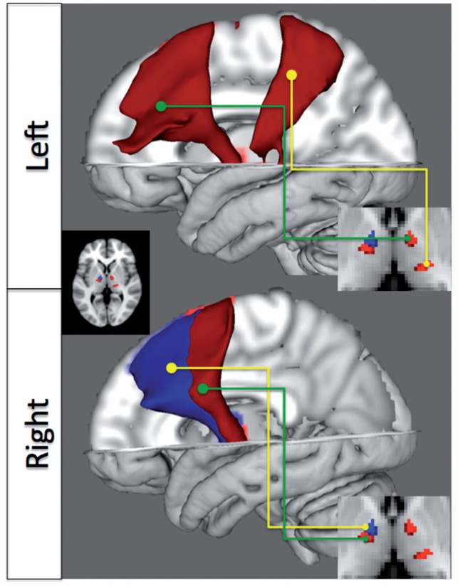
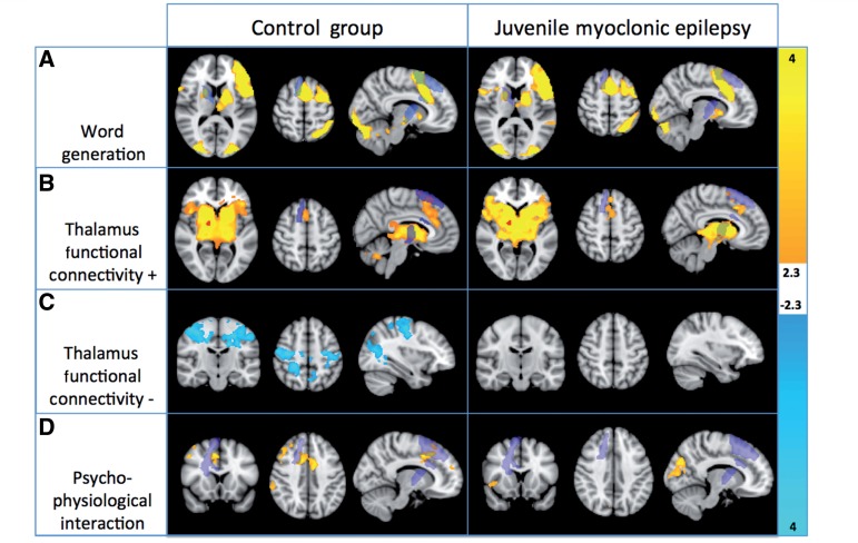
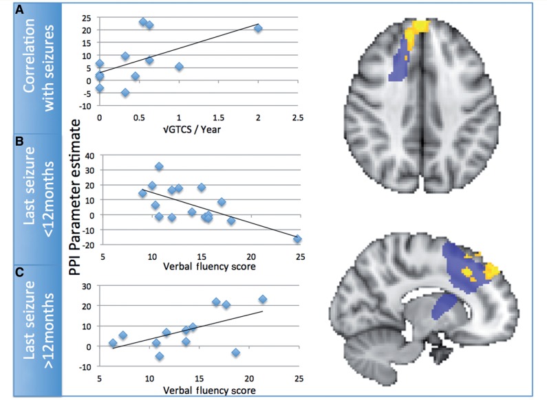
Juvenile myoclonic epilepsy is the most common idiopathic generalized epilepsy, characterized by frequent myoclonic jerks, generalized tonic-clonic seizures and, less commonly, absences. Neuropsychological and, less consistently, anatomical studies have indicated frontal lobe dysfunction in the disease. Given its presumed thalamo-cortical basis, we investigated thalamo-cortical structural connectivity, as measured by diffusion tensor imaging, in a cohort of 28 participants with juvenile myoclonic epilepsy and detected changes in an anterior thalamo-cortical bundle compared with healthy control subjects. We then investigated task-modulated functional connectivity from the anterior thalamic region identified using functional magnetic resonance imaging in a task consistently shown to be impaired in this group, phonemic verbal fluency. We demonstrate an alteration in task-modulated connectivity in a region of frontal cortex directly connected to the thalamus via the same anatomical bundle, and overlapping with the supplementary motor area. Further, we show that the degree of abnormal connectivity is related to disease severity in those with active seizures. By integrating methods examining structural and effective interregional connectivity, these results provide convincing evidence for abnormalities in a specific thalamo-cortical circuit, with reduced structural and task-induced functional connectivity, which may underlie the functional abnormalities in this idiopathic epilepsy.
Figures
References
-
- Aghakhani Y, Bagshaw AP, Benar CG, Hawco C, Andermann F, Dubeau F, et al. fMRI activation during spike and wave discharges in idiopathic generalized epilepsy. Brain. 2004;127:1127–44. - PubMed
-
- Alexander GE, Crutcher MD. Functional architecture of basal ganglia circuits: neural substrates of parallel processing. Trends Neurosci. 1990;13:266–71. - PubMed
-
- Andersson JLR, Jenkinson M, Smith SM. Nonlinear registration, aka Spatial normalisation. FMRIB Technical Report TR07JA2, FMRIB Analysis Group of the University of Oxford, 2007. www.fmrib.ox.ac.uk/analysis/techrep.
-
- Ashburner J, Friston KJ. Unified segmentation. NeuroImage. 2005;26:839–51. - PubMed
Publication types
MeSH terms
Grants and funding
LinkOut - more resources
Full Text Sources
Medical
