

Medial perirhinal cortex disambiguates confusable objects
- PMID: 23250887
- PMCID: PMC3525054
- DOI: 10.1093/brain/aws277
Medial perirhinal cortex disambiguates confusable objects
Abstract
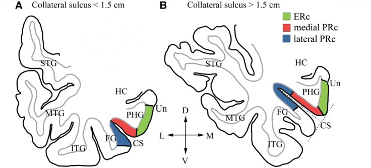
Our brain disambiguates the objects in our cluttered visual world seemingly effortlessly, enabling us to understand their significance and to act appropriately. The role of anteromedial temporal structures in this process, particularly the perirhinal cortex, is highly controversial. In some accounts, the perirhinal cortex is necessary for differentiating between perceptually and semantically confusable objects. Other models claim that the perirhinal cortex neither disambiguates perceptually confusable objects nor plays a unique role in semantic processing. One major hurdle to resolving this central debate is the fact that brain damage in human patients typically encompasses large portions of the anteromedial temporal lobe, such that the identification of individual substructures and precise neuroanatomical locus of the functional impairments has been difficult. We tested these competing accounts in patients with Alzheimer's disease with varying degrees of atrophy in anteromedial structures, including the perirhinal cortex. To assess the functional contribution of each anteromedial temporal region separately, we used a detailed region of interest approach. From each participant, we obtained magnetic resonance imaging scans and behavioural data from a picture naming task that contrasted naming performance with living and non-living things as a way of manipulating perceptual and semantic confusability; living things are more similar to one another than non-living things, which have more distinctive features. We manually traced neuroanatomical regions of interest on native-space cortical surface reconstructions to obtain mean thickness estimates for the lateral and medial perirhinal cortex and entorhinal cortex. Mean cortical thickness in each region of interest, and hippocampal volume, were submitted to regression analyses predicting naming performance. Importantly, atrophy of the medial perirhinal cortex, but not lateral perirhinal cortex, entorhinal cortex or hippocampus, significantly predicted naming performance on living relative to non-living things. These findings indicate that one specific anteromedial temporal lobe region-the medial perirhinal cortex-is necessary for the disambiguation of perceptually and semantically confusable objects. Taken together, these results support a hierarchical account of object processing, whereby the perirhinal cortex at the apex of the ventral object processing system is required to bind properties of not just perceptually, but also semantically confusable objects together, enabling their disambiguation from other similar objects and thus comprehension. Significantly, this model combining a hierarchical object processing architecture with a semantic feature statistic account explains why category-specific semantic impairments for living things are associated with anteromedial temporal lobe damage, and pinpoints the root of this syndrome to perirhinal cortex damage.
Figures

Similar articles
-
False positives to confusable objects predict medial temporal lobe atrophy.Hippocampus. 2013 Sep;23(9):832-41. doi: 10.1002/hipo.22137. Epub 2013 May 30. Hippocampus. 2013. PMID: 23609914
-
Objects and categories: feature statistics and object processing in the ventral stream.J Cogn Neurosci. 2013 Oct;25(10):1723-35. doi: 10.1162/jocn_a_00419. Epub 2013 May 10. J Cogn Neurosci. 2013. PMID: 23662861 Free PMC article.
-
Object-specific semantic coding in human perirhinal cortex.J Neurosci. 2014 Apr 2;34(14):4766-75. doi: 10.1523/JNEUROSCI.2828-13.2014. J Neurosci. 2014. PMID: 24695697 Free PMC article.
-
Structural magnetic resonance imaging for the early diagnosis of dementia due to Alzheimer's disease in people with mild cognitive impairment.Cochrane Database Syst Rev. 2020 Mar 2;3(3):CD009628. doi: 10.1002/14651858.CD009628.pub2. Cochrane Database Syst Rev. 2020. PMID: 32119112 Free PMC article.
-
The anatomy of object processing: the role of anteromedial temporal cortex.Q J Exp Psychol B. 2005 Jul-Oct;58(3-4):361-77. doi: 10.1080/02724990544000013. Q J Exp Psychol B. 2005. PMID: 16194974 Review.
Cited by
-
Assessing mild cognitive impairment using object-location memory in immersive virtual environments.Hippocampus. 2022 Sep;32(9):660-678. doi: 10.1002/hipo.23458. Epub 2022 Aug 2. Hippocampus. 2022. PMID: 35916343 Free PMC article.
-
Cats and Apples: Semantic Fluency Performance for Living Things Identifies Patients with Very Early Alzheimer's Disease.Arch Clin Neuropsychol. 2021 Jul 19;36(5):838-843. doi: 10.1093/arclin/acaa109. Arch Clin Neuropsychol. 2021. PMID: 33237317 Free PMC article.
-
Semantic feature norms: a cross-method and cross-language comparison.Behav Res Methods. 2024 Sep;56(6):5788-5797. doi: 10.3758/s13428-023-02311-1. Epub 2023 Dec 20. Behav Res Methods. 2024. PMID: 38123826 Free PMC article.
-
Animacy and real-world size shape object representations in the human medial temporal lobes.Hum Brain Mapp. 2018 Sep;39(9):3779-3792. doi: 10.1002/hbm.24212. Epub 2018 Jun 26. Hum Brain Mapp. 2018. PMID: 29947037 Free PMC article.
-
The 12 Years Preceding Mild Cognitive Impairment Due to Alzheimer's Disease: The Temporal Emergence of Cognitive Decline.J Alzheimers Dis. 2015;48(4):1095-107. doi: 10.3233/JAD-150137. J Alzheimers Dis. 2015. PMID: 26402083 Free PMC article.
References
-
- American Psychiatric Association. Diagnostic and statistical manual of mental disorders. 4th edn. Washington, DC: American Psychiatric Association; 1994.
-
- Barense MD, Gaffan D, Graham KS. The human medial temporal lobe processes online representations of complex objects. Neuropsychologia. 2007;45:2963–74. - PubMed
-
- Braak H, Braak E. Neuropathological stageing of Alzheimer-related changes. Acta Neuropathol. 1991;82:239–59. - PubMed
Publication types
MeSH terms
Grants and funding
LinkOut - more resources
Full Text Sources
Medical
Miscellaneous
