. 2013 Mar;81(3):673-83.
doi: 10.1128/IAI.01043-12. Epub 2012 Dec 17.

The B subunit of an AB5 toxin produced by Salmonella enterica serovar Typhi up-regulates chemokines, cytokines, and adhesion molecules in human macrophage, colonic epithelial, and brain microvascular endothelial cell lines

Affiliations

The B subunit of an AB5 toxin produced by Salmonella enterica serovar Typhi up-regulates chemokines, cytokines, and adhesion molecules in human macrophage, colonic epithelial, and brain microvascular endothelial cell lines

Hui Wang et al. Infect Immun. 2013 Mar.

Abstract

The principal function of bacterial AB5 toxin B subunits is to interact with glycan receptors on the surfaces of target cells and mediate the internalization of holotoxin. However, B subunit-receptor interactions also have the potential to impact cell signaling pathways and, in so doing, contribute to pathogenesis independently of the catalytic (toxic) A subunits. Various Salmonella enterica serovars, including Salmonella enterica serovar Typhi, encode an AB5 toxin (ArtAB), the A subunit of which is an ADP-ribosyltransferase related to the S1 subunit of pertussis toxin. However, although the A subunit is able to catalyze ADP-ribosylation of host G proteins, a cytotoxic phenotype has yet to be identified for the holotoxin. We therefore examined the capacity of the purified B subunit (ArtB) from S. Typhi to elicit cytokine, chemokine, and adhesion molecule responses in human macrophage (U937), colonic epithelial (HCT-8) cell, and brain microvascular endothelial cell (HBMEC) lines. Secretion of the chemokines monocyte chemotactic protein 1 (MCP-1) and interleukin 8 (IL-8) was increased in all three tested cell lines, with macrophage inflammatory protein 1α (MIP-1α), MIP-1β, and granulocyte colony-stimulating factor (G-CSF) also significantly increased in U937 cells. ArtB also upregulated the cytokines tumor necrosis factor alpha (TNF-α) and IL-6 in HBMECs and HCT-8 cells, but not in U937 cells, while intercellular adhesion molecule 1 (ICAM-1) was upregulated in HCT-8 and U937 cells and vascular cell adhesion molecule 1 (VCAM-1) was upregulated in HBMECs. Thus, ArtB may contribute to pathogenesis independently of the A subunit by promoting and maintaining a strong inflammatory response at the site of infection.


Figures

Fig 1
Fig 1
Effect of ArtB on apoptosis and necrosis. U937 cells were incubated with ArtB or ArtAB (10 μg/ml) for 24 h. The cells were then labeled live with annexin V and PI and analyzed by FACS. (A) Dot plots of annexin V versus PI relative fluorescence intensities (RFI). Cells in the lower left quadrant (annexin V and PI double negative) were classified as living; cells in the lower right (LR) quadrant (only annexin V positive) were classified as apoptotic; cells in the upper right (UR) quadrant (annexin V and PI double positive) were considered necrotic. (B) Bar chart showing percent apoptosis and necrosis derived from 10,000 cell counts; the data are representative of two independent experiments.
Fig 2
Fig 2
ArtB-induced changes in secreted chemokines/cytokines at 24 h. HCT-8 or U937 cells or HBMEC were treated with 10 μg/ml ArtB for 24 h or untreated. Levels of secreted chemokines or cytokines (as indicated) were assayed using a cytometric bead array (see Materials and Methods). The data are presented as RFI for each chemokine/cytokine (means and standard errors of the mean [SEM] from at least three experiments). ***, P < 0.001; **, P < 0.01; and *, P < 0.05 relative to control cells (Student's unpaired two-tailed t test).
Fig 3
Fig 3
ArtB dose-dependent changes in secreted chemokines/cytokines. HCT-8 or U937 cells or HBMEC were treated with the indicated doses of ArtB for 24 h, and secreted chemokines or cytokines were assayed using a cytometric bead array (see Materials and Methods). The data are presented as RFI for each chemokine/cytokine (means and SEM from at least three experiments). ***, P < 0.001; **, P < 0.01; and *, P < 0.05 relative to control cells (Student's unpaired two-tailed t test).
Fig 4
Fig 4
ArtB dose-dependent changes in cell-bound chemokines/cytokines in HCT-8 (A) and U937 (B) cells. HCT-8 or U397 cells were treated with the indicated doses of ArtB for 24 h. Cell-bound chemokines or cytokines were measured by direct probing with biotin-conjugated specific antibodies, followed by streptavidin-PE, and analyzed by FACS. The data are the mean chemokine or cytokine RFI, which were acquired from at least 5,000 cells. The results are representative of two independent experiments.
Fig 5
Fig 5
ArtB-induced changes in chemokine/cytokine mRNA in HCT-8 and U937 cells. HCT-8 or U937 cells were treated with 10 μg/ml ArtB for 4 h or left untreated (A); alternatively, U937 cells were treated with 10 μg/ml ArtB or ArtAB for 4 h (B). Total RNA was then extracted from the cells, and chemokine/cytokine mRNA (as indicated) was quantitated by PCR arrays (A) or real-time RT-PCR (B) (see Materials and Methods). The results are expressed as the fold change in [mRNA] relative to levels in control cells. The data are shown as the means and SD of three independent experiments; ***, P < 0.001; **, P < 0.01; and *, P < 0.05 relative to control cells (Student's unpaired two-tailed t test).
Fig 6
Fig 6
ArtB-induced changes in secreted adhesion molecules. (A) HCT-8 or U937 cells or HBMEC were treated with 10 μg/ml ArtB for 24 h or left untreated. (B) HCT-8 or U937 cells or HBMEC were treated with the indicated dose of ArtB for 24 h. Levels of secreted adhesion molecules (as indicated) were determined using a cytometric bead array (see Materials or Methods). The data are presented as RFI for each adhesion molecule (means and SEM from at least three experiments). **, P < 0.01, and *, P < 0.05 relative to control cells (Student's unpaired two-tailed t test).
Fig 7
Fig 7
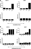
ArtB-induced changes in cell-bound adhesion molecules. (A) HCT-8 or U937 cells were treated with ArtB (10 μg/ml) for 1, 4, or 24 h or left untreated. (B) HCT-8 or U937 cells were treated with the indicated dose of ArtB for 24 h. The cell-bound adhesion molecules ICAM-1 and VCAM-1 were measured by direct probing with PE-conjugated adhesion molecule antibodies and analyzed by FACS. The data are the mean RFI, which were acquired from at least 5,000 cells. The results are representative of two independent experiments.
Fig 8
Fig 8
ArtB-induced changes in adhesion molecule mRNA. HCT-8 or U937 cells were treated with ArtB (10 μg/ml) for 4 h or left untreated. Total RNA was then extracted from cells, and adhesion molecule mRNAs (as indicated) were quantitated using PCR arrays (see Materials and Methods). The results are expressed as the fold change in [mRNA] relative to levels in control cells. The data are shown as the means of three independent experiments; *, significant difference relative to control cells (P < 0.05).

References

    1. Beddoe T, Paton AW, Le Nours J, Rossjohn J, Paton JC. 2010. Structure, biological functions and applications of the AB5 toxins. Trends Biochem. Sci. 35:411–418 - PMC - PubMed
    1. Paton AW, Srimanote P, Talbot UM, Wang H, Paton JC. 2004. A new family of potent AB5 cytotoxins produced by Shiga toxigenic Escherichia coli. J. Exp. Med. 200:35–46 - PMC - PubMed
    1. Paton AW, Beddoe T, Thorpe CM, Whisstock JC, Wilce MC, Rossjohn J, Talbot UM, Paton JC. 2006. AB5 subtilase cytotoxin inactivates the endoplasmic reticulum chaperone BiP. Nature 443:548–552 - PubMed
    1. Wolfson JJ, May KL, Thorpe CM, Jandhyala DM, Paton JC, Paton AW. 2008. Subtilase cytotoxin activates PERK, IRE1 and ATF6 endoplasmic reticulum stress-signalling pathways. Cell Microbiol. 10:1775–1786 - PMC - PubMed
    1. May KL, Paton JC, Paton AW. 2010. Escherichia coli subtilase cytotoxin induces apoptosis regulated by host Bcl-2 family proteins, Bax/Bak. Infect. Immun. 78:4691–4696 - PMC - PubMed

Publication types

MeSH terms