. 2013 Feb 15;112(4):618-32.
doi: 10.1161/CIRCRESAHA.112.300735. Epub 2012 Dec 18.

AKT2 confers protection against aortic aneurysms and dissections

Affiliations

AKT2 confers protection against aortic aneurysms and dissections

Ying H Shen et al. Circ Res. .

Abstract

Rationale: Aortic aneurysm and dissection (AAD) are major diseases of the adult aorta caused by progressive medial degeneration of the aortic wall. Although the overproduction of destructive factors promotes tissue damage and disease progression, the role of protective pathways is unknown.

Objective: In this study, we examined the role of AKT2 in protecting the aorta from developing AAD.

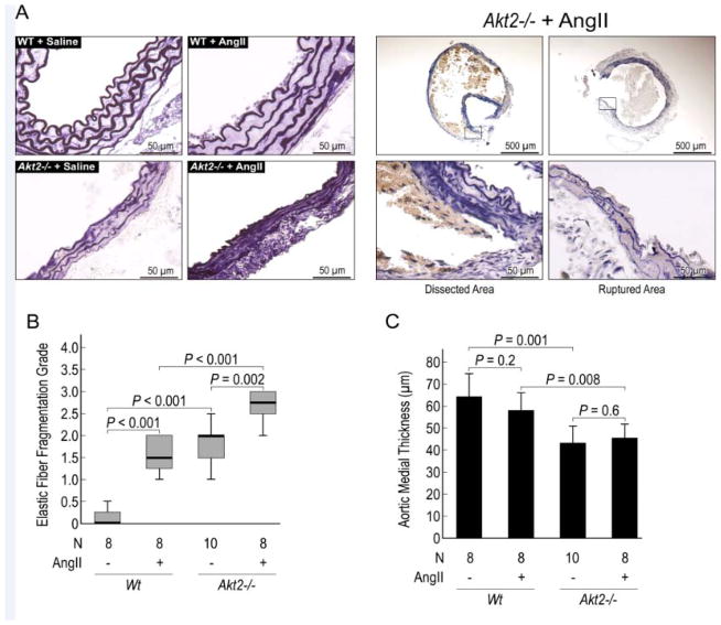
Methods and results: AKT2 and phospho-AKT levels were significantly downregulated in human thoracic AAD tissues, especially within the degenerative medial layer. Akt2-deficient mice showed abnormal elastic fibers and reduced medial thickness in the aortic wall. When challenged with angiotensin II, these mice developed aortic aneurysm, dissection, and rupture with features similar to those in humans, in both thoracic and abdominal segments. Aortas from Akt2-deficient mice displayed profound tissue destruction, apoptotic cell death, and inflammatory cell infiltration that were not observed in aortas from wild-type mice. In addition, angiotensin II-infused Akt2-deficient mice showed significantly elevated expression of matrix metalloproteinase-9 (MMP-9) and reduced expression of tissue inhibitor of metalloproteinase-1 (TIMP-1). In cultured human aortic vascular smooth muscle cells, AKT2 inhibited the expression of MMP-9 and stimulated the expression of TIMP-1 by preventing the binding of transcription factor forkhead box protein O1 to the MMP-9 and TIMP-1 promoters.

Conclusions: Impaired AKT2 signaling may contribute to increased susceptibility to the development of AAD. Our findings provide evidence of a mechanism that underlies the protective effects of AKT2 on the aortic wall and that may serve as a therapeutic target in the prevention of AAD.


Figures

Figure 1
Figure 1
Decreased phospho-AKT levels in the aortic wall of human aortic aneurysm and dissection (AAD) tissues. (A) Phospho-AKT, total-AKT, AKT1, and AKT2 proteins in human control aortas, thoracic aortic aneurysm (TAA) tissues, and thoracic aortic dissection (TAD) tissues were detected by Western blot by using anti phospho-AKT (Ser 473) and anti-AKT antibodies. Representative blots and quantification of the mean intensities of phospho-AKT, total AKT, AKT1, and AKT2 bands (normalized with those of β-actin) show decreased levels of AKT2 and phospho-AKT, as well as decreased ratios of phospho-AKT/total AKT in aortas from TAA and TAD patients. (B) Phospho-AKT (Ser 473) and total AKT proteins in control, TAA, and TAD tissues were detected by immunostaining. Representative images are shown. For each sample, the positive-staining areas for phospho-AKT and total AKT were measured in five randomly selected microscopic fields (magnification, 400×) and normalized with the total number of cells/nuclei in the counted area. (C) The mean normalized staining signals for phospho-AKT, total AKT, and the ratio of phospho-AKT to total AKT are shown.
Figure 2
Figure 2
Development of aortic aneurysms and dissections in Akt2/− mice infused with AngII. WT and Akt2/− mice were infused with saline or angiotensin II (AngII) for 4 weeks. (A) Representative excised aortas show that Akt2/− mice challenged with AngII developed aneurysms involving the thoracic and abdominal aorta. (B) The incidence of all aortic aneurysms and dissections in Akt/− and WT mice (P<0.001). (C) Histologically confirmed aortic dissection, with a characteristic true lumen (TL) and false lumen (FL), in AngII-infused Akt2−/− mice. (D) In an AngII-infused Akt2−/− mouse that suddenly died, median sternotomy (left panel) revealed that blood surrounded the heart and great vessels, which was caused by the rupture of an ascending aortic aneurysm (arrow, right panel). (E) Comparison of aortic diameter among saline or AngII-infused WT and Akt2/− mice. (F) The types of aortic lesions, according to a modified classification system based on that of Daugherty and colleagues, are shown.
Figure 3
Figure 3
Significantly increased elastic fiber destruction and reduced medial thickness in angiotensin II (AngII)-infused Akt2/− mice. (A) Representative histologic sections of the aorta (Verhoeff elastic staining) demonstrating abnormalities in elastic lamellar architecture in saline-infused Akt2/− mice and marked elastin destruction in AngII-infused Akt2/− mice. Comparisons of (B) elastic fiber fragmentation scores (Grade 0 = none; Grade 1 = minimal; Grade 2 = moderate; Grade3 = severe) and (C) medial thickness among groups are shown.
Figure 4
Figure 4
Significantly increased apoptosis and inflammatory cell infiltration in angiotensin II (AngII)-infused Akt2/− mice. (A) Representative terminal deoxynucleotidyl transferase dUTP nick end labeling (TUNEL)-stained images and comparison of the number of TUNEL-positive cells normalized to the total number of cells are shown. The number of TUNEL-positive cells was significantly increased in the lesion areas of AngII-infused Akt2/− mice, particularly in dissected areas. Cleaved caspase 3 (B) and apoptosis inducing factor (AIF) (C) were detected the aortas of AngII-infused Akt2/− mice. (D) Representative images of immunofluorescence staining and quantification (mean positive staining area normalized with evaluated aortic area) of CD68-positive macrophages in the aortic wall show a significant increase in macrophages in AngII-infused Akt2/− mice.
Figure 5
Figure 5
Significantly increased MMP-9 expression and MMP-9/TIMP-1 ratio in angiotensin II (AngII)-infused Akt2/− mice. (A) Representative images of immunofluorescence staining and quantification of the mean positive staining area normalized with the evaluated aortic area show increased MMP-9 expression in aortas from AngII-infused Akt2/− mice. Double staining for MMP-9 and CD68 or MMP-9 and SM22α shows increased MMP-9 expression in (B) CD68-positive macrophages and in (C) SM22α-positive SMCs in the aortas of AngII-infused Akt2/− mice. (D) Representative images of immunofluorescence staining and quantification of TIMP-1 expression (the mean positive-staining area normalized with the evaluated area) shows lower TIMP-1 expression in aortas from AngII-infused Akt2/− mice than in AngII-infused WT mice. (E) Double staining for MMP-9 and TIMP-1 in an aorta from an AngII-infused Akt2/−mouse. Comparison shows increased MMP-9/TIMP-1 ratios in aortas from AngII-infused Akt2/− mice. (F) Representative zymography images show increased MMP activity in aortas from AngII-infused Akt2/− mice.
Figure 6
Figure 6
Effects of AKT2 on TIMP-1 and MMP-9 expression. (A) Western blot analysis of TIMP-1 and MMP-9 protein levels in aortic vascular smooth muscle cells (VSMCs) that were transfected with wild-type AKT2 (WT-AKT2), or AKT2 shRNA. Representative blots from 3 independent experiments are shown. Quantification of the mean intensities of TIMP-1 and MMP-9 bands (normalized with those of β-actin) are shown. (B) TIMP-1 and MMP-9 mRNA levels were examined by reverse transcription and quantitative real-time PCR in VSMCs transfected with WT-AKT2 or AKT2 shRNA. TIMP-1 and MMP-9 mRNA expression was normalized with that of β-actin and is expressed as the percentage of the control. Data represent the mean ± SD (N=3).
Figure 7
Figure 7
The effects of FOXO1 on MMP-9 and TIMP-1 expression. Aortic vascular smooth muscle cells (VSMCs) were transfected with plasmids expressing wild-type (WT)-FOXO1, constitutively active (CA)-FOXO1, dominant negative (DN)-FOXO1, or FOXO1 siRNA, and (A) MMP-9 and TIMP-1 protein expression was examined by Western blot. Representative blots from 3 independent experiments are shown. Quantification of the mean intensities of TIMP-1 and MMP-9 bands (normalized with those of β-actin) are shown. (B) MMP-9 and TIMP-1 mRNA levels were examined by reverse transcription and quantitative real-time PCR and were normalized with levels of β-actin mRNA. Levels of mRNA are expressed as the percentage of the control. Data represent the mean ± SD (N=3). (C) Diagram showing the location of FOXO binding sites in the human MMP-9 promoter. (D) ChIP analysis showing that FOXO-1 bound to FOXO binding site 2 of the MMP-9 promoter in aortic VSMCs transfected with WT-FOXO1 and CA-FOXO1 but not DN-FOXO1. FOXO1-DNA complexes were cross-linked by formaldehyde and immunoprecipitated with anti-FOXO1 antibody. Sites in the MMP-9 promoter bound by FOXO1 were detected by PCR and normalized with input DNA. Representative blots from 2 independent experiments are shown. (E) Diagram showing the location of FOXO and GATA1 binding sites in the human TIMP-1 promoter. (F) ChIP analysis showing that FOXO-1 bound to the TIMP-1 promoter in aortic VSMCs transfected with WT-FOXO1 and CA-FOXO1 but not DN-FOXO1. FOXO1-DNA complexes were cross-linked and immunoprecipitated with anti-FOXO1 antibody and anti-GATA1 antibody. Sites in the TIMP-1 promoter bound by FOXO1 and GATA1 were detected by PCR and normalized with input DNA. Representative blots from 2 independent experiments are shown.
Figure 8
Figure 8
Inhibition of FOXO1-mediated MMP-9 expression by AKT2. Vascular smooth muscle cells were transfected with wild-type (WT)-FOXO1 in the presence of WT-AKT2 and AKT2 shRNA. (A) MMP-9 and TIMP-1 protein levels were examined by Western blot. Representative blots from 3 independent experiments are shown. Quantification of the mean intensities of TIMP-1 and MMP-9 bands (normalized with those of β-actin) are shown. (B) MMP-9 and TIMP-1 mRNA levels were examined by reverse transcription and quantitative real-time PCR and normalized with those of β-actin mRNA. The relative levels of mRNA are expressed as the percentage of the control. Data represent the mean ± SD (N=3). (C) FOXO1 binding to the MMP-9 promoter was examined by ChIP analysis. (D) FOXO1 and GATA1 binding to the TIMP-1 promoter was examined by ChIP analysis. Representative blots from 2 independent experiments are shown. (E) Immunostaining showed increased FOXO1 nuclear translocation in SMCs transfected with Akt2 siRNA or treated with Wortmannin. Immunostaining showed increased FOXO1 expression and nuclear localization in (F) aortas of AngII-infused Akt2/− mice and (G) degenerative aortic media in human thoracic aortic aneurysm (TAA) tissue.

Comment in

References

    1. Kochanek KD, Kirmeyer SE, Martin JA, Strobino DM, Guyer B. Annual summary of vital statistics: 2009. Pediatrics. 2012;129:338–348. - PMC - PubMed
    1. Lopez-Candales A, Holmes DR, Liao S, Scott MJ, Wickline SA, Thompson RW. Decreased vascular smooth muscle cell density in medial degeneration of human abdominal aortic aneurysms. Am J Pathol. 1997;150:993–1007. - PMC - PubMed
    1. Schlatmann TJ, Becker AE. Pathogenesis of dissecting aneurysm of aorta. Comparative histopathologic study of significance of medial changes. Am J Cardiol. 1977;39:21–26. - PubMed
    1. Manning BD, Cantley LC. Akt/pkb signaling: Navigating downstream. Cell. 2007;129:1261–1274. - PMC - PubMed
    1. Shiojima I, Walsh K. Role of akt signaling in vascular homeostasis and angiogenesis. Circ Res. 2002;90:1243–1250. - PubMed

Publication types

MeSH terms