. 2013 Jan 2;110(1):318-23.
doi: 10.1073/pnas.1212708110. Epub 2012 Dec 18.

Atomic model of the human cardiac muscle myosin filament

Affiliations

Atomic model of the human cardiac muscle myosin filament

Hind A Al-Khayat et al. Proc Natl Acad Sci U S A. .

Abstract

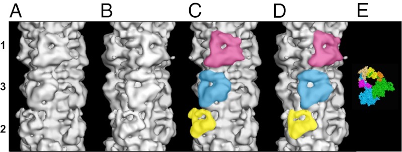
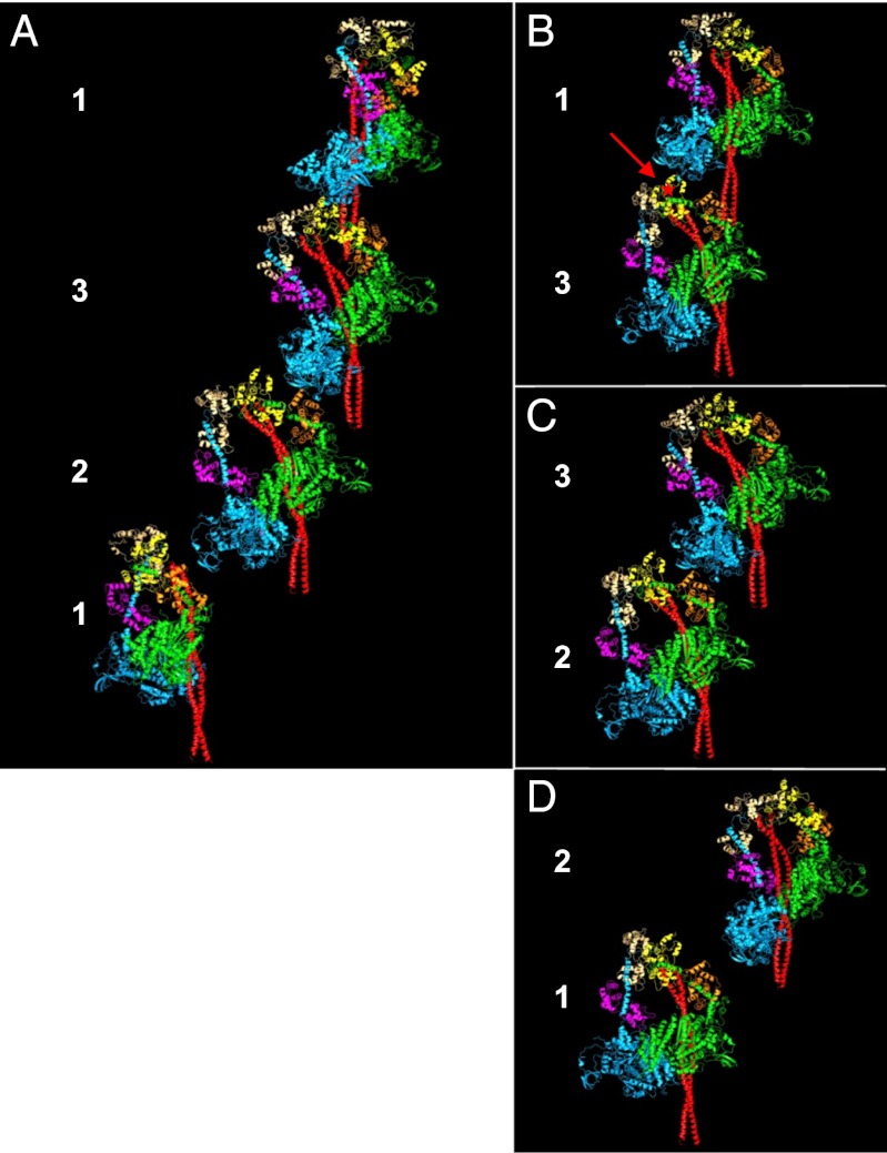
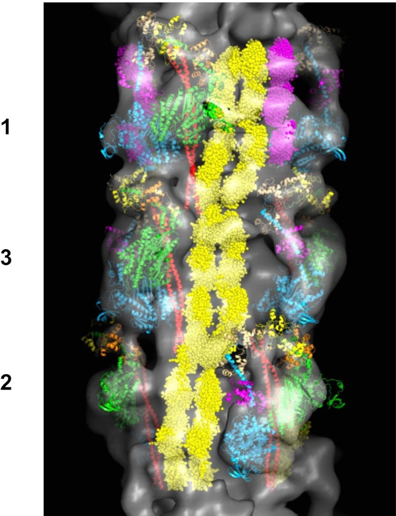
Of all the myosin filaments in muscle, the most important in terms of human health, and so far the least studied, are those in the human heart. Here we report a 3D single-particle analysis of electron micrograph images of negatively stained myosin filaments isolated from human cardiac muscle in the normal (undiseased) relaxed state. The resulting 28-Å resolution 3D reconstruction shows axial and azimuthal (no radial) myosin head perturbations within the 429-Å axial repeat, with rotations between successive 132 Å-, 148 Å-, and 149 Å-spaced crowns of heads close to 60°, 35°, and 25° (all would be 40° in an unperturbed three-stranded helix). We have defined the myosin head atomic arrangements within the three crown levels and have modeled the organization of myosin subfragment 2 and the possible locations of the 39 Å-spaced domains of titin and the cardiac isoform of myosin-binding protein-C on the surface of the myosin filament backbone. Best fits were obtained with head conformations on all crowns close to the structure of the two-headed myosin molecule of vertebrate chicken smooth muscle in the dephosphorylated relaxed state. Individual crowns show differences in head-pair tilts and subfragment 2 orientations, which, together with the observed perturbations, result in different intercrown head interactions, including one not reported before. Analysis of the interactions between the myosin heads, the cardiac isoform of myosin-binding protein-C, and titin will aid in understanding of the structural effects of mutations in these proteins known to be associated with human cardiomyopathies.


Conflict of interest statement

The authors declare no conflict of interest.

Figures

Fig. 1.
Fig. 1.
(AD) Surface views of the final 3D reconstruction of the human cardiac myosin filament obtained by single-particle EM analysis and displayed using PyMOL showing a length of a full 429-Å repeat. The reconstruction is shown in two views (A and C; B and D) related by a 25° rotation about the filament axis, such that in A and C, a myosin head pair on level 1 is facing the viewer, and in B and D, a myosin head pair on level 3 is facing the viewer. Note that the two views in A and C and in B and D are quite distinct because of the different perturbations in the crowns. The triangular-shaped configurations for the three head pairs on each crown level are shown in yellow, blue, and pink for levels 2, 3, and 1, respectively, in C and D. The bare zone is at the bottom of the map in all of the four views. (E) Crystal structure for the head pair (11). The blocked and free heads are color-coded as in refs. –. Blocked head: motor domain, green; essential light chain, orange; regulatory light chain, yellow. Free head: motor domain, cyan; essential light chain, pink; regulatory light chain, beige.
Fig. 2.
Fig. 2.
(A) Three-dimensional map of the view in Fig. 1B showing a full 429-Å repeat together with fitting of the atomic model of myosin heads in the off state to the head motifs in crowns 2, 3, and 1 after splitting and fitting separately the two heads from Alamo et al. (10). The majority of the atomic structures for the head pairs is well-contained within the density map on crowns 3 and 1 but less on crown 2, especially for the two lever-arm parts. The S2 is also fitted, as described in the text. (B) Same view as in A, but shown without the back part of the map to illustrate the fitting of the heads and S2 within the densities on levels 2, 3, and 1. S2 is shown in red and the bare-zone direction is toward the bottom.
Fig. 3.
Fig. 3.
(A) Fitted crystal structures showing head pairs and S2 on successive crowns along one strand of the myosin filament. Head pairs in the 2,3,1 crown sequence are quite closely connected, whereas there is a distinct gap between crowns 1 and 2. (B) Head pairs on crowns 3 and 1 are connected through an interaction between the RLC of the blocked head on crown 3 and the motor domain of the free head on crown 1. The red arrowed star indicates the predicted position of serine 15 in human myosin RLC. (C) A different type of intermolecular connection is observed between crowns 3 and 2 that is mediated by an interaction between the ELC of the blocked head on crown 2 and the motor domain of the free head on crown 3. (D) No intermolecular interaction is seen between the head pairs in crowns 1 and 2. S2 is shown in red, and the bare zone is oriented vertically down in all views.
Fig. 4.
Fig. 4.
Final 3D map (Fig. 1) showing a full 429-Å repeat, shown with myosin head pairs, S2 (red), titin (yellow), and the three C-terminal domains of cMyBP-C (C8–C10) (magenta). The bare-zone direction is toward the bottom.

References

    1. Huxley HE. Electron microscope studies on the structure of natural and synthetic protein filaments from striated muscle. J Mol Biol. 1963;7:281–308. - PubMed
    1. Squire JM. General model of myosin filament structure. II. Myosin filaments and cross-bridge interactions in vertebrate striated and insect flight muscles. J Mol Biol. 1972;72(1):125–138. - PubMed
    1. Labeit S, Kolmerer B. Titins: Giant proteins in charge of muscle ultrastructure and elasticity. Science. 1995;270(5234):293–296. - PubMed
    1. Trinick J. Titin as a scaffold and spring. Cytoskeleton. Curr Biol. 1996;6(3):258–260. - PubMed
    1. Offer G, Moos C, Starr R. A new protein of the thick filaments of vertebrate skeletal myofibrils. Extractions, purification and characterization. J Mol Biol. 1973;74(4):653–676. - PubMed

Publication types