Peripartum cardiomyopathy presenting with predominant left ventricular diastolic dysfunction: efficacy of bromocriptine
- PMID: 23251175
- PMCID: PMC3521613
- DOI: 10.1155/2012/476903
Peripartum cardiomyopathy presenting with predominant left ventricular diastolic dysfunction: efficacy of bromocriptine
Abstract
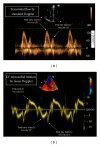
Management of patients with peripartum cardiomyopathy (PPCM) is still a major clinical problem, as only half of them or slightly more show complete recovery of left ventricular (LV) function despite conventional evidence-based treatment for heart failure. Recent observations suggested that bromocriptine might favor recovery of LV systolic function in patients with PPCM. However, no evidence exists regarding its effect on LV diastolic dysfunction, which is commonly observed in these patients. Tissue Doppler (TD) is an echocardiographic technique that provides unique information on LV diastolic performance. We report the case of a 37-year-old white woman with heart failure (NYHA class II), moderate LV systolic dysfunction (ejection fraction 35%), and severe LV diastolic dysfunction secondary to PPCM, who showed no improvement after 2 weeks of treatment with ramipril, bisoprolol, and furosemide. At 6-week followup after addition of bromocriptine, despite persistence of LV systolic dysfunction, normalization of LV diastolic function was shown by TD, together with improvement in functional status (NYHA I). At 18-month followup, the improvement in LV diastolic function was maintained, and normalization of systolic function was observed. This paper might support the clinical utility of bromocriptine in patients with PPCM by suggesting a potential benefit on LV diastolic dysfunction.
Figures


Similar articles
-
Cabergoline treatment promotes myocardial recovery in peripartum cardiomyopathy.ESC Heart Fail. 2023 Feb;10(1):465-477. doi: 10.1002/ehf2.14210. Epub 2022 Oct 27. ESC Heart Fail. 2023. PMID: 36300679 Free PMC article.
-
Initial Right Ventricular Dysfunction Severity Identifies Severe Peripartum Cardiomyopathy Phenotype With Worse Early and Overall Outcomes: A 24-Year Cohort Study.J Am Heart Assoc. 2018 Apr 23;7(9):e008378. doi: 10.1161/JAHA.117.008378. J Am Heart Assoc. 2018. PMID: 29686029 Free PMC article.
-
Clinical features differentiating Takotsubo cardiomyopathy in the peripartum period from peripartum cardiomyopathy.Heart Vessels. 2020 May;35(5):665-671. doi: 10.1007/s00380-019-01537-4. Epub 2019 Nov 9. Heart Vessels. 2020. PMID: 31705186
-
Clinical aspects of left ventricular diastolic function assessed by Doppler echocardiography following acute myocardial infarction.Dan Med Bull. 2001 Nov;48(4):199-210. Dan Med Bull. 2001. PMID: 11767125 Review.
-
A Systematic Review of the Utility of Bromocriptine in Acute Peripartum Cardiomyopathy.Cureus. 2021 Sep 24;13(9):e18248. doi: 10.7759/cureus.18248. eCollection 2021 Sep. Cureus. 2021. PMID: 34603902 Free PMC article. Review.
Cited by
-
Peripartum cardiomyopathy and dilated cardiomyopathy: different at heart.Front Physiol. 2015 Jan 15;5:531. doi: 10.3389/fphys.2014.00531. eCollection 2014. Front Physiol. 2015. PMID: 25642195 Free PMC article. Review.
-
Bromocriptine Use in Peripartum Cardiomyopathy: Review of Cases.AJP Rep. 2018 Oct;8(4):e335-e342. doi: 10.1055/s-0038-1675832. Epub 2018 Nov 21. AJP Rep. 2018. PMID: 30473907 Free PMC article.
-
Peripartum cardiomyopathy: a review.Heart Fail Rev. 2021 Nov;26(6):1287-1296. doi: 10.1007/s10741-020-10061-x. Epub 2021 Jun 17. Heart Fail Rev. 2021. PMID: 34138401 Free PMC article. Review.
-
Analysis of Clinical Profiles and Echocardiographic Cardiac Outcomes in Peripartum Cardiomyopathy (PPCM) vs. PPCM with Co-Existing Hypertensive Pregnancy Disorder (HPD-PPCM) Patients: A Systematic Review and Meta-Analysis.J Clin Med. 2023 Aug 15;12(16):5303. doi: 10.3390/jcm12165303. J Clin Med. 2023. PMID: 37629345 Free PMC article. Review.
References
-
- Elkayam U. Clinical characteristics of peripartum cardiomyopathy in the United States: diagnosis, prognosis, and management. Journal of the American College of Cardiology. 2011;58(7):659–670. - PubMed
-
- Sliwa K, Fett J, Elkayam U. Peripartum cardiomyopathy. Lancet. 2006;368(9536):687–693. - PubMed
-
- Biteker M, Ilhan E, Biteker G, Duman D, Bozkurt B. Delayed recovery in peripartum cardiomyopathy: an indication for long-term follow-up and sustained therapy. European Journal of Heart Failure. 2012;14(8):895–901. - PubMed
-
- Goland S, Bitar F, Modi K, et al. Evaluation of the clinical relevance of baseline left ventricular ejection fraction as a predictor of recovery or persistence of severe dysfunction in women in the United States with peripartum cardiomyopathy. Journal of Cardiac Failure. 2011;17(5):426–430. - PubMed
LinkOut - more resources
Full Text Sources

